NCERT Solutions for Class 11 Biology Chapter 17 Breathing and Exchange of Gases
These Solutions are part of NCERT Solutions for Class 11 Biology. Here we have given NCERT Solutions for Class 11 Biology Chapter 17 Breathing and Exchange of Gases.
Question 1.
Define vital capacity. What is its significance?
Solution:
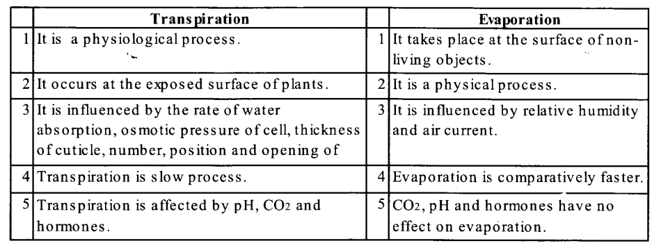
The maximum volume of air a person can breathe in after a forced expiration it is about 4000mL in a normal adult person. Vital capacity is higher in athletes and singers. Cigarette smokers have a lower vital capacity of the lungs. This includes ERC, TV, and IRV, or the maximum volume of air a person can breathe out after a forced inspiration.
Question 2.
State the volume of air remaining in the lungs after a normal breathing.
Solution:
The volume of air remaining in the lungs after normal respiration is called functional residual capacity. It includes ERV + RV = Expiratory reserve volume + residual volume
Question 3.
volume Diffusion of gases occurs in the alveolar region only and not in the other parts of the respiratory system. Why?
Solution:
Alveoli are the primary sites of the exchange of gases. The alveolar region is having enough pressure gradient to facilitate the diffusion of gases. Other regions of the respiratory system don’t have the required pressure gradient. Additionally, the membrane of alveoli is thin enough to facilitate the exchange of gases in a convenient manner.
Question 4.
What are the major transport mechanisms for CO2? Explain.
Solution:
Transport of carbon dioxide: About 4 ml of carbon dioxide is transported by every 100 ml of blood.
C02 is transported in three forms in the blood.
(i) In the dissolved form in plasma about 7% of C02 dissolves in the plasma of blood, just as it gets dissolved in water.
(ii) As bicarbonates
Erythrocytes have a high concentration of the enzyme, carbonic anhydrase which catalyzes the following reactions;
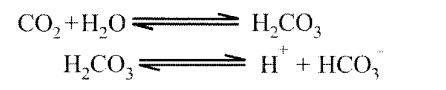
About 70% of C02 is transported as bicarbonates.
(iii) As carbaminohaemoglobin
C02 combines with the globin part of haemoglobin and forms carbamino haemoglobin. About 23% of CO2 transported in this manner.
Question 5.
What will be the pO2 and pCO2 in the atmospheric air compared to those in the alveolar air?
(i) pO2 lesser, pCO2higher
(ii) pO2 higher, pCO2lesser
(iii) pO2 higher, pCO2 higher
(iv) pO2 lesser, pCO2lesser
Solution:
(ii) pO2 higher, pCO2 lesser
pO2 higher will create the pressure gradient to facilitate the movement of O2 from atmosphere to alveoli and pCO2 lesser will create the movement of CO2 from alveoli to atmosphere.
Question 6.
Explain the process of inspiration under normal conditions.
Solution:
The intake of air into the lungs is known as inspiration. Inspiration occurs when the pressure within the lungs is less than the atmospheric pressure. It is initiated by the contraction of the diaphragm which increases the volume of the thoracic chamber in the anteroposterior axis. The contraction of external intercostal muscles lifts up the ribs and the sternum causing an increase in the volume of the thoracic chamber in the dorsoventral axis. The overall increase in the thoracic volume causes a similar increase in pulmonary volume which decreases the intra-pulmonary pressure to less than the atmospheric pressure which forces the air from outside to move into the lungs.
Question 7.
How is respiration regulated?
Solution:
- Respiratory rhythm centre, present in medulla region of brain is responsible for respiration regulation.
- Its function can be moderate by pneumotaxic centre, present in pons region of brain.
- A chemosensitive area present adjacent to rhythm centre, is highly sensitive to C02 and H+.
- Chemosenstive centre due to increase in C02 and H+ can signal the rhythm centre to make adjustment to eliminate these substances.
- Receptors associated with aortic arch and carotid artery also can recognise changes in CO2 and H+ concentration and send necessary signals to rhythm centre for remedial actions.
Question 8.
What is the effect of pC02 on oxygen transport?
Solution:
At low pC02, blood can carry the maximum amount of oxygen as oxyhemoglobin. At high pCO2, the affinity for oxygen decreases and oxyhaemoglobin dissociates to free oxygen.
So at high pC02, oxygen transport is inhibited.
Question 9.
What happens to the respiratory process in a man going up a hill?
Solution:
There is a fall of PO2 level at high altitudes. This lowers alveolar PO2 and consequently reduces the diffusion of oxygen from the alveolar air to the blood. So oxygenation of the blood is decreased progressively.
After some time, the affected person gets adjusted to the surroundings due to which the heart rate is accelerated, RBC count in the blood is increased, haemoglobin level and oxygen-carrying capacity are also increased.
Question 10.
What is the site of gaseous exchange in an insect?
Solution:
Insects have a complex system of intercommunicating air tubes called tracheae to enable them to exchange gases between the environment and the body cells (tracheal respiration).
Question 11.
Define oxygen dissociation curve. Can you suggest any reason for its sigmoidal pattern?
Solution:
The curve in which the percentage saturation of hemoglobin with O2 is plotted against the partial pressure of oxygen (PO2) is called the oxygen dissociation curve. At a PO2 of 100 mm Hg, 100 percent saturation of Hb takes place 90% saturation of Hb takes place even at a P02 of 60mm Hg. An I fall of PCX, from 100 to 60mm Hg will cause only 10% decrease in saturation of Hb. Hence the curve takes the shape of a sigmoid.
Question 12.
Have you heard about hypoxia? Try to gather information about it, and discuss it with your friends.
Solution:
Hypoxia is the condition in which there is a deficiency of oxygen at the tissue level.
- Arterial hypoxia: It’s because of low level of oxygen in the blood. It occurs when the atmosphere does not contain enough oxygen and there is obstruction in the respiratory passage.
- Anaemic hypoxia: It is due to very low level of haemoglobin in the blood.
- Stagnant hypoxia: It is due to the inadequate blood flow to deliver oxygen to the tissue.
- Histoxic hypoxia: It is due to the presence of toxic substances in the oxygen inhaled, e.g.: Cyanide poisoning.
Question 13.
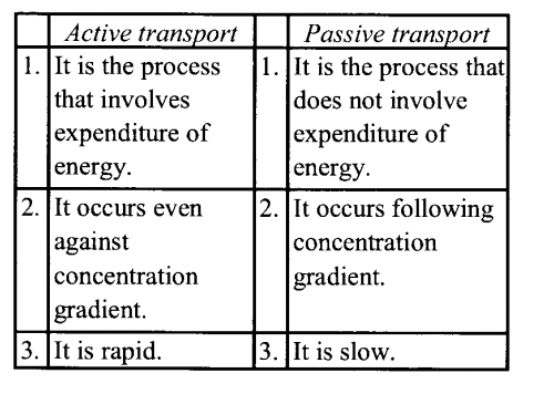
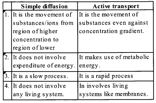
Distinguish between
(a) IRV and ERV
(b) Inspiratory capacity and Expiratory capacity.
(c) Vital capacity and Total lung capacity.
Solution:
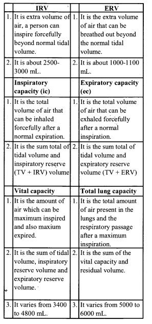
Question 14.
What is tidal volume? Find out the tidal volume (approximate value) for a healthy human in an hour.
Solution:
The volume of air inspired or expired/breath during normal respiration is approx. 500 ml., i.e., a healthy man can inspire or expire approximately 6000 to 8000 ml of air per minute.
VERY SHORT ANSWER QUESTIONS
Question 1.
Name the enzyme which catalyses the bicarbonate formation in RBCs.
Solution:
Enzyme carbonic anhydrase.
Question 2.
What is carbamino haemoglobin?
Solution:
It is a complex formed by the combination of carbon dioxide with the globin part of haemoglobin.
Question 3.
What is tidal volume?
Solution:
The volume of air inspired or expired with every normal breath during effortless respiration is called tidal volume.
Question 4.
What term is used for the volume of air left in the lungs even after the most powerful expiration?
Solution:
Residual volume.
Question 5.
Name the respiratory organ of
(a) Butterfly
(b) Frog larva.
Solution:
(i) Butterfly – trachea
(ii) Frog larva – gills.
Question 6.
What is the role of oxyhaemoglobin after releasing molecular oxygen in the tissue?
Solution:
Oxyhaemoglobin after releasing oxygen collects carbon dioxide from the tissue and form carbaminohaemoglobin.
Question 7.
What are the two factors that contribute to the dissociation of oxyhaemoglobin in the atrial blood to release’ molecular oxygen in active tissue?
Solution:
The two factors are :
(a) Lowp02
(b) HighpC02
Question 8.
Name the double-walled sac which covers the lungs in mammals.
Solution:
Pleura.
Question 9.
What prevents the collapsing of our trachea during breathing ?
Solution:
C-shaped cartilages at regular intervals.
Question 10.
Define inspiratory reserve volume.
Solution:
The extra volume of air that can be inspired beyond the normal tidal volume, is called inspiratory reserve volume.
Question 11.
Which part(s) of the brain control(s) breathing movements?
Solution:
Medulla and pons.
Question 12.
What is oxyhaemoglobin?
Solution:
Oxyhaemoglobin is a complex formed when oxygen combies with the Fe2+ part of haemoglobin.
Question 13.
How much of oxygen is transported by 100 ml of blood under normal physiological conditions?
Solution:
About 5 mL.
Question 14.
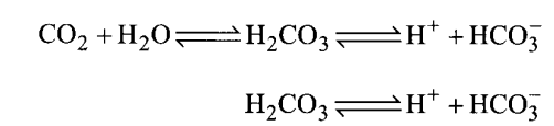
Write the chemical reaction catalysed by enzyme carbonic anhydrase.
Solution:
Carbonic anhydrase catalyses the following reaction:
Question 15.
How does penumotaxic centre alter the respiratory rate?
Solution:
Pneumotaxic centre can reduce the duration of inspiration and alter the respiratory rate.
Question 16.
What is the percentage of C02 transported as sodium bicarbonate?
Solution:
70% of C02 is transported as sodium bicarbonate.
Question 17.
What will happen if the human blood becomes acidic?
Solution:
Oxygen carrying capacity of haemoglobin will decrease.
SHORT ANSWER QUESTIONS
Question 1.
Write four conditions necessary to facilitates efficient gaseous exchange between human respiratory surface and the environment.
Solution:
Conditions for efficient gas exchange are as follows:
(a) The membrane should be thin.
(b) It should be highly vascularized.
(c) It should be highly permeable to gases.
(d) There should a partial pressure difference on both sides of lung.
Question 2.
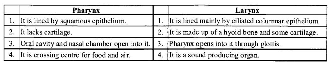
Differentiate between pharynx and larynx.
Solution:
The main differences between the pharynx and larynx are as follows:
Question 3.
Why is hemoglobin called a conjugated protein? What happens to the molecule at a high and low partial pressure of oxygen?
Solution:
Hemoglobin: It is called conjugated protein because it consists of a basic protein globin and a non-protein heme.
The haemoglobin when exposed to the high partial pressure of oxygen combines with oxygen to form oxyhaemoglobin which carries 4 molecules of oxygen loosely bound to the four Fe2+ ions. When this oxyhaemoglobin reaches the tissues where there is low oxygen pressure oxyhaemoglobin dissociates into oxygen and deoxyhemoglobin.
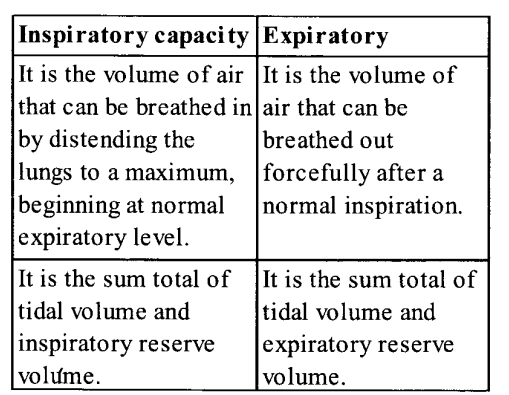
Question 4.
Differentiate between inspiratory capacity and expiratory capacity.
Solution:
The differences between inspiratory and expiratory capacity are :
Question 5.
Diffusion of gases occurs in the alveolar region only and not in the other parts of the respiratory system. Why?
Solution:
The alveoli have very thin walls consisting of squamous epithelium. The alveolar wall is provided with an extensive network of blood capillaries; due to the intimate contact of the blood capillaries and alveolar wall, there is an exchange of gases taking place easily. In the other part the membrane/wall is not so thin to allow for diffusion.
Question 6.
What percentage of oxygen is transported by erythrocytes in the blood? What happens to the remaining?
Solution:
About 97% of the oxygen is transported by erythrocytes. The remaining 3% is transported in dissolved form in the plasma.
Question 7.
What is asthma? Explain.
Solution:
Asthma: It is the hypersensitivity of bronchioles to any foreign substance, characterized by the spasm of the smooth muscles of the walls of the bronchioles.
Question 8.
What is emphysema? What is its major cause?
Solution:
Emphysema: Emphysema is a chronic disorder is which alveolar walls are damaged and hence the surface area for exchange of. gases is reduced. It is caused mainly by cigarette smoking.
Question 9.
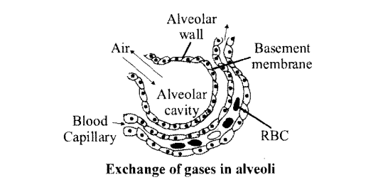
Draw a labelled diagram of a section of an alveolus with a pulmonary capillary.
Solution:
Question 10.
What will happen if the patient has been inhaling polluted air containing high content of CO?
Solution:
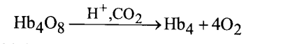
Hemoglobin has much more affinity about 250 times for CO than oxygen. It readily combines with CO to form the most stable compound called carboxyhemoglobin. It may be fatal for the patient.
Question 11.
What is pneumonia? What are its causes?
Solution:
Pneumonia is a respiratory disease in which oxygen has difficulty in diffusing through the flammed alveoli and the blood O2 may be drastically reduced and blood PCO2 remains normal. It is caused by streptococcus pneumonia. Its symptoms are trembling, pain in the chest, fever, cough, etc. It is mostly observed in children and old age.
LONG ANSWER QUESTIONS
Question 1.
What do you mean by occupational lung disease? Enumerate the prevention measure that should be adopted by a person likely to be exposed to substances that cause occupational diseases?
Solution:
Occupational lung disease as the name suggests it is the disease of lung due to the occupation of the human.
Cause: These are caused by harmful substances, such as gas fumes or dust, present in the environment where a person works. Silicosis and asbestoses are common examples, which occur due to chronic exposure of silica and asbestos dust in the mining industry.
Symptoms: It is characterized by the proliferation of fibrous connective tissue (fibrosis) of upper part of lung, causing inflammation.
Prevention: The occupational disease expresses symptoms after chronic exposure (i.e., 10-15 years or even more). Most of the occupational diseases including silicosis and asbestosis is are incurable. Therefore, the person which is exposed to such irritants should adopt preventive measures. These protective measures are as follows:
(i) Minimize the exposure of harmful dust at the workplace.
(ii) Workers should be informed about the harm of exposure to such dust.
(iii) Workers must have protective gear and clothing at the workplace.
(iv) health of the workers should be regularly checked up.
Question 2.
Explain the regulation of respiration by the nervous system.
Solution:
Regulation of respiratory rhythm
- The ability to maintain and moderate the respiratory rhythm according to the demand of the body tissues is due to neural control.
- The respiratory rhythm centre located in the medulla of the brain, is primarily responsible for this regulation, f Pneumotaxic centre present in the brain functions as the ‘switch off point for regulation; by altering the duration of inspiration, it can alter the respiratory rate.
- A chemosensitive area is situated adjacent to the rhythm centre; it is highly sensitive to carbon dioxide and hydrogen ions.
- An increase in the concentration of these substances activates this centre which in turn sends signals to the rhythm centre to make necessary adjustments in the respiratory process.
- Receptors associated with aortic arch and carotid artery also are sensitive to carbon di oxide and H+ ions; they too send signals to the respiratory rhythm centre.
- Oxygen plays only an insignificant role in the regulation of respiratory rhythm.
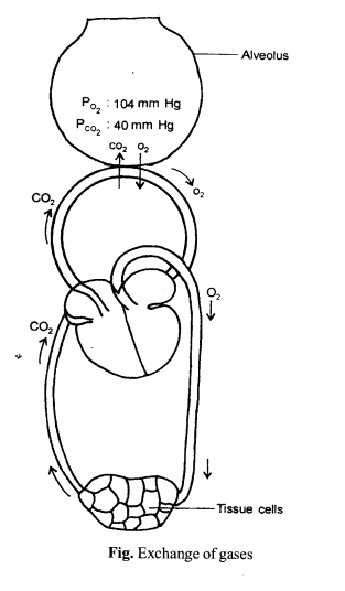
Question 3.
How does the exchange of respiratory gases take place in the alveoli or lungs?
Solution:
Gaseous exchange in alveoli:
- The alveolar wall is very thin and contains a rich network of interconnected capillaries.
- Due to this, the alveolar wall seems to be a sheet of flowing blood and is called the respiratory membrane.
- It consists mainly of the alveolar epithelium, epithelial basement membrane, a thin interstitial space, capillary basement membrane, and capillary endothermal membrane. All these layers cumulatively form a membrane of 0.2 mm thickness.
- The respiratory membrane has a limit of gas exchange between alveoli and pulmonary blood. It is called diffusing capacity. It is dependent on the solubility of respiratory gases.
- The partial pressure of oxygen (p02) in the alveoli is higher (104 mm Hg) than that in the deoxygenated blood in the capillaries of the pulmonary arteries (40 mm Hg). As the gases diffuse from higher to a lower concentration, the movement of oxygen is from the alveoli to the blood. The reverse is the case in relation to carbon dioxide.
- The partial pressure of carbon dioxide (pC02) is higher in deoxygenated blood (45 mm Hg), than in alveoli (40 mm Hg), therefore, CO2 passes from the blood to the alveoli.
Question 4.
How are inspiration and expiration take place in humans?
Solution:
The inflow (inspiration) and outflow (expiration) of air occur between the atmosphere and lungs by the expansion and contraction of lungs.
Inspiration: It is the process by which fresh air enters the lungs.
- The external intercostal muscles present between the ribs contract and pull these ribs and sternum upward and outward increasing the volume to the thoracic cavity.
- The diaphragm becomes flats and gets lowered by the contraction of its muscles thereby increasing the volume of the thoracic cavity.
- The abdominal muscles relax and allow the compression of the abdominal organ by the diaphragm.
- As the volume of the thoracic cavity increases and as a result, there is a decrease in air pressure in the lungs. The greater pressure outside the body causes air to flow rapidly into the nostrils through the respiratory tract to the lungs.
Expiration: It is a process by which foul air (CO2) is expelled out from the lungs.
- Internal intercostal muscles contract so that they pull in ribs downward and inward decreasing the size of the thoracic cavity.
- The muscle fibres of the diaphragm relax making it convex, decreasing the volume of the thoracic cavity.
- Contraction of abdominal muscles compresses this abdomen and pushes it towards the diaphragm.
The overall volume of the thoracic cavity decrease and foul air goes outside from the cavities of alveoli through the respiratory tract.
We hope the NCERT Solutions for Class 11 Biology at Work Chapter 17 Breathing and Exchange of Gases, help you. If you have any query regarding NCERT Solutions for Class 11 Biology at Work Chapter 17 Breathing and Exchange of Gases, drop a comment below and we will get back to you at the earliest.