NCERT Solutions for Class 11 Biology Chapter 21 Neural control and co-ordination
These Solutions are part of NCERT Solutions for Class 11 Biology. Here we have given NCERT Solutions for Class 11 Biology Chapter 21 Neural control and co-ordination.
Question 1.
Briefly describe the structure of the following:
(a) Brain
(b) Eye
(c) Ear
Solution:
Structure of brain:
- The brain is the central information processing organ of the body,
- The human brain is well protected by the skull.
- Inside the skull, cranial meninges cover the brain. These are tough tissue layers.
Meninges consist of 3 layers which are as follows :
- The outermost – layer is the dura mater
- The middle layer is arachnoid
- The inner layer is pia mater.
The brain can be divided into three major parts which are given below:
- forebrain
- midbrain
- hindbrain
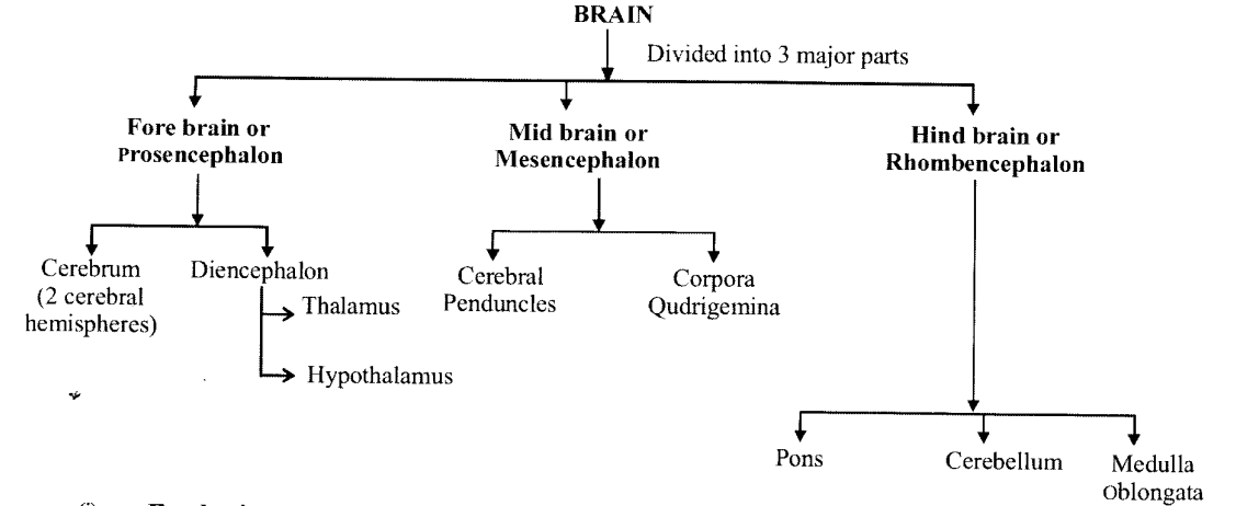
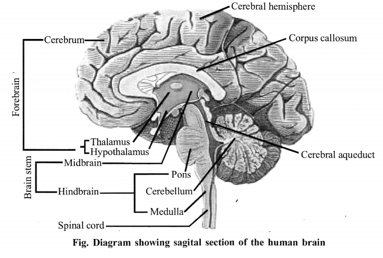
Forebrain:
1. Cerebrum consists two cerebral hemisphere on the dorsal surface. It is connected by a tract of nerve fibres called corpus callosum. Cerebral hemispheres are covered by the layer of cells called cerebral cortex and are thrown into prominent folds referred as grey matter. Inner part of the cerebral hemisphere is white matter.
2. Diencephlon is the posterior part of fore-brain. It consists of thalamus and hypothalamus.
- Thalamus is a major co-ordinating centre for sensory and motor signaling. It forms 80 % of diencephalon.
- Hypothalamus contains a number of centres which control many functions Like – hunger, thirst, sleep, sweating, body temperature and emotions
(ii) Midbrain:
It is located between the thalamus/ hypothalamus of the forebrain and pons of the hindbrain.
It forms the brain stem with the hindbrain. Anterior part of mid-brain contains two cerebral peduncles, which controls the muscle of limbs and Posterior part of mid-brain in four optic lobs called corpora quadri
gemiana i.e. two upper and two lower.
(iii) Hindbrain: Hindbrain consists of pons. Cerebellum and medulla of longata.
- Pons is present below the midbrain and upper side of medulla oblongata. It possesses pneumotaxic area of respiratory centre.
- Cerebellum is the 2nd largest part of brain, which lies behind cerebrum and provides the additional space for many neuron and maintains equilibrium or posture of the body.
- Medulla oblongata lies below cerebellum and continues into the spinal-cord. It contains a respiratory centre for regulating breatheing, Cardiac centre for regulating heartbeat and blood pressure, and also has reflex centre for swallowing, coughing, sneezing, etc.
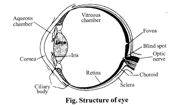
(b) Structure of eye :
- Our paired eyes are located in sockets of the skull called orbits.
- The adult human eyeball is nearly a spherical structure.
- The wall of the eyeball is composed of three layers which are given below :
- .The external layer is composed of a dense connective tissue and is called the sclera. The anterior portion of this layer is called the cornea.
- The middle layer is called the choroid contains many blood vessels and looks bluish.
- The choroid layer is thin over the posterior two-thirds of the eyeball, but it becomes thick in the anterior part to form the ciliary body.
- The ciliary body itself continues forward to form a pigmented and opaque structure called the iris which is the visible coloured portion of the eye.
- The eyeball contains a transparent crystalline lens which is held in place by ligaments attached to the ciliary body.
- In front of the lens, the aperture surrounded by the iris is called the pupil. The diameter of the pupil is regulated by the muscle fibres of iris.
The inner layer is the retina and contains three layers of cells- ganglion cells, bipolar cells and photoreceptor cells.

- In the human eye, there are three types of cones which possess their own characteristic photopigments that respond to red, green and blue lights.
- The sensations of different colours are produced by various combinations of these cones and their photopigments. When these cones are stimulated equally, a sensation of white light is produced.
- The optic nerves leave the eye and the retinal blood vessels enter it at a point medial to and slightly above the posterior pole of the eyeball.
- Photoreceptor cells are not present in that region and hence it is called the blind spot. At the posterior pole of the eye lateral to the blind sport, there is a yellowish pigmented spot called macula lutea with a central pit called the fovea.
- The fovea is a thinned-out portion of the retina where only the cones are densely packed.
- It is the point where the visual acuity (resolution) is the greatest.
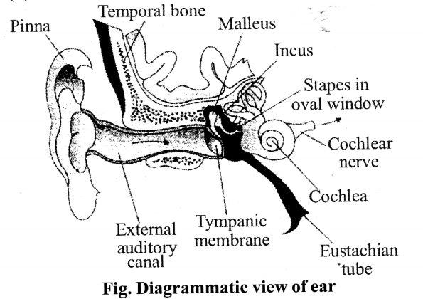
(c) Structure of ear:
Anatomically, the ear can be divided into three major sections called the outer ear, the middle ear and the inner ear.
Outer ear. The outer ear consists of the pinna and external auditory meatus (canal). The pinna collects the vibrations in the air which produce sound.
- The external auditory meatus leads inwards and extends up to the tympanic membrane (the ear dfum).
- There are very fine hairs and wax-secreting sebaceous glands in the skin of the pinna and the meatus.
- The tympanic membrane is composed of connective tissues covered with skin outside and with mucus membrane inside.
- Middle ear : The middle ear contains three ossicles called malleus, incus and stapes which are attached to one another in a chain-like fashion. These ossicles transmit sound waves further inside the ear.
- An Eustachian tube connects the middle ear cavity with the pharynx. The Eustachian tube helps in equalizing the pressures on either sides of the eardrum. Inner ear the fluid-filled inner ear called labyrinth consists of two parts, the bony and the membranous labyrinths. The bony labyrinth is a series of channels.
- Inside these channels lies the membranous labyrinth, which is surrounded by a fluid called perilymph.
The membranous labyrinth is filled with a fluid called endolymph. - The coiled portion of the labyrinth is called cochlea. The membranes constituting cochlea, the Meissner’s membrane and basilar membrane, divide the surrounding perilymph filled bony labyrinth into an upper scala vestibuli and a lower scala tympani.
- The organ of corti is a structure located on the basilar membrane which contains hair cells that act as auditory receptors. The hair cells are present in rows on the internal side of the organ of Corti.
- The inner ear also contains a complex system called vestibular apparatus, located above the cochlea. The vestibular apparatus is composed of three semi-circular canals and the otolith organ consisting of the saccule and utricle.
- The base of canals is swollen and is called ampulla, which contains a projecting ridge called crista ampullaris which has hair cells. The saccule and utricle contain a projecting ridge called macula.
- The crista and macula are the specific receptors of the vestibular apparatus responsible for maintenance of balance of the body and posture.
Question 2.
Compare the following:
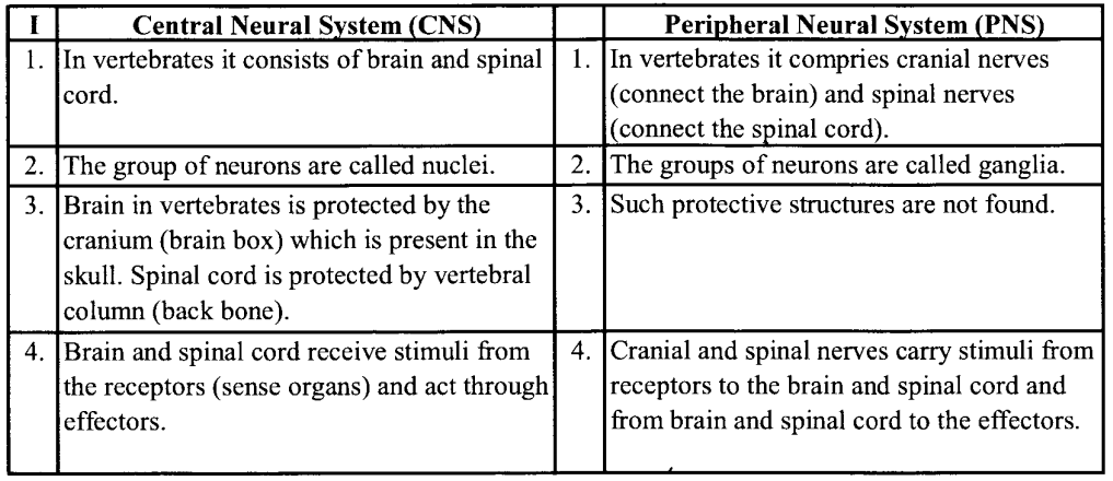
(i) Central Neural System (CNS) and Peripheral neural system (PNS)
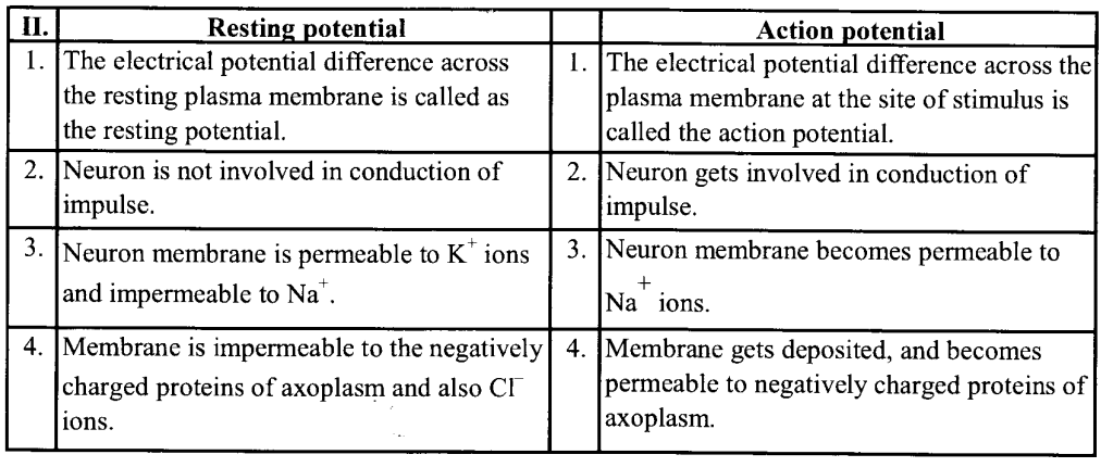
(ii) Resting potential and action potential
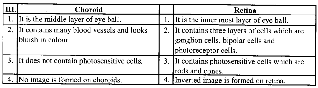
(iii) Choroid and retina
Solution:
Differences between central neural system and peripheral nervous system are as follows :
Differences between resting potential and action potential are as follows:
Differences between choroid and retina are as follows :
Question 3.
Explain the following processes:
(a) Polarization of the membrane of a nerve fibre
(b) Depolarization of the membrane of a nerve fibre
(c) Conduction of a nerve impulse along a nerve fibre
(d) Transmission of a nerve impulse across a chemical synapse
Solution:
(a) When a neuron is not conducting any impulse; i.e resting, the axoplasm inside the axon contains high concentration of K+ and negatively charged proteins and low concentration of Na+. In contrast, the fluid outside the axon contains a low concentration of K+, a high concentration of Na+ and thus forms a concentration gradient. These ionic gradients across the resting membrane are maintained by the active transport of ions by the sodium-potassium pump which transports 3 Na+ outwards for 2K+ into the cell. As a result, the outer surface of the axonal membrane possesses a positive charge while its inner surface becomes negatively charged and therefore is polarised.
(b) When a stimulus is applied at a site, (eg: site A) on the polarised membrane, the membrane at site A becomes freely permeable to Na+. This leads to a rapid influx of Na+ followed by the reversal of the polarity at that site, i.e the outer surface of the membrane becomes negatively charged and the inner side becomes positively charged. The polarity of the membrane at site A is thus reversed and hence depolarised.
(c) Conduction of nerve impulse along a nerve fibre:
- When a stimulus is applied at a site on the polarised membrane the membrane at the site A becomes freely permeable to Na+.
- Asa result polarity gets reversed by a rapid inflow of Na+.
- After the reversal of polarity of the membrane, the membrane becomes depolarised.
- The electrical potential difference across the plasma membrane at the site A is called the action potential; which is termed as nerve impulse.
- The axon membrane (site B) has a positive charge on the outer surface and a negative charge on its inner surface. As a result, a current flows on the inner surface from site A to site B.
- On the outer surface current flows from site B to site A, to complete the circuit of current flow.
- Hence, the polarity at the site is reversed, and an action potential is generated at site B.
- Thus, the impulse generated at site A arrives at site B. The sequence is repeated along the length of the axon and consequently impulse is conducted.
- The rise in the stimulus-induced permeability to Na+ is extremely short-lived.
- It is quickly followed by a rise in permeability to K+. Within a fraction of a second, K+ diffuses outside the membrane and restores the resting potential of the membrane at the site of excitation and the fibre becomes once more responsive to further stimulation.
(d) Transmission of a nerve impulse across chemical synapse:
- A nerve impulse is transmitted from one neuron to another through junction called synapses.
- Electrical current can flow directly from one neuron to the other across these synapses.
- The membrane of the pre-and post-synaptic neurons are separated by fluid-filled space called synaptic deft.
- Chemicals called neurotransmitters are involved in the transmission of impulses at these synapses.
- The axon terminals contain vesicles filled these neurotransmitters.
- When an impulse (action potential) arrives at the axon terminal, it stimulates the movement of the synaptic vesicles towards the membrane where they fuse with the plasma membrane and release their neurotransmitters in the synaptic cleft. The released neurotransmitters bind to their specific receptors, present on the post- synaptic membrane.
- This binding opens ion channels allowing the entry of ions which can generate a new potential in the post- synaptic neuron. The new potential developed may be either excitatory or inhibitory.
Question 4.
Draw labelled diagrams of the following :
(a) Neuron
(b) Brain
(c) Eye
(d) Ear
Solution:
(a) Neuron
(b) Brain
(c) Eye
(d)Ear
Question 5.
Write short notes on the following
(a) Neural coordination
(b) Forebrain
(c) Midbrain
(d) Hindbrain
(e) Retina
(f) Ear ossicles
(g) Cochlea
(h) Organ of Corti
(i) Synapse
Solution:
(a) Neural co-ordination:
- The functions of the organs/ organ systems in our body must be coordinated to maintain homeostasis. Coordination is the process through which two or more organs interact and complement the functions of one another. For example, when we do physical exercises, the energy demand is increased for maintaining an increased muscular activity.
- The supply of oxygen is also increased. The increased supply of oxygen necessitates an increase in the rate of respiration, heartbeat and increased blood flow via blood vessels.
- When physical exercise is stopped, the activities of nerves, lungs, heart and kidney gradually return to their normal conditions.
- Thus, the functions of muscles, lungs, heart, blood vessels, kidney and other organs are coordinated while performing physical exercises.
- In our body the neural system and the endocrine system jointly coordinate and integrate all the activities of the organs so that they function in a synchronised fashion.
(b) Forebrain :
- The forebrain consists of cerebrum, thalamus and hypothalamus. Cerebrum forms the major part of the human brain. A deep cleft divides the cerebrum longitudinally into two halves, which are termed as the left and right cerebral hemispheres.
- The hemispheres are connected by a tract of nerve fibres called corpus callosum.
The cerebral cortex contains motor areas, sensory areas and large regions that are neither clearly sensory nor motor in function. - These regions called as the association areas are responsible for complex functions like intersensory associations, memory and communication.
- The cerebrum wraps around a structure called thalamus, which is a major coordinating centre for sensory and motor signaling. Another very important part of the brain called hypothalamus lies at the base of the thalamus.
- The hypothalamus contains a number of centres which control body temperature, urge for eating and drinking. It also contains several groups of neurosecretory cells, which secrete hormones called hypothalamic hormones. The inner parts of cerebral hemispheres and a group of associated deep structures like amygdala, hippocampus, etc., form a complex structure called the limbic lobe or limbic system.
- Along with the hypothalamus, it is involved in the regulation of sexual behaviour, expression of emotional reactions (e.g., excitement, pleasure, rage and fear), and motivation.
(c) Midbrain:
- It is located between the thalamus hypothalamus of the forebrain and pons of the hindbrain.
- The dorsal portion of the midbrain consists of four small lobes called as corpora quadrigemina.
- A canal called the cerebral aqueduct passes through the midbrain. Neural control and co-ordination.
(d) Hindbrain:
- It consists of pons, cerebellum and medulla oblongata.
- Cerebellum has very convoluted surface to provide the additional space for many more neurons.
- The medulla is the part that continues as a spinal cord.
- The medulla contains centres which control respiration, cardiovascular reflexes and gastric secretions.
(e) Retina :
- The retina contains three layers of cells – from inside to outside ganglion cells, bipolar cells and photoreceptor cells.
- There are two types of photoreceptor cells, namely, rods and cones. These cells contain the light-sensitive proteins called the photopigments.
- The daylight (photopic) vision and colour vision are functions of cones and the twilight (scotopic) vision is the function of the rods.
- The rods contain a purplish-red protein called the rhodopsin or visual purple, which contains a derivative of vitamin A.
- In the human eye, there are three types of cones that possess their own characteristic photopigments that respond to red, green and blue lights.
- The sensations of different colours are produced by various combinations of these cones and their photopigments. When these cones are stimulated equally, a sensation of white light is produced.
(f) Ear ossicles –
- The middle ear contains three ossicles called malleus, incus and stapes which are attached to one another in a chain-like fashion.
- Malleus is attached to the tympanic membrane and the stapes is attached to the oval window of the cochlea.
- The ear ossicles increase the efficiency of transmission of sound waves to the inner ear.
(g) Cochlea :
- The fluid-filled inner ear called labyrinth consists of two parts, the bony and the membranous labyrinths. The coiled portion of the labyrinth is called cochlea.
- The membranes constituting cochlea, the reissner’s and basilar, divide the surounding perilymph filled bony labyrinth into an upper scala vestibuli and a lower scala tympani.
- The space within cochlea called scala media is filled with endolymph. At ‘ the base of the cochlea, the scala vestibuli ends at the oval window, while the scala tympani terminates at the round window which opens to the middle ear.
(h) Organ of corti:
- The organ of corti is a structure located on the basilar membrane which contains hair cells that act as auditory receptors.
- The hair cells are present in rows on the internal side of the organ of corti.
- The basal end of the hair cell is in close contact with the afferent nerve fibres.
- A large number of processes called stereo cilia are projected from the apical part of each hair cell.
- Above the rows of the hair cells is a thin elastic membrane called tectorial membrane.
(i) Synapse:
- A nerve impulse is transmitted from one neuron to another through junctions called synapses.
- A synapse is formed by the membranes of a pre-synaptic neuron and a post-synaptic neuron, which may or may not be separated by a gap called synaptic cleft.
- There are two types of synapses, namely, electrical synapses and chemical synapses.
Electrical synapse:
- At electrical synapses, the membranes of pre-and post-synaptic neurons are in very close proximity.
- Electrical current can flow directly from one neuron into the other across these synapses.
- Transmission of an impulse across electrical synapses is very similar to impulse conduction along a single axon.
- Impulse transmission across an electrical synapse is always faster than that across a chemical synapse.
- Electrical synapses are rare in our system.
Chemical synapse:
- At a chemical synapse, the membranes of the pre-and post-synaptic neurons are separated by a fluid-filled space called synaptic cleft.
- Chemicals called neurotransmitters are involved in the transmission of impulses at these synapses.
The axon terminals contain vesicles filled with these neurotransmitters. - When an impulse (action potential) arrives at the axon terminal, it stimulates the movement of the synaptic vesicles towards the membrane where they fuse with the plasma membrane and release their neurotransmitters in the synaptic cleft.
- The released neurotransmitters bind to their specific receptors, present on the post-synaptic membrane.
- This binding opens ion channels allowing the entry of ions which can generate a new potential in the post-synaptic neuron. The new potential developed may be either excitatory or inhibitory.
Question 6.
Give a brief account of :
(a) Mechanism of synaptic transmission
(b) Mechanism of vision
(c) Mechanism of hearing
Solution:
(a) Synaptic transmission can be of two types :
(i) Transmission of nerve impulse through electrical synapse.
(ii) Transmission of nerve impulse through chemical synapse.
Electrical synaptic transmission: At electrical synapses, the membranes of pre- and post- synaptic neurons are in very close proximity.
Electrical current can flow directly from one neuron into the.other across these synapses. Transmission of an impulse is very similar to impulse conduction along a single axon.
Chemical synaptic transmission : The membranes of the pre- and post-synaptic neurons are separated by a fluid-filled space called synaptic cleft. Neurotransmitters are involved in the transmission of impulses.
When an impulse (action potential) arrives at the axon terminal, it stimulates the movement of the synaptic vesicles towards the membrane where they fuse with the plasma membrane and release their neurotransmitters in the synaptic cleft.
The released neurotransmitters bind to their specific receptors, present on the post-synaptic membrane.
This binding opens ion-channels allowing the entry of ions which can generate a new potential in the post-synaptic neuron.
(b) Mechanism of vision
- The light rays in visible wavelength focussed on the retina through the cornea and lens generate potentials (impulses) in rods and cones.
- The photosensitive compounds (photopigments) in the human eyes is composed of opsin (a protein) and retinal (an aldehyde of vitamin A).
- Light induces dissociation of the retinal from opsin resulting in changes in the structure of the opsin. This causes membrane permeability changes.
- As a result, potential differences are generated in the photoreceptor cells. This produces a signal that generates action potentials in the ganglion cells through the bipolar cells.
- These action potentials (impulses) are transmitted by the optic nerves to the visual cortex area of the brain, where the neural impulses are analysed and the image formed on the retina is recognised based on earlier memory and experience.
Mechanism of hearing
- The external ear receives sound waves and directs them to the ear drum. The ear drum vibrates in response to the sound waves and these vibrations are transmitted through the ear ossicles (malleus, incus and stapes) to the oval window.
- The vibrations are passed through the oval window on the fluid of the cochlea, where they generate waves in the lymphs.
- The waves in the lymphs induce a ripple in the basilar membrane.
- These movements of the basilar membrane bend the hair cells, pressing them against the tectorial membrane.
- As result, nerve impulses are generated in the associated afferent neurons. These impulses are transmitted by the afferent fibres via auditory nerves to the auditory cortex of the brain, where the impulses are analysed and the sound is recognised.
Question 7.
Answer briefly:
(a) How do you perceive the colour of an object?
(b) Which part of our body helps us in maintaining the body balance?
(c) How does the eye regulate the amount of light that falls on the retina?
Solution:
(a) Rods and cones are photoreceptor cells that contain light-sensitive proteins called photopigments. The daylight vision and colour vision are functions of cones. There are three types of cones which respond to red, green and blue lights. The sensations of different colours are produced by various combinations of these cones and their photopigments. When these cones are stimulated equally, a sensation of white light is produced.
(b) The crista and macula are the specific receptors of the vestibular apparatus responsible for the maintenance of the balance of the body.
(c) The pupil in the eye functions as an aperture. This dilates in case of low light and constricts in case of intense light thereby regulating the amount of light falling on the retina.
Question 8.
Explain the following:
(a) Role of Na+ in the generation of the action potential.
(b) Mechanism of generation of light-induced impulse in the retina.
(c) Mechanism through which a sound produces a nerve impulse in the inner ear.
Solution:
(a) When a stimulus is applied at a site, (eg: site A) on the polarised membrane, the membrane at site A becomes freely permeable to Na+. This leads to a rapid influx of Na+ followed by the reversal of the polarity at that site, i.e., the outer surface of the membrane becomes negatively charged and the inner side becomes positively charged. The polarity of the membrane at site A is thus reversed and hence depolarised. Thus action potential is generated across the plasma membrane.
(b) Mechanism of vision: The rays in visible wavelength focussed on the retina through the cornea and lens generate potentials (impulse) in rods and cones. The photo-sensitive compounds (photopigments) in the human eyes is composed of opsin and retinal. Light induces dissociation of the retinal from opsin resulting in changes in the structure of the opsin. This causes membrane permeability to change.
As a result, potential differences are generated in the photoreceptor cells. This produces a signal that generates action potentials in the ganglion cells through the bipolar cells. These action potentials are transmitted by the optic nerves to the visual cortex area of the brain, where the neural impulses are analysed and the image formed on the retina is recognized based on earlier memory and experience.
(c) Mechanism of hearing: The external ear receives sound waves and directs them to the eardrum. The eardrum vibrates in response to the sound waves and these vibrations are transmitted through the ear ossicles to the oval window. The vibrations are passed through the oval window on to the fluid of the cochlea, where they generate waves in the lymphs.
The waves in the lymphs induce a ripple in the basilar membrane. These movements of the basilar membrane bend the hair cells, pressing them against the tectorial membrane. As a result, nerve impulses are generated in the associated afferent neurons. These impulses are transmitted by the afferent fibres via auditory nerves to the auditory cortex of the brain, where the impulses are analysed and the sound is recognised.
Question 9.
Differentiate between:
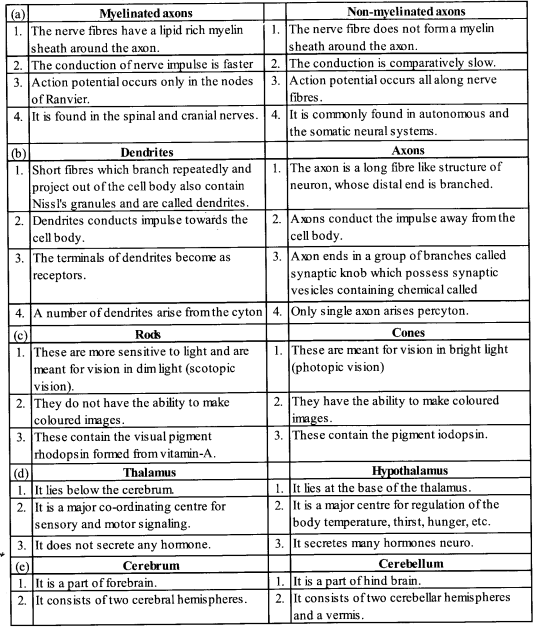
(a) Myelinated and non-myelinated axons
(b) Dendrites and axons
(c) Rods and cones
(d) Thalamus and Hypothalamus
(e) Cerebrum and Cerebellum
Solution:
Differences:
Question 10.
Answer the following:
(a) Which part of the ear determines the pitch of a sound ?
(b) Which part of the human brain is the most developed?
(c) Which part of our central neural system acts as a master clock ?
Solution:
(a) Pitch represents the perceived fundamental frequency of a sound. Pitch is a subjective sensation in which a listener assigns perceived tones to relative positions on a musical scale based primarily on the frequency of vibration. As spund is finally perceived by the temporal lobe of the cerebral cortex so it can be said that cerebral cortex perceives the pitch of the sound.
(b) Cerebral cortex (c) Hindbrain
Question 11.
The region of the vertebrate eye, where the optic nerve passes out of the retina, is called the
(a) fovea
(b) iris
(c) blind spot
(d) optic chiasma
Solution:
(c) Blindspot
Question 12.
Distinguish between
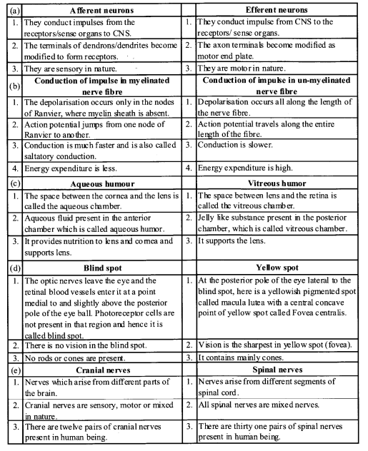
(a) afferent neurons and efferent neurons
(b) impulse conduction in a myelinated nerve fibre and unmyelinated nerve fibre
(c) aqueous humor and vitreous humor
(d) blind spot and yellow spot
(e) cranial nerves and spinal nerves.
Solution:
Differences:
VERY SHORT ANSWER QUESTIONS
Question 1.
Name the band of nerve fibres that joins the cerebral hemispheres in mammals.
Solution:
The hemispheres are connected by a tract of nerve fibres called corpus callosum.
Question 2.
How many types of nerve fibres do PNS have ? Name them.
Solution:
The nerve fibres of the PNS are of two types :
(a) afferent fibres
(b) efferent fibres
Question 3.
Name the functional unit of nervous system.
Solution:
The functional unit of nervous system is neuron.
Question 4.
How many types of axons are present in CNS? Name them.
Solution:
There are two types of axons, namely, myelinated and non-myelinated.
Question 5.
Name the functional junction between two neurons.
Solution:
A synapse is the functional junction between two neurons.
Question 6.
What does synaptic vesicles contain ?
Solution:
Synaptic vesicles contain chemicals called neurotransmitters.
Question 7.
Name the fluid in which membranous labyrinth of the inner ear floats.
Solution:
The fluid in which membranous labyrinth of the inner ear floats is perilymph.
Question 8.
Why is blind spot devoid of the ability of vision?
Solution:
Blind spot have no photoreceptor cells (rods & cones) and hence it is devoid of vision.
Question 9.
Name the canal which passes through midbrain.
Solution:
Cerebral aqueduct is the canal which passes through midbrain.
Question 10.
Name the type of neuron which carries the signal from CNS to the effector organs.
Solution:
Efferent neuron carries the signal from CNS to the effector/organs.
Question 11.
Name the fluid which fills anterior chamber of eye.
Solution:
An aqueous fluid which fills anterior chamber of eye is aqueous humor.
Question 12.
Name the cells which are responsible for photopic (daylight) and colour vision.
Solution:
Cones are responsible for photopic (day light) and colour vision.
Question 13.
Name the part through which middle ear communicates with the internal ear.
Solution:
Oval window helps the middle ear communicates with the internal ear.
Question 14.
Name the specific receptors of the vestibular apparatus.
Solution:
The crista and macula are the specific receptors of the vestibular apparatus.
Question 15.
Give the technical names of the auditory ossicles in their natural sequence.
Solution:
The technical names of the auditory ossicles are malleus, incus, staps.
Question 16.
Name the membranes constituting the cochlea.
Solution:
Reissner’s membrane and basilar membrane constitute the cochlea.
Question 17.
What is brain stem ?
Solution:
Brain stem is part of brain that lies in continuation of spinal cord, viz., medulla oblongata, pons and mid brain (with or without diencephalon of forebrain)
Question 18.
What is optic chiasma?
Solution:
A cross like structure found on anterior surface of hypothalamus is called optic chiasma.
Question 19.
Name the largest and longest cranial nerve?
Solution:
The largest cranial nerve is Trigeminal nerve and the longest cranial nerve is vagus nerve.
SHORT ANSWER QUESTIONS
Question 1.
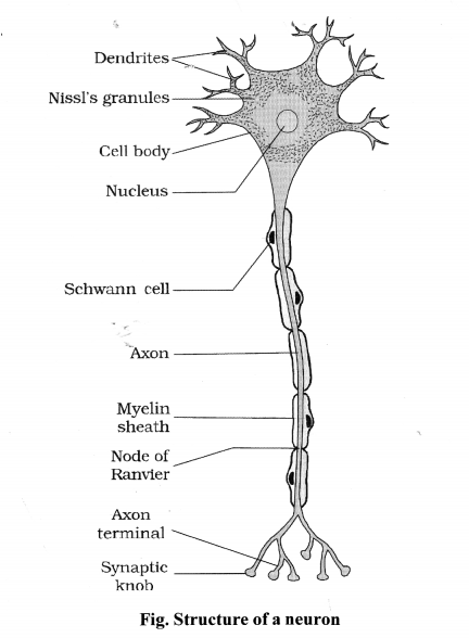
What are Nissl’s granules.
Solution:
The cell body contains cytoplasm with typical cell organelles and certain granular bodies called Nissl’s granules.
Question 2.
Describe the location and the role of ciliary body in human eye.
Solution:
Ciliary body is thick vascular, less pigmented ring shaped muscular structure occuring at the junction of choroid and iris. Ciliary body controls the size of pupil and in this way controls the amount of light entering the eye.
Question 3.
Explain the structural and functional significance of fovea in human eye.
Solution:
Fovea
– The fovea is a thinned-out portion of the retina where only the cones are densely packed, ft is the point where the visual acuity (resolution) is the greatest.
– It is a slightly depressed, tiny circular area found in the retina, just above the blind spot.
Question 4.
What is a reflex action?
Solution:
A sudden withdrawal of a body part which comes in contact with objects that are extremely hot, cold pointed or animals that are scary or poisonous. The entire process of response to a peripheral nervous stimulation, that occurs involuntarily, i.e., without conscious effort or thought and requires the involvement of a part of the central nervous system is called a reflex action.
Question 5.
Where are synaptic vesicles found? Name their chemical contents? What is the function of these contents?
Solution:
Synaptic vesicles are found in the bulbous expansion called synaptic knob, at the nerve terminal.
- Each synaptic vesicle contains as many as 10,000 molecules of a neurotransmitter substance, that is responsible for transmission of nerve impulse across the synapse.
- When a wave of depolarisation reaches the presynaptic membrane, the voltage-gated calcium channels concentrated at the synapse open and Ca ions diffuse into the terminal from the surrounding fluid.
- The Ca++ ions stimulate the synaptic vesicles to move to the terminal membrane, fuse with it and then rupture by exocytosis into the cleft.
- This neurotransmitter diffuses across the synapse and stimulates the membrane of the next neuron.
Question 6.
Write short notes on hindbrain.
Solution:
The hindbrain comprises pons, cerebellum and medulla (also called the medulla oblongata). Pons consists of fibre tracts that interconnect different regions of the brain. Cerebellum has very convoluted surface in order to provide the additional space for tnany more neurons. The medulla of the brain is connected to the spinal cord. The medulla contains centres which control respiration, cardiovascular reflexes and gastric secretions.
Question 7.
Enumerate the functions of hypothalamus.
Solution:
Functions of hypothalamus are as follows:
(i) Hypothalamus maintains homeostasis i. e., internal equilibrium of the body.
(ii) It has centres for regulation of hunger, thirst, emotions.
(iii) It organises behaviour like fighting, feeling etc., related to survival of species.
(iv) It maintains a constant body temperature.
(v) It secretes neurohormones, some of which 2.
control the functioning of pituitary glands called hypothalamic hormones.
Question 8.
Write a brief note on autonomic nervous system.
Solution:
Autonomic nervous is a part of peripheral nervous system. It controls activities occur in our body that are normally in voluntary such as heart beat, gut peristalsis, etc. Most of the actions of this system is controlled within the spinal cord or brain by reflexes known as visceral reflexes. It is maintanied by centre in medulla and hypothalamas. It maintains homeostasis. These are divided into two systems sympathetic and parasympathetic nervous system. The sympathetic nervous system mainly functions in quick responses and parasympathetic nervous system functions in actions which do not require immediate response.
Question 9.
What are nodes of ranvier?
Solution:
These are the periodic gaps or breakes in the 3. myelin sheath these breaks helps in the conduction of electricity in neurons the resistance to current flow between the axoplasm and fluid outside the cell is low these nodes set
up the local circuits to flow current inside neurons as a result, the action potential jump from node to node and passes along the myelineted axon faster compared to the series of small local circuits in a non-myelinated axon.
Long ANSWER QUESTIONS
Question 1.
Explain briefly the structure and functions of middle ear.
Solution:
The middle ear is an air-filled chamber on the inner side of eardrum. Its cavity communicates with an air-filled tube called eustachian tube, which maintains a balanced air pressure on either side of the tympanum.
The small bones called auditory ossicles are present in the middle ear. The malleus is attached to the ear-drum on one side and to the incus on the other side. The incus intum articulates with the stapes. The stapes is attached to the membrane over an oval-window between the middle ear and the internal ear.
Functions:
- The auditory ossicles transmit the sound- induced vibrations of the ear-drum to the endolymph in the internal ear.
- The eustachian tube balances and maintains a constant pressure on either side of the ear-drum.
Question 2.
Write a note on the retina.
Solution:
Retina
- The innermost layer of the wall of eyeball is the retina. It is composed of several layers of cells
- the photoreceptor layer contains rods and cones, the intermediate layer has bipolar neurons, which synapse with retinal ganglion cells and their axons bundle to form optic nerve.
- The rod cells contain rhodopsin while the cone cells contain iodopsin.
- The point in the retina where the optic nerve leaves the eye, is called as blind spot.
- Lateral to the blind spot, there is a yellowish pigmented spot called macula lutea with a central pit called the fovea.
- Fovea is the region where cones are densely packed and the vision is the sharpest.
Question 3.
Make a brief note on forebrain.
Solution:
Forebrain consists of cerebrum, thalamus and hypothalamus.
- Cerebrum occurs as two cerebral hemispheres that are joined together by corpus callosum.
- Each cerebral hemisphere is divided by other grooves into four lobes – frontal, parietal, temporal and occipital.
- The convolutions and fissures greatly enlarge the surface area of the cortex.
- The outer surface of cerebrum (cortex) has grey matter and inner to the cortex is white matter.
- Thalamus lies just underthe cerebrum, i.e., cerebrum wraps around the thalamus.
- Thalamus is the major coordinating centre for sensory and motor signals.
- Hypothalamus lies at the base of the thalamus. .
- It has centres to control body temperature, hunger, thirst, etc.
- It contains several groups of neuro secretory cells which secrete hormones.
- Limbic system is constituted by the inner parts of cerebral hemispheres and a group of structures called amygdala and hippocampus.
- Along with the hypothalamus, limbic system is involved in the regulation of sexual behaviour, expression of emotions, motivation, etc.
Question 4.
What is a synapse? How is the nerve impulse transmitted across a synapse?
Solution:
Synapse: The functional/intercommunicating, junction between two neurons, the axon of one neuron and the dendron/dendrite/soma of another neuron, through which impulse is conducted, is called a synapse.
Conduction of nerve impulse across a synapse:
- When a nerve impulse reaches the pre- synaptic membrane (membrane of synaptic button), the voltage-gated calcium channels, concentrated in the synapse, open.
- Calcium ions from the fluid in the synapse diffuse into the synaptic button and stimulate the synaptic vesicles to move to the terminal membrane, fuse with it and then rupture (exocytosis) to release the neurotransmitter.
- The neurotransmitter quickly diffuses across the synaptic cleft in the fluid and stimulates certain specific receptor molecules on the post-synaptic membrane (membrane of the next dendron/dendrite) and causes sparking and electrical current, passing the signal.

We hope the NCERT Solutions for Class 11 Biology at Work Chapter 21 Neural control and co-ordination, help you. If you have any query regarding NCERT Solutions for Class 11 Biology at Work Chapter 21 Neural control and co-ordination, drop a comment below and we will get back to you at the earliest.