NCERT Solutions for Class 11 Biology Chapter 18 Body Fluids and Circulation

NCERT Solutions for Class 11 Biology Chapter 18 Body Fluids and Circulation

These Solutions are part of NCERT Solutions for Class 11 Biology. Here we have given NCERT Solutions for Class 11 Biology Chapter 18 Body Fluids and Circulation.

Question 1.
Name the components of formed elements in the blood and mention one major function of each of them.
Solution:
Blood is a mobile connective tissue composed of a fluid, the plasma and formed elements.
Formed elements includes erythrocytes, leucocytes and platelets.
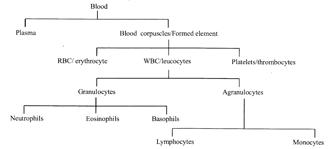
NCERT Solutions for Class 11 Biology Chapter 18 Body Fluids and Circulation 1
Blood corpuscles.
(a) Erythrocytes are the most prevalent corpuscles (approx. 5 to5.5 million/mm3 of blood). They contain carbonic anhydrase enzymes which help in the transportation ofCO2 and haemoglobin pigment which helps in the transportation of O2.
(b) Leucocytes lack any pigment and most active motile constituents of blood (approx. 6000-8000/ mm3 of blood). They may be of two types.
1. Granulocytes are the cells containing granules and a polymorphic nucleus.

  • Neutrophils: responsible for protection against infection.
  • Eosinophils: play important role in an allergic reaction.
  • Basophils: significant in inflammatory reaction.

2. Agranulocytes are cells which lack granules

  • Lymphocytes play a key role in immunological reactions.
  • Monocytes are phagocytic in nature.

(c) Platelets: There are involved in the coagulation or clotting of blood.

Question 2.
What is the importance of plasma proteins?
Solution:
Fibrinogen, globulins and albumins are the major plasma proteins. Fibrinogen are needed for clotting or coagulation of blood. Globulins primarily are involved in defense mechanisms of the body. Albumins help in osmotic balance.

Question 3.
Match Column I with Column II:
Column I                                           Column II
(a) Eosinophils                              (i) Coagulation
(b) RBC                                      (ii) Universal recipient
(c) AB blood Group                          (iii) Resist infection
(d) Platelets                               (iv) Contraction of heart
(e) Systole                                  (v) Gas transport
Solution:
Column I                                                           Column II
(a) Eosinophils                                  (iii) Resist infections
(b) RBC                                          (v) Gas transport
(c) AB blood Group                                (ii) Universal Recipient
(d) Platelets                                      (i) Coagulation
(e) Systole                                        (iv) Contraction of heart

Question 4.
Why do we consider blood as a connective tissue?
Solution:
Blood is a special connective tissue consisting of a fluid matrix, plasma, and the formed elements. It is circulated throughout the body.

Question 5.
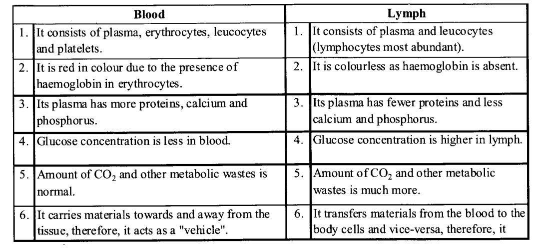
What is the difference between lymph and blood?
Solution:
Differences between blood and lymph are as following :
NCERT Solutions for Class 11 Biology Chapter 18 Body Fluids and Circulation 2

Question 6.
What is meant by double circulation? What is its significance?
Solution:
The blood pumped by the right ventricle enters the pulmonary artery whereas the left ventricle pumps blood into the aorta. The deoxygenated blood pumped into the pulmonary artery is passed on to the lungs from where the oxygenated blood is carried by the pulmonary veins into the left atrium. This pathway constitutes pulmonary circulation. The oxygenated blood entering the aorta is carried by a network of arteries, arterioles, and capillaries to the tissues from where the deoxygenated blood is collected by a system of venules, veins, and vena cava and emptied into the right atrium.

This is the systemic circulation (fig 18.1). The systemic circulation provides nutrients, O2, and other essential substances to the tissues and takes CO2, and other harmful substances away for elimination. A unique vascular connection exists between the digestive tract and liver called the hepatic portal system. The hepatic portal vein carries blood from the intestine to the liver before it is delivered to the systemic circulation. A special coronary system of blood vessels is present in our body exclusively for the circulation of blood to and from the cardiac musculature.

Question 7.
Write the differences between
(a) Blood and lymph
(b) OPen and closed system of circulation
(c) Systole and Diastole
(d) P-wave and T-wave
Solution:
(a) Differences between Blood and lymph NCERT Solutions for Class 11 Biology Chapter 18 Body Fluids and Circulation 2
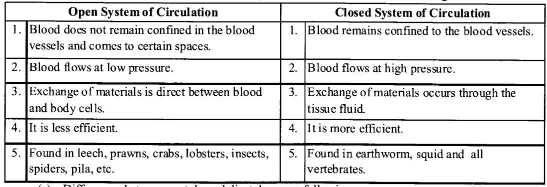
(b) Differences between open and closed systems of circulation are as following :
NCERT Solutions for Class 11 Biology Chapter 18 Body Fluids and Circulation 3

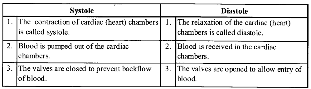
(c) Differences between systole and diastole are as following :
NCERT Solutions for Class 11 Biology Chapter 18 Body Fluids and Circulation 4
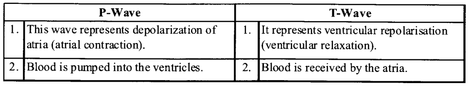
(d) Differences between P-wave and T-wave are as following :
NCERT Solutions for Class 11 Biology Chapter 18 Body Fluids and Circulation 5

Question 8.
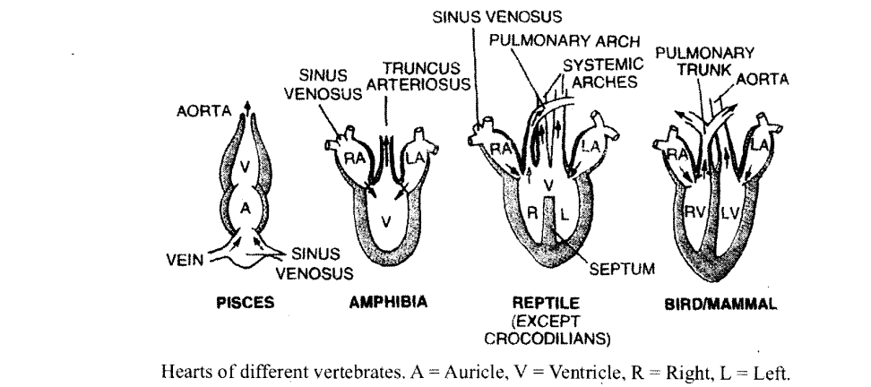
Describe the evolutionary change in the pattern of heart among the vertebrates.
Solution:

  • As it is clear from the following diagram the heart of fish has two chambers.
  • This means there is no separate circulation for oxygenated and deoxygenated blood.
  • There is separation of two chambers in the atrium of amphibians.
  • This has further evolved to partial separation of ventricle in reptiles.
  • Finally in birds there is complete separation of oxygenated and deoxygenated blood circulation with advent of four chambers in the heart.
  • Mammal heart is the most developed having the most efficient double circulatory system.
    NCERT Solutions for Class 11 Biology Chapter 18 Body Fluids and Circulation 6


Question 9.
Why do we call our heart myogenic?
Solution:
Normal activities of the heart are regulated intrinsically i.e., auto regulated by specialised muscles, hence our heart is called myogenic.

Question 10.
The sino-atrial node is called the pacemaker of our heart. Why?
Solution:
The SA node is located in the wall of right auricle slightly below the opening of the superior vena cava. It has a unique property of self-excitation which enables it to act as the pacemaker of the heart. It spontaneously initiates a wave of contraction which spreads over both the auricles more or less simultaneously along the muscle fibres.

Question 11.
What is the significance of the atrioventricular node and atrioventricular bundle in the functioning of the heart?
Solution:
Another mass of model tissue is seen in the lower left comer of the right atrium close to the atrioventricular septum called the atrioventricular node (AVN). A bundle of nodal fibers, atrioventricular bundle (AV bundle) continues from the AVN which passes through the atrioventricular septa to emerge on the top of the interventricular septum and immediately divides into a right and left bundle. These branches give rise to minute fibers throughout the ventricular musculature of the respective sides and are called Purkinje fibers. These fibers along with the right and left bundle are known as the bundle of HIS. The nodal musculature has the ability to generate action potentials without any external stimuli i.e., it is auto excitable. However, the number of action potentials that could be generated in a minute varies at different parts of the nodal system.

Question 12.
Define a cardiac cycle and the cardiac output.
Solution:
Cardiac cycle.   A regular sequence of three events :
(i) auricular systole,
(ii) ventricular systole, and
(iii) joint diastole or complete cardiac diastole (relaxation ofboth auricles and ventricles) during the completion of one heart beat is known as heart cycle or cardiac cycle.
Cardiac output. The amount of blood pumped by heart per minute is called cardiac output or heart output. Heart beats 72 times per minute and pumps out about 70 ml of blood during each beat. Therefore, 72 x 70 or 5040 ml (roughly 5 liters) is the cardiac output.

Question 13.
Explain heart sounds.
Solution:
During each cardiac cycle, two prominent sounds are produced which can be easily heard through a stethoscope. The first heart sound (dub) is associated with the closure of the tricuspid and bicuspid valves whereas the second heart sound (dub) is associated with the closure of the semilunar valves. These sounds are of clinical diagnostic significance.

Question 14.
Draw a standard ECG and explain the different segments in it.
Solution:
The recording of electrical potential generated by the spread of cardiac impulse is called an electrocardiogram (ECG).

ECG is the graphic record of electronic current produced by the excitation of cardiac muscles.

A normal electrocardiogram is composed of P wave, QRS complex and T wave, P wave indicate the depolarisation of the atria. QRS complex expresses ventricular depolarisation. T wave indicates repolarization of ventricles.
NCERT Solutions for Class 11 Biology Chapter 18 Body Fluids and Circulation 7

VERY SHORT ANSWER QUESTIONS

Question 1.
What is the Coagulation of Blood?
Solution:
When blood has been shed, it quickly becomes sticky and soon sets as a red jelly. This jelly or clot contracts or shrinks, and a straw-colored fluid called serum is squeezed out from it.

Question 2.
What is meant by an open circulatory system?
Solution:
When the blood does not remain confined to the blood vessels and flows into open spaces called sinus. It is termed as the open circulatory system.

Question 3.
What is the role of basophils in human body?
Solution:
Basophils are significant in allergic reactions.

Question 4.
Write down the main function of neutrophils.
Solution:
Neutrophils are mainly responsible for protection against infection.

Question 5.
What are the functions of lymphocytes?
Solution:
Lymphocytes play an important role in cell- mediated immunity.

Question 6.
What is tunica externa?
Solution:
Tunica externa is the outermost layer of blood vessels (artery and vein) made up of fibrous connective tissue with collagen fibres.

Question 7.
Mention the function of pericardial fluid.
Solution:
Pericardial fluid keeps the surface of heart moist and prevents the friction between heart wall and surrounding tissues.

Question 8.
What does the QRS complex indicate?
Solution:
QRS complex represent the ventricular depolarizations.

Question 9.
Name the most common disorder of blood circulatory system.
Solution:
Hypertension.

Question 10.
Where is tricuspid valve located? (April 87)
Solution:
The tricuspid valve is located between the right Atrium (auricle) and right ventricle.

Question 11.
Name the type of granulocytes that play an important role in detoxification.
Solution:
Eosinophils.

Question 12.
What transmits the cardiac impulse from the atria to the ventricles?
Solution:
Atrio – ventricular bundle from the atrioventricular node transmits the cardiac impulse from the atria to ventricles.

Question 13.
What is RBCs density in the blood of an adult human?
Solution:
About 5.0 – 5.5 millions/mm3 ofblood.

Question 14.
Name the reptile that has a four-chambered heart
Solution:
Crocodile.

Question 15.
What are Purkinje fibres?
Solution:
The minute branches of the right and left AV-bundles, that are found throughout the ventricular musculature of the respective sides, are called Purkinje fibres.

Question 16.
Name two vital organs affected by high blood
1. pressure or hypertension.

Solution:
Brain, kidney

Question 17.
What is the main symptom of heart failure?
Solution:
Congestion of the lungs.

Question 18.
Which vein carries oxygenated blood?
Solution:
Pulmonary vein.

Question 19.
Which is the valve present in between the left auricle and left ventricle? (April 97, M.Q.P.)
Solution:
The Bicuspid or mitral valve.

Question 20.
Which human organ is known as the “graveyard of RBCs”?
Solution:
Spleen.

SHORT ANSWER QUESTIONS

Question 1.
What are thrombocytes? Where are they produced in the human body?
Solution:
Thrombocytes or blood platelets are colourless formed elements of blood which appear round, or biconvex, or irregular and helps in clotting of blood. They are produced from the megakaryocytes (special cells in the bone marrow).

Question 2.
What is a serum?
Solution:
Serum is straw coloured fluid left after the clotting of blood. It is also called blood serum.

Question 3.
Name the different types of granulocytes. Give the function of the one of which constitutes the maximum percentage of total leucocytes.
Solution:
Granulocytes are of three types neutrophils, eosinophils, basophils.
Neutrophils constitute the maximum percentage of the total leucocyte, mainly responsible for protection against infection. They engulf the foreign substances by phagocytosis.

Question 4.
Why the closed circulatory system is more efficient than an open circulatory system?
Solution:
Advantage of closed circulatory system:
(i) Flow of blood is faster in the closed circulatory system as compared to open circulatory system. Sufficiently high blood pressure can be maintained.

  • Blood does not come in contact with the tissues/organs.
  • The volume of blood flowing to a particular tissue/organ can be regulated according to the need.

(ii) The blood flows under pressure so that all parts of the body receive blood with equal efficiency.
(iii) It transports materials efficiently.
(iv) There are checks for the regulation of the amount and speed of blood passing into an organ according to the requirement of that organs.

Question 5.
Name a portal system present in man. Write its one function.
Solution:
Hepatic portal system.
Significance: The blood which comes from the alimentary canal contains digested food like glucose and amino acids. The excess of glucose is converted into glycogen which is stored in the liver for later use.

Question 6.
Write down the functions of lymph.
Solution:
Functions of lymph :
(i) Lymph acts as a ‘middle man’ which transports oxygen, food materials, hormones etc. to the body cells and brings carbon dioxide and other metabolic wastes from body cells to blood.
(ii) It keeps the body cell moist.
(iii) It maintains the volume of blood.
(iv) It absorbs and transports fat and fat-soluble vitamins from the intestine.

Question 7.
Explain the chemical events taking place during clotting /coagulation of blood.
(Foreign 1997)
Solution:
An injury or a trauma stimulates the plate- 195 lets in the blood to release certain factors which activate the mechanism of coagulation. Clot or coagulam informed mainly of network threads called fibrils in which dead and damaged formed elements of blood are trapped. Fibrins are formed by the conversion of inactive fibrinogens in the plasma by the enzyme thrombin.

Thrombins, in turn are formed from another inactive substance present in the plasma called prothrombin. An enzyme complex, thrombokinase, is required for the above reaction which is formed by a series of linked enzymic reactions involving a number of factors present in the plasma in an inactive state. Calcium ions play a very important role in clotting.

Question 8.
What is an average number of thrombocytes in human blood? What is their function?
Solution:
1,50,000 to 3,50,000 platelets/mm3 of blood
– They release substances that are concerned with the clotting of blood.

Question 9.
How many chambers are present in the heart of a fish? Name them.
Solution:
There are two chambers one atrium and one ventricle. ,

Question 10.
What is meant by single circulation? Give an example.
Solution:
Single circulation

  • Single circulation is the phenomenon in which the heart of an animal receives and pumps blood once for e.g. fish.
  • The heart of fish is two-chambered with an atrium and a ventricle.
  • The heart pumps only deoxygenated blood, to the gills for oxygenation.

Question 11.
Where and from which cells do platelets originate? What is their life span? How do they act when blood vessels get injured?
Solution:
Platelets originate from the megakaryocytes in the bone marrow.

  • They live for about seven days.
  • They release thromboplastins, which help convert prothrombin of the plasma into thrombin and thus they are involved in clotting of blood.

Question 12.
Explain any three disorders of the circulatory system.
Solution:
(i) High Blood Pressure (Hypertension):
Hypertension is the term for blood pressure that is higher than normal (120/80). High blood pressure leads to heart diseases and also affects vital organs like the brain and kidney.

(ii) Coronary Artery Disease (CAD):
Coronary Artery Disease, often referred to as atherosclerosis, affects the vessels that supply blood to the heart muscle. It is caused by deposits of calcium, fat, cholesterol, and fibrous tissues, which makes the lumen of arteries narrower.

(iii) Heart Failure:
Heart failure means the state of the heart when it is not pumping blood effectively enough to meet the needs of the body. The main symptoms are congestion of the lungs.

Question 13.
What is Arteriosclerosis? What are its causes?
Solution:
It is thickening, hardening and loss of elasticity of the wall of arteries. This process progressively
restricts the blood flow to one’s organs and tissues and can lead to severe health risks. It is caused by the build-up of fatty plaque, cholestrol and some other substances in and on the artery wall.

Question 14.
Define Vagus escape.
Solution:
The stimulation of vagus nerve decreases the heart rate but its continuous stimulation shows no further decrease. This is known as vagus escape.

Question 15.
What is erythroblastosis foetalis? How it occurs?
Solution:
It is a type of haemolytic disease of new-borns due to ABO blood type A, B, or O is not compatible with blood group of foetus. It develops in a foetus, when IgG molecules produced by the mother passes through the placenta.

Question 16.
Why capillaries are known as exchange vessels?
Solution:
These have very thin walls which allows the passage of nutrients from blood into body tissues. It also allows the passage of waste product came from body tissues. So, capillaries are known as exchange vessels.

Long ANSWER QUESTIONS

Question 1.
Briefly explain the cardiac cycle.
Solution:
The sequential event in the heart which is cyclically repeated is called the cardiac cycle and it consists of systole and diastole of both the atria and ventricles. Initially, all the four chambers of the heart are in a relaxed state, i.e., joint diastole. Tricuspid and bicuspid valves are open resulting in the inflow of blood into the left and right ventricles from pulmonary veins and vena cava. Semilunar valves are closed at this stage. SAN (sino-atrial node) generates an action poten­tial resulting in simultaneous contraction of both atria – the atrial systole.

The action potential is conducted to the ventricular side by the AVN and AV bundle from where the bundle of His transmits it through the entire ventricular musculature. This causes contrac­tion of ventricles (ventricular systole) and relaxation of atria (diastole). Ventricular Systole increases the ventricular pressure causing the closure of tricuspid and bicuspid valves. As pressure increases, the semilunar valves of the pulmonary artery and aorta are forced open, allowing blood to flow through them into circulatory pathways.

The ventricles now relax (ventricular diastole) and semilunar valves close. As the ventricular pressure declines further, tricuspid and bicuspid valves are pushed open by the pressure in the atria. The blood now once again moves freely to the ventricles. The ventricles and atria are now again in a relaxed (joint diastole) state, as ear­lier and the process continue. The duration of a cardiac cycle is 0.8 sec­onds and during this period each ventricle pumps about 70 ml of blood.

Question 2.
(a) Draw the L. S. of a human heart showing the internal structure. Label the parts of the left side of the heart and the blood vessels that enter and leave the chambers of the same side.
(b) What is the significance of the remnant of sinus venosus in the mammalian heart?
Solution:
(a)
NCERT Solutions for Class 11 Biology Chapter 18 Body Fluids and Circulation 8

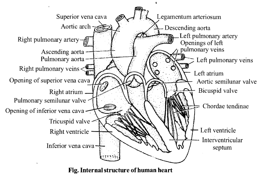
(b) It is believed that the remnant of sinus venous is modified into a sino-atrial node (SA Node) in the mammalian heart which is nothing but a mass of neuromuscular tissue laying in the wall of right atrium near the openings of the superior vena cava. It originates impulses for the regulation of the heartbeat. Thus acts as the pacemaker of heart.

Question 3.
Describe briefly the steps involved in the coagulation of blood at the site of injury of a blood vessel.
Solution:

  • Blood exhibits coagulation or clotting in response to an injury or trauma.
  • This is a mechanism to prevent excessive loss of blood from the body. We would have observed a dark reddish-brown scum formed at the site of a cut or an injury over a period of time.
  • It is a clot or coagulum formed mainly of a network of threads called fibrils in which dead and damaged formed elements of blood are trapped.
  • Fibrins are formed by the conversion of inactive fibrinogens in the plasma by the enzyme thrombin.
  • Thrombins, in turn, are formed from another inactive substance present in the plasma called prothrombin.
  • An enzyme complex, thrombokinase, is required for the above reaction.
  • This complex is formed by a series of linked enzymic reactions (cascade process) involving a number of factors present in the plasma in an inactive state.
  • An injury or a trauma stimulates the platelets in the blood to release certain factors which activate the mechanism of coagulation.
  • Certain factors released by the tissues at the site of injury also can initiate coagulation. Calcium ions play a very important role in clotting.

Question 4.
Why is it necessary to check the Rh-factor of the blood of a pregnant woman?
Solution:
Rh-Factor

  • Rh-antigen is present on the surface of erythrocytes in about 80-85% of human beings.
  • The individuals who possess this antigen are called Rh-positive and those who do not have it are called Rh-negative.
  • An Rh-negative person, when exposed to Rh-positive blood, develops anti-Rh- antibodies.
  • If a pregnant woman who is Rh-negative, bears an Rh-positive foetus, will develop anti-Rh-antibodies during the first delivery when the foetal blood comes in contact with her blood.
  • These antibodies linger in the blood for sufficiently long periods.
  • If she carries a second foetus, that is Rh-positive, the anti-Rh-antibodies in her blood enter the foetal circulation and cause damage to the foetal RBCs, and could become fatal.
  • This condition is called erythroblastosis foetalis.

We hope the NCERT Solutions for Class 11 Biology at Work Chapter 18 Body Fluids and Circulation, help you. If you have any query regarding NCERT Solutions for Class 11 Biology at Work Chapter 18 Body Fluids and Circulation, drop a comment below and we will get back to you at the earliest.

NCERT Solutions for Class 11 English Hornbill Poem 4 Childhood

Here we are providing NCERT Solutions for Class 11 English Hornbill Poem 4 Childhood. Students can get Class 11 English Childhood  NCERT Solutions, Questions and Answers designed by subject expert teachers.

Childhood NCERT Solutions for Class 11 English Hornbill Poem 4

Childhood NCERT Text Book Questions and Answers

Childhood Think it out

Question 1.
Identify the stanza that talks of each of the following.
Answer:
Individuality rationalism hypocrisy
Stanza I deals with rationalism.
Stanza II deals with hypocrisy.
Stanza III deals with individuality.

Question 2.
What according to the poem is involved in the process of growing up?
Answer:
According to the poet, the process of growing up primarily means loss of innocence and simplicity. It means losing trust in people, noticing their faults and becoming more individualistic. It essentially means loss of sincerity, faith, trust and acceptance.

Question 3.
What is the poet’s feeling towards childhood?
Answer:
The poet feels that childhood is a period of innocence when one accepts people at face value and trusts ffiem. It is that time of life when one trusts others without dwelling on the insincerity in the world. It is a phase of acceptance and humility.

Question 4.
Which do you think are the most poetic lines? Why?
Answer:
The last stanza of the poem is most poetic as it summarises the poem with sensitivity and seeks solace in the fact that the virtues related with childhood are not dead and gone. However, they are just found in children who the poet still considers virtuous.

NCERT Solutions for Class 11 Biology Chapter 21 Neural control and co-ordination

NCERT Solutions for Class 11 Biology Chapter 21 Neural control and co-ordination

These Solutions are part of NCERT Solutions for Class 11 Biology. Here we have given NCERT Solutions for Class 11 Biology Chapter 21 Neural control and co-ordination.

Question 1.
Briefly describe the structure of the following:
(a) Brain
(b) Eye

(c) Ear
Solution:
Structure of brain:

  • The brain is the central information processing organ of the body,
  • The human brain is well protected by the skull.
  • Inside the skull, cranial meninges cover the brain. These are tough tissue layers.

Meninges consist of 3 layers which are as follows :

  • The outermost – layer is the dura mater
  • The middle layer is arachnoid
  • The inner layer is pia mater.

The brain can be divided into three major parts which are given below:

  • forebrain
  • midbrain
  • hindbrain

NCERT Solutions for Class 11 Biology Chapter 21 Neural control and co-ordination 1
Forebrain:
1. Cerebrum consists two cerebral hemisphere on the dorsal surface. It is connected by a tract of nerve fibres called corpus callosum. Cerebral hemispheres are covered by the layer of cells called cerebral cortex and are thrown into prominent folds referred as grey matter. Inner part of the cerebral hemisphere is white matter.
2. Diencephlon is the posterior part of fore-brain. It consists of thalamus and hypothalamus.

  • Thalamus is a major co-ordinating centre for sensory and motor signaling. It forms 80 % of diencephalon.
  • Hypothalamus contains a number of centres which control many functions Like – hunger, thirst, sleep, sweating, body temperature and emotions

(ii) Midbrain:
It is located between the thalamus/ hypothalamus of the forebrain and pons of the hindbrain.
It forms the brain stem with the hindbrain. Anterior part of mid-brain contains two cerebral peduncles, which controls the muscle of limbs and Posterior part of mid-brain in four optic lobs called corpora quadri
gemiana i.e. two upper and two lower.

(iii) Hindbrain: Hindbrain consists of pons. Cerebellum and medulla of longata.

  • Pons is present below the midbrain and upper side of medulla oblongata. It possesses pneumotaxic area of respiratory centre.
  • Cerebellum is the 2nd largest part of brain, which lies behind cerebrum and provides the additional space for many neuron and maintains equilibrium or posture of the body.
  • Medulla oblongata lies below cerebellum and continues into the spinal-cord. It contains a respiratory centre for regulating breatheing, Cardiac centre for regulating heartbeat and blood pressure, and also has reflex centre for swallowing, coughing, sneezing, etc.

(b) Structure of eye :

  1. Our paired eyes are located in sockets of the skull called orbits.
  2. The adult human eyeball is nearly a spherical structure.
  3. The wall of the eyeball is composed of three layers which are given below :
  4. .The external layer is composed of a dense connective tissue and is called the sclera. The anterior portion of this layer is called the cornea.
  5. The middle layer is called the choroid contains many blood vessels and looks bluish.
  6. The choroid layer is thin over the posterior two-thirds of the eyeball, but it becomes thick in the anterior part to form the ciliary body.
  7. The ciliary body itself continues forward to form a pigmented and opaque structure called the iris which is the visible coloured portion of the eye.
  8. The eyeball contains a transparent crystalline lens which is held in place by ligaments attached to the ciliary body.
  9.  In front of the lens, the aperture surrounded by the iris is called the pupil. The diameter of the pupil is regulated by the muscle fibres of iris.

The inner layer is the retina and contains three layers of cells- ganglion cells, bipolar cells and photoreceptor cells.
NCERT Solutions for Class 11 Biology Chapter 21 Neural control and co-ordination 2

  • In the human eye, there are three types of cones which possess their own characteristic photopigments that respond to red, green and blue lights.
  • The sensations of different colours are produced by various combinations of these cones and their photopigments. When these cones are stimulated equally, a sensation of white light is produced.
  • The optic nerves leave the eye and the retinal blood vessels enter it at a point medial to and slightly above the posterior pole of the eyeball.
  • Photoreceptor cells are not present in that region and hence it is called the blind spot. At the posterior pole of the eye lateral to the blind sport, there is a yellowish pigmented spot called macula lutea with a central pit called the fovea.
  • The fovea is a thinned-out portion of the retina where only the cones are densely packed.
  • It is the point where the visual acuity (resolution) is the greatest.

(c) Structure of ear:
Anatomically, the ear can be divided into three major sections called the outer ear, the middle ear and the inner ear.
Outer ear. The outer ear consists of the pinna and external auditory meatus (canal). The pinna collects the vibrations in the air which produce sound.

  • The external auditory meatus leads inwards and extends up to the tympanic membrane (the ear dfum).
  • There are very fine hairs and wax-secreting sebaceous glands in the skin of the pinna and the meatus.
  • The tympanic membrane is composed of connective tissues covered with skin outside and with mucus membrane inside.
  • Middle ear : The middle ear contains three ossicles called malleus, incus and stapes which are attached to one another in a chain-like fashion. These ossicles transmit sound waves further inside the ear.
  • An Eustachian tube connects the middle ear cavity with the pharynx. The Eustachian tube helps in equalizing the pressures on either sides of the eardrum. Inner ear the fluid-filled inner ear called labyrinth consists of two parts, the bony and the membranous labyrinths. The bony labyrinth is a series of channels.
  • Inside these channels lies the membranous labyrinth, which is surrounded by a fluid called perilymph.
    The membranous labyrinth is filled with a fluid called endolymph.
  • The coiled portion of the labyrinth is called cochlea. The membranes constituting cochlea, the Meissner’s membrane and basilar membrane, divide the surrounding perilymph filled bony labyrinth into an upper scala vestibuli and a lower scala tympani.
  • The organ of corti is a structure located on the basilar membrane which contains hair cells that act as auditory receptors. The hair cells are present in rows on the internal side of the organ of Corti.
  • The inner ear also contains a complex system called vestibular apparatus, located above the cochlea. The vestibular apparatus is composed of three semi-circular canals and the otolith organ consisting of the saccule and utricle.
  • The base of canals is swollen and is called ampulla, which contains a projecting ridge called crista ampullaris which has hair cells. The saccule and utricle contain a projecting ridge called macula.
  • The crista and macula are the specific receptors of the vestibular apparatus responsible for maintenance of balance of the body and posture.

Question 2.
Compare the following:
(i) Central Neural System (CNS) and Peripheral neural system (PNS)
(ii) Resting potential and action potential
(iii) Choroid and retina
Solution:
Differences between central neural system and peripheral nervous system are as follows :
NCERT Solutions for Class 11 Biology Chapter 21 Neural control and co-ordination 3
Differences between resting potential and action potential are as follows:
NCERT Solutions for Class 11 Biology Chapter 21 Neural control and co-ordination 4
Differences between choroid and retina are as follows :
NCERT Solutions for Class 11 Biology Chapter 21 Neural control and co-ordination 5

Question 3.
Explain the following processes:
(a) Polarization of the membrane of a nerve fibre
(b) Depolarization of the membrane of a nerve fibre
(c) Conduction of a nerve impulse along a nerve fibre
(d) Transmission of a nerve impulse across a chemical synapse
Solution:
(a) When a neuron is not conducting any impulse; i.e resting, the axoplasm inside the axon contains high concentration of K+ and negatively charged proteins and low concentration of Na+. In contrast, the fluid outside the axon contains a low concentration of K+, a high concentration of Na+ and thus forms a concentration gradient. These ionic gradients across the resting membrane are maintained by the active transport of ions by the sodium-potassium pump which transports 3 Na+ outwards for 2K+ into the cell. As a result, the outer surface of the axonal membrane possesses a positive charge while its inner surface becomes negatively charged and therefore is polarised.

(b) When a stimulus is applied at a site, (eg: site A) on the polarised membrane, the membrane at site A becomes freely permeable to Na+. This leads to a rapid influx of Na+ followed by the reversal of the polarity at that site, i.e the outer surface of the membrane becomes negatively charged and the inner side becomes positively charged. The polarity of the membrane at site A is thus reversed and hence depolarised.

(c) Conduction of nerve impulse along a nerve fibre:

  • When a stimulus is applied at a site on the polarised membrane the membrane at the site A becomes freely permeable to Na+.
  • Asa result polarity gets reversed by a rapid inflow of Na+.
  • After the reversal of polarity of the membrane, the membrane becomes depolarised.
  • The electrical potential difference across the plasma membrane at the site A is called the action potential; which is termed as nerve impulse.
  • The axon membrane (site B) has a positive charge on the outer surface and a negative charge on its inner surface. As a result, a current flows on the inner surface from site A to site B.
  • On the outer surface current flows from site B to site A, to complete the circuit of current flow.
  • Hence, the polarity at the site is reversed, and an action potential is generated at site B.
  • Thus, the impulse generated at site A arrives at site B. The sequence is repeated along the length of the axon and consequently impulse is conducted.
  • The rise in the stimulus-induced permeability to Na+ is extremely short-lived.
  • It is quickly followed by a rise in permeability to K+. Within a fraction of a second, K+ diffuses outside the membrane and restores the resting potential of the membrane at the site of excitation and the fibre becomes once more responsive to further stimulation.

(d) Transmission of a nerve impulse across chemical synapse:

  • A nerve impulse is transmitted from one neuron to another through junction called synapses.
  • Electrical current can flow directly from one neuron to the other across these synapses.
  • The membrane of the pre-and post-synaptic neurons are separated by fluid-filled space called synaptic deft.
  • Chemicals called neurotransmitters are involved in the transmission of impulses at these synapses.
  • The axon terminals contain vesicles filled these neurotransmitters.
  • When an impulse (action potential) arrives at the axon terminal, it stimulates the movement of the synaptic vesicles towards the membrane where they fuse with the plasma membrane and release their neurotransmitters in the synaptic cleft. The released neurotransmitters bind to their specific receptors, present on the post- synaptic membrane.
  • This binding opens ion channels allowing the entry of ions which can generate a new potential in the post- synaptic neuron. The new potential developed may be either excitatory or inhibitory.

Question 4.
Draw labelled diagrams of the following :
(a) Neuron
(b) Brain
(c) Eye
(d) Ear

Solution:
(a) Neuron
NCERT Solutions for Class 11 Biology Chapter 21 Neural control and co-ordination 6

(b) Brain
NCERT Solutions for Class 11 Biology Chapter 21 Neural control and co-ordination 7

(c) Eye
NCERT Solutions for Class 11 Biology Chapter 21 Neural control and co-ordination 8

(d)Ear
NCERT Solutions for Class 11 Biology Chapter 21 Neural control and co-ordination 9

Question 5.
Write short notes on the following
(a) Neural coordination
(b) Forebrain

(c) Midbrain
(d) Hindbrain

(e) Retina
(f) Ear ossicles

(g) Cochlea
(h) Organ of Corti

(i) Synapse
Solution:
(a) Neural co-ordination:

  • The functions of the organs/ organ systems in our body must be coordinated to maintain homeostasis. Coordination is the process through which two or more organs interact and complement the functions of one another. For example, when we do physical exercises, the energy demand is increased for maintaining an increased muscular activity.
  • The supply of oxygen is also increased. The increased supply of oxygen necessitates an increase in the rate of respiration, heartbeat and increased blood flow via blood vessels.
  • When physical exercise is stopped, the activities of nerves, lungs, heart and kidney gradually return to their normal conditions.
  • Thus, the functions of muscles, lungs, heart, blood vessels, kidney and other organs are coordinated while performing physical exercises.
  • In our body the neural system and the endocrine system jointly coordinate and integrate all the activities of the organs so that they function in a synchronised fashion.

(b) Forebrain :

  • The forebrain consists of cerebrum, thalamus and hypothalamus. Cerebrum forms the major part of the human brain. A deep cleft divides the cerebrum longitudinally into two halves, which are termed as the left and right cerebral hemispheres.
  • The hemispheres are connected by a tract of nerve fibres called corpus callosum.
    The cerebral cortex contains motor areas, sensory areas and large regions that are neither clearly sensory nor motor in function.
  • These regions called as the association areas are responsible for complex functions like intersensory associations, memory and communication.
  • The cerebrum wraps around a structure called thalamus, which is a major coordinating centre for sensory and motor signaling. Another very important part of the brain called hypothalamus lies at the base of the thalamus.
  • The hypothalamus contains a number of centres which control body temperature, urge for eating and drinking. It also contains several groups of neurosecretory cells, which secrete hormones called hypothalamic hormones. The inner parts of cerebral hemispheres and a group of associated deep structures like amygdala, hippocampus, etc., form a complex structure called the limbic lobe or limbic system.
  • Along with the hypothalamus, it is involved in the regulation of sexual behaviour, expression of emotional reactions (e.g., excitement, pleasure, rage and fear), and motivation.

(c) Midbrain:

  • It is located between the thalamus hypothalamus of the forebrain and pons of the hindbrain.
  • The dorsal portion of the midbrain consists of four small lobes called as corpora quadrigemina.
  • A canal called the cerebral aqueduct passes through the midbrain. Neural control and co-ordination.

(d) Hindbrain:

  • It consists of pons, cerebellum and medulla oblongata.
  • Cerebellum has very convoluted surface to provide the additional space for many more neurons.
  • The medulla is the part that continues as a spinal cord.
  • The medulla contains centres which control respiration, cardiovascular reflexes and gastric secretions.

(e) Retina :

  • The retina contains three layers of cells – from inside to outside ganglion cells, bipolar cells and photoreceptor cells.
  • There are two types of photoreceptor cells, namely, rods and cones. These cells contain the light-sensitive proteins called the photopigments.
  • The daylight (photopic) vision and colour vision are functions of cones and the twilight (scotopic) vision is the function of the rods.
  • The rods contain a purplish-red protein called the rhodopsin or visual purple, which contains a derivative of vitamin A.
  • In the human eye, there are three types of cones that possess their own characteristic photopigments that respond to red, green and blue lights.
  • The sensations of different colours are produced by various combinations of these cones and their photopigments. When these cones are stimulated equally, a sensation of white light is produced.

(f) Ear ossicles –

  • The middle ear contains three ossicles called malleus, incus and stapes which are attached to one another in a chain-like fashion.
  • Malleus is attached to the tympanic membrane and the stapes is attached to the oval window of the cochlea.
  • The ear ossicles increase the efficiency of transmission of sound waves to the inner ear.

(g) Cochlea :

  • The fluid-filled inner ear called labyrinth consists of two parts, the bony and the membranous labyrinths. The coiled portion of the labyrinth is called cochlea.
  • The membranes constituting cochlea, the reissner’s and basilar, divide the surounding perilymph filled bony labyrinth into an upper scala vestibuli and a lower scala tympani.
  • The space within cochlea called scala media is filled with endolymph. At ‘ the base of the cochlea, the scala vestibuli ends at the oval window, while the scala tympani terminates at the round window which opens to the middle ear.

(h) Organ of corti:

  • The organ of corti is a structure located on the basilar membrane which contains hair cells that act as auditory receptors.
  • The hair cells are present in rows on the internal side of the organ of corti.
  • The basal end of the hair cell is in close contact with the afferent nerve fibres.
  • A large number of processes called stereo cilia are projected from the apical part of each hair cell.
  • Above the rows of the hair cells is a thin elastic membrane called tectorial membrane.

(i) Synapse:

  • A nerve impulse is transmitted from one neuron to another through junctions called synapses.
  • A synapse is formed by the membranes of a pre-synaptic neuron and a post-synaptic neuron, which may or may not be separated by a gap called synaptic cleft.
  • There are two types of synapses, namely, electrical synapses and chemical synapses.

Electrical synapse:

  • At electrical synapses, the membranes of pre-and post-synaptic neurons are in very close proximity.
  • Electrical current can flow directly from one neuron into the other across these synapses.
  • Transmission of an impulse across electrical synapses is very similar to impulse conduction along a single axon.
  • Impulse transmission across an electrical synapse is always faster than that across a chemical synapse.
  • Electrical synapses are rare in our system.

Chemical synapse:

  • At a chemical synapse, the membranes of the pre-and post-synaptic neurons are separated by a fluid-filled space called synaptic cleft.
  • Chemicals called neurotransmitters are involved in the transmission of impulses at these synapses.
    The axon terminals contain vesicles filled with these neurotransmitters.
  • When an impulse (action potential) arrives at the axon terminal, it stimulates the movement of the synaptic vesicles towards the membrane where they fuse with the plasma membrane and release their neurotransmitters in the synaptic cleft.
  • The released neurotransmitters bind to their specific receptors, present on the post-synaptic membrane.
  • This binding opens ion channels allowing the entry of ions which can generate a new potential in the post-synaptic neuron. The new potential developed may be either excitatory or inhibitory.

Question 6.
Give a brief account of :
(a) Mechanism of synaptic transmission
(b) Mechanism of vision
(c) Mechanism of hearing
Solution:
(a) Synaptic transmission can be of two types :
(i) Transmission of nerve impulse through electrical synapse.
(ii) Transmission of nerve impulse through chemical synapse.

Electrical synaptic transmission: At electrical synapses, the membranes of pre- and post- synaptic neurons are in very close proximity.
Electrical current can flow directly from one neuron into the.other across these synapses. Transmission of an impulse is very similar to impulse conduction along a single axon.
Chemical synaptic transmission : The membranes of the pre- and post-synaptic neurons are separated by a fluid-filled space called synaptic cleft. Neurotransmitters are involved in the transmission of impulses.
When an impulse (action potential) arrives at the axon terminal, it stimulates the movement of the synaptic vesicles towards the membrane where they fuse with the plasma membrane and release their neurotransmitters in the synaptic cleft.
The released neurotransmitters bind to their specific receptors, present on the post-synaptic membrane.
This binding opens ion-channels allowing the entry of ions which can generate a new potential in the post-synaptic neuron.

(b) Mechanism of vision

  • The light rays in visible wavelength focussed on the retina through the cornea and lens generate potentials (impulses) in rods and cones.
  • The photosensitive compounds (photopigments) in the human eyes is composed of opsin (a protein) and retinal (an aldehyde of vitamin A).
  • Light induces dissociation of the retinal from opsin resulting in changes in the structure of the opsin. This causes membrane permeability changes.
  • As a result, potential differences are generated in the photoreceptor cells. This produces a signal that generates action potentials in the ganglion cells through the bipolar cells.
  • These action potentials (impulses) are transmitted by the optic nerves to the visual cortex area of the brain, where the neural impulses are analysed and the image formed on the retina is recognised based on earlier memory and experience.

Mechanism of hearing

  • The external ear receives sound waves and directs them to the ear drum. The ear drum vibrates in response to the sound waves and these vibrations are transmitted through the ear ossicles (malleus, incus and stapes) to the oval window.
  • The vibrations are passed through the oval window on the fluid of the cochlea, where they generate waves in the lymphs.
  • The waves in the lymphs induce a ripple in the basilar membrane.
  • These movements of the basilar membrane bend the hair cells, pressing them against the tectorial membrane.
  •  As result, nerve impulses are generated in the associated afferent neurons. These impulses are transmitted by the afferent fibres via auditory nerves to the auditory cortex of the brain, where the impulses are analysed and the sound is recognised.

Question 7.
Answer briefly:
(a) How do you perceive the colour of an object?
(b) Which part of our body helps us in maintaining the body balance?
(c) How does the eye regulate the amount of light that falls on the retina?
Solution:
(a) Rods and cones are photoreceptor cells that contain light-sensitive proteins called photopigments. The daylight vision and colour vision are functions of cones. There are three types of cones which respond to red, green and blue lights. The sensations of different colours are produced by various combinations of these cones and their photopigments. When these cones are stimulated equally, a sensation of white light is produced.

(b) The crista and macula are the specific receptors of the vestibular apparatus responsible for the maintenance of the balance of the body.

(c) The pupil in the eye functions as an aperture. This dilates in case of low light and constricts in case of intense light thereby regulating the amount of light falling on the retina.

Question 8.
Explain the following:
(a) Role of Na+ in the generation of the action potential.
(b) Mechanism of generation of light-induced impulse in the retina.
(c) Mechanism through which a sound produces a nerve impulse in the inner ear.
Solution:
(a) When a stimulus is applied at a site, (eg: site A) on the polarised membrane, the membrane at site A becomes freely permeable to Na+. This leads to a rapid influx of Na+ followed by the reversal of the polarity at that site, i.e., the outer surface of the membrane becomes negatively charged and the inner side becomes positively charged. The polarity of the membrane at site A is thus reversed and hence depolarised. Thus action potential is generated across the plasma membrane.

(b) Mechanism of vision: The rays in visible wavelength focussed on the retina through the cornea and lens generate potentials (impulse) in rods and cones. The photo-sensitive compounds (photopigments) in the human eyes is composed of opsin and retinal. Light induces dissociation of the retinal from opsin resulting in changes in the structure of the opsin. This causes membrane permeability to change.

As a result, potential differences are generated in the photoreceptor cells. This produces a signal that generates action potentials in the ganglion cells through the bipolar cells. These action potentials are transmitted by the optic nerves to the visual cortex area of the brain, where the neural impulses are analysed and the image formed on the retina is recognized based on earlier memory and experience.

(c) Mechanism of hearing: The external ear receives sound waves and directs them to the eardrum. The eardrum vibrates in response to the sound waves and these vibrations are transmitted through the ear ossicles to the oval window. The vibrations are passed through the oval window on to the fluid of the cochlea, where they generate waves in the lymphs.

The waves in the lymphs induce a ripple in the basilar membrane. These movements of the basilar membrane bend the hair cells, pressing them against the tectorial membrane. As a result, nerve impulses are generated in the associated afferent neurons. These impulses are transmitted by the afferent fibres via auditory nerves to the auditory cortex of the brain, where the impulses are analysed and the sound is recognised.

Question 9.
Differentiate between:
(a) Myelinated and non-myelinated axons
(b) Dendrites and axons
(c) Rods and cones
(d) Thalamus and Hypothalamus
(e) Cerebrum and Cerebellum
Solution:
Differences:
NCERT Solutions for Class 11 Biology Chapter 21 Neural control and co-ordination 10

Question 10.
Answer the following:
(a) Which part of the ear determines the pitch of a sound ?
(b) Which part of the human brain is the most developed?
(c) Which part of our central neural system acts as a master clock ?
Solution:
(a) Pitch represents the perceived fundamental frequency of a sound. Pitch is a subjective sensation in which a listener assigns perceived tones to relative positions on a musical scale based primarily on the frequency of vibration. As spund is finally perceived by the temporal lobe of the cerebral cortex so it can be said that cerebral cortex perceives the pitch of the sound.
(b) Cerebral cortex (c) Hindbrain

Question 11.
The region of the vertebrate eye, where the optic nerve passes out of the retina, is called the
(a) fovea
(b) iris
(c) blind spot
(d) optic chiasma

Solution:
(c) Blindspot

Question 12.
Distinguish between
(a) afferent neurons and efferent neurons
(b) impulse conduction in a myelinated nerve fibre and unmyelinated nerve fibre
(c) aqueous humor and vitreous humor
(d) blind spot and yellow spot
(e) cranial nerves and spinal nerves.
Solution:
Differences:
NCERT Solutions for Class 11 Biology Chapter 21 Neural control and co-ordination 11

VERY SHORT ANSWER QUESTIONS

Question 1.
Name the band of nerve fibres that joins the cerebral hemispheres in mammals.
Solution:
The hemispheres are connected by a tract of nerve fibres called corpus callosum.

Question 2.
How many types of nerve fibres do PNS have ? Name them.
Solution:
The nerve fibres of the PNS are of two types :
(a) afferent fibres
(b) efferent fibres

Question 3.
Name the functional unit of nervous system.
Solution:
The functional unit of nervous system is neuron.

Question 4.
How many types of axons are present in CNS? Name them.
Solution:
There are two types of axons, namely, myelinated and non-myelinated.

Question 5.
Name the functional junction between two neurons.
Solution:
A synapse is the functional junction between two neurons.

Question 6.
What does synaptic vesicles contain ?
Solution:
Synaptic vesicles contain chemicals called neurotransmitters.

Question 7.
Name the fluid in which membranous labyrinth of the inner ear floats.
Solution:
The fluid in which membranous labyrinth of the inner ear floats is perilymph.

Question 8.
Why is blind spot devoid of the ability of vision?
Solution:
Blind spot have no photoreceptor cells (rods & cones) and hence it is devoid of vision.

Question 9.
Name the canal which passes through midbrain.
Solution:
Cerebral aqueduct is the canal which passes through midbrain.

Question 10.
Name the type of neuron which carries the signal from CNS to the effector organs.
Solution:
Efferent neuron carries the signal from CNS to the effector/organs.

Question 11.
Name the fluid which fills anterior chamber of eye.
Solution:
An aqueous fluid which fills anterior chamber of eye is aqueous humor.

Question 12.
Name the cells which are responsible for photopic (daylight) and colour vision.
Solution:
Cones are responsible for photopic (day light) and colour vision.

Question 13.
Name the part through which middle ear communicates with the internal ear.
Solution:
Oval window helps the middle ear communicates with the internal ear.

Question 14.
Name the specific receptors of the vestibular apparatus.
Solution:
The crista and macula are the specific receptors of the vestibular apparatus.

Question 15.
Give the technical names of the auditory ossicles in their natural sequence.
Solution:
The technical names of the auditory ossicles are malleus, incus, staps.

Question 16.
Name the membranes constituting the cochlea.
Solution:
Reissner’s membrane and basilar membrane constitute the cochlea.

Question 17.
What is brain stem ?
Solution:
Brain stem is part of brain that lies in continuation of spinal cord, viz., medulla oblongata, pons and mid brain (with or without diencephalon of forebrain)

Question 18.
What is optic chiasma?
Solution:
A cross like structure found on anterior surface of hypothalamus is called optic chiasma.

Question 19.
Name the largest and longest cranial nerve?
Solution:
The largest cranial nerve is Trigeminal nerve and the longest cranial nerve is vagus nerve.

SHORT ANSWER QUESTIONS

Question 1.
What are Nissl’s granules.
Solution:
The cell body contains cytoplasm with typical cell organelles and certain granular bodies called Nissl’s granules.

Question 2.
Describe the location and the role of ciliary body in human eye.
Solution:
Ciliary body is thick vascular, less pigmented ring shaped muscular structure occuring at the junction of choroid and iris. Ciliary body controls the size of pupil and in this way controls the amount of light entering the eye.

Question 3.
Explain the structural and functional significance of fovea in human eye.
Solution:
Fovea
– The fovea is a thinned-out portion of the retina where only the cones are densely packed, ft is the point where the visual acuity (resolution) is the greatest.
– It is a slightly depressed, tiny circular area found in the retina, just above the blind spot.

Question 4.
What is a reflex action?
Solution:
A sudden withdrawal of a body part which comes in contact with objects that are extremely hot, cold pointed or animals that are scary or poisonous. The entire process of response to a peripheral nervous stimulation, that occurs involuntarily, i.e., without conscious effort or thought and requires the involvement of a part of the central nervous system is called a reflex action.

Question 5.
Where are synaptic vesicles found? Name their chemical contents? What is the function of these contents?
Solution:
Synaptic vesicles are found in the bulbous expansion called synaptic knob, at the nerve terminal.

  • Each synaptic vesicle contains as many as 10,000 molecules of a neurotransmitter substance, that is responsible for transmission of nerve impulse across the synapse.
  • When a wave of depolarisation reaches the presynaptic membrane, the voltage-gated calcium channels concentrated at the synapse open and Ca ions diffuse into the terminal from the surrounding fluid.
  • The Ca++ ions stimulate the synaptic vesicles to move to the terminal membrane, fuse with it and then rupture by exocytosis into the cleft.
  • This neurotransmitter diffuses across the synapse and stimulates the membrane of the next neuron.

Question 6.
Write short notes on hindbrain.
Solution:
The hindbrain comprises pons, cerebellum and medulla (also called the medulla oblongata). Pons consists of fibre tracts that interconnect different regions of the brain. Cerebellum has very convoluted surface in order to provide the additional space for tnany more neurons. The medulla of the brain is connected to the spinal cord. The medulla contains centres which control respiration, cardiovascular reflexes and gastric secretions.

Question 7.
Enumerate the functions of hypothalamus.
Solution:
Functions of hypothalamus are as follows:
(i) Hypothalamus maintains homeostasis i. e., internal equilibrium of the body.
(ii) It has centres for regulation of hunger, thirst, emotions.
(iii) It organises behaviour like fighting, feeling etc., related to survival of species.
(iv) It maintains a constant body temperature.
(v) It secretes neurohormones, some of which 2.
control the functioning of pituitary glands called hypothalamic hormones.

Question 8.
Write a brief note on autonomic nervous system.
Solution:
Autonomic nervous is a part of peripheral nervous system. It controls activities occur in our body that are normally in voluntary such as heart beat, gut peristalsis, etc. Most of the actions of this system is controlled within the spinal cord or brain by reflexes known as visceral reflexes. It is maintanied by centre in medulla and hypothalamas. It maintains homeostasis. These are divided into two systems sympathetic and parasympathetic nervous system. The sympathetic nervous system mainly functions in quick responses and parasympathetic nervous system functions in actions which do not require immediate response.

Question 9.
What are nodes of ranvier?
Solution:
These are the periodic gaps or breakes in the 3. myelin sheath these breaks helps in the conduction of electricity in neurons the resistance to current flow between the axoplasm and fluid outside the cell is low these nodes set
up the local circuits to flow current inside neurons as a result, the action potential jump from node to node and passes along the myelineted axon faster compared to the series of small local circuits in a non-myelinated axon.

Long ANSWER QUESTIONS

Question 1.
Explain briefly the structure and functions of middle ear.
Solution:
The middle ear is an air-filled chamber on the inner side of eardrum. Its cavity communicates with an air-filled tube called eustachian tube, which maintains a balanced air pressure on either side of the tympanum.
The small bones called auditory ossicles are present in the middle ear. The malleus is attached to the ear-drum on one side and to the incus on the other side. The incus intum articulates with the stapes. The stapes is attached to the membrane over an oval-window between the middle ear and the internal ear.
Functions:

  • The auditory ossicles transmit the sound- induced vibrations of the ear-drum to the endolymph in the internal ear.
  • The eustachian tube balances and maintains a constant pressure on either side of the ear-drum.

Question 2.
Write a note on the retina.
Solution:
Retina

  • The innermost layer of the wall of eyeball is the retina. It is composed of several layers of cells
  • the photoreceptor layer contains rods and cones, the intermediate layer has bipolar neurons, which synapse with retinal ganglion cells and their axons bundle to form optic nerve.
  • The rod cells contain rhodopsin while the cone cells contain iodopsin.
  • The point in the retina where the optic nerve leaves the eye, is called as blind spot.
  • Lateral to the blind spot, there is a yellowish pigmented spot called macula lutea with a central pit called the fovea.
  • Fovea is the region where cones are densely packed and the vision is the sharpest.

Question 3.
Make a brief note on forebrain.
Solution:
Forebrain consists of cerebrum, thalamus and hypothalamus.

  • Cerebrum occurs as two cerebral hemispheres that are joined together by corpus callosum.
  • Each cerebral hemisphere is divided by other grooves into four lobes – frontal, parietal, temporal and occipital.
  • The convolutions and fissures greatly enlarge the surface area of the cortex.
  • The outer surface of cerebrum (cortex) has grey matter and inner to the cortex is white matter.
  • Thalamus lies just underthe cerebrum, i.e., cerebrum wraps around the thalamus.
  • Thalamus is the major coordinating centre for sensory and motor signals.
  • Hypothalamus lies at the base of the thalamus. .
  • It has centres to control body temperature, hunger, thirst, etc.
  • It contains several groups of neuro secretory cells which secrete hormones.
  • Limbic system is constituted by the inner parts of cerebral hemispheres and a group of structures called amygdala and hippocampus.
  •  Along with the hypothalamus, limbic system is involved in the regulation of sexual behaviour, expression of emotions, motivation, etc.

Question 4.
What is a synapse? How is the nerve impulse transmitted across a synapse?
Solution:
Synapse: The functional/intercommunicating, junction between two neurons, the axon of one neuron and the dendron/dendrite/soma of another neuron, through which impulse is conducted, is called a synapse.
Conduction of nerve impulse across a synapse:

  • When a nerve impulse reaches the pre- synaptic membrane (membrane of synaptic button), the voltage-gated calcium channels, concentrated in the synapse, open.
  • Calcium ions from the fluid in the synapse diffuse into the synaptic button and stimulate the synaptic vesicles to move to the terminal membrane, fuse with it and then rupture (exocytosis) to release the neurotransmitter.
  • The neurotransmitter quickly diffuses across the synaptic cleft in the fluid and stimulates certain specific receptor molecules on the post-synaptic membrane (membrane of the next dendron/dendrite) and causes sparking and electrical current, passing the signal.
    NCERT Solutions for Class 11 Biology Chapter 21 Neural control and co-ordination 12

We hope the NCERT Solutions for Class 11 Biology at Work Chapter 21 Neural control and co-ordination, help you. If you have any query regarding NCERT Solutions for Class 11 Biology at Work Chapter 21 Neural control and co-ordination, drop a comment below and we will get back to you at the earliest.

NCERT Solutions for Class 11 English Hornbill Poem 3 The Voice of the Rain

Here we are providing NCERT Solutions for Class 11 English Hornbill Poem 3 The Voice of the Rain. Students can get Class 11 English The Voice of the Rain NCERT Solutions, Questions and Answers designed by subject expert teachers.

The Voice of the Rain NCERT Solutions for Class 11 English Hornbill Poem 3

The Voice of the Rain NCERT Text Book Questions and Answers

The Voice of the Rain Think it out

Question 1.
There are two voices in the poem. Who do they belong to? Which lines indicate this?
Answer:
The two voices in the poem are that of the poet or narrator and of the rain. The first two lines indicate the two speakers. The poet questions the gentle rain “And who art thou?” and much to his surprise it gives him an answer, that he translates.

Question 2.
What does the phrase “strange to tell” mean?
Answer:
The phrase “strange to tell” could suggest

  • that the poet is surprised to receive an answer from the rain,
  • the poet is surprised at the answer. It is something that he had not really given a thought to.

Question 3.
There is a parallel drawn between rain and music. Which words indicate this? Explain the similarity between the two.
Answer:
There is a similarity between rain and music; it is indicated in the last two lines of the poem. The rain takes a full circle and comes back to its creator, just like the song that originates from its birthplace and after travelling around the earth, comes back to its place of origin.

Question 4.
How is the cyclic movement of rain brought out in the poem? Compare it with what you have learnt in science.
Answer:
In the poem, the rain chalks out its course from the sea or earth to the heaven and then back. In the process, it changes form. The water cycle follows the same course—evaporation, condensation and precipitation.

Question 5.
Why are the last two lines put within brackets?
Answer:
The last two lines are put within brackets because they are not a part of the dialogue between the narrator and rain. It is the similarity that the poet establishes between a song and rain.

Question 6.
List the pairs of opposites found in the poem.
Answer:

  • rise – descend
  • changed – same
  • land – sea
  • reck’d – unreck’d
  • vaguely – formed
  • lave – droughts
  • wandering – returns
  • latent/unbom – life

II. Notice the following sentence patterns.

1. And who art thou? said I to the soft-failing shower.
2. I am the Poem of Earth, said the voice of the rain.
3. Eternal I rise
4. For song…duly with love returns Rewrite the above sentences in prose.
Answer:
Rewrite the above sentences in prose.

  1. I asked the gently falling rain, “Who are you?”
  2. The voice of the rain said, “I am the poetry of the Earth.”
  3. I ascend from the earth without end.
  4. Like the song that originates from its birthplace and after completion, travels around the earth, whether one takes heed of it or not, and then comes back to its place of origin with love.

III. Look for some more poems on the rain and see how this one is different from them.

Langston Hughes poem “April Rain Song” similarly chooses to discuss the impact of rain on life. It beautifully describes rain as “silver liquid drops” playing a sleep song on the roof. While Hughes’s poem is a celebration of rainfall, and expresses his love for rain, Whitman’s poem describes a dialogue between the poet and the rain, where rain articulates its cycle of life beginning and ending with its origin.
(Answers will vary)

NCERT Solutions for Class 11 Biology Chapter 5 Morphology of Flowering Plants

NCERT Solutions for Class 11 Biology Chapter 5 Morphology of Flowering Plants

These Solutions are part of NCERT Solutions for Class 11 Biology. Here we have given NCERT Solutions for Class 11 Biology Chapter 5 Morphology of Flowering Plants.

Question 1.
What is meant by modification of root? What type of modification of root is found in the:
(a) Banyan tree
(b) Turnip
(c) Mangrove trees.
Solution:
Root in some plants change their shape and structure and become modified to perform functions other than absorption and conduction of water and minerals. This change is called the modification of root.
(a) Banyan Tree has roots called prop roots which are hanging structures that help in support.
(b) Turnip has tap roots which get swollen and store food.
(c) Mangrove trees are found in a marshy area. The roots get modified into pneumatic structures providing extra passage to allow additional oxygen to the plant.

Question 2.
Justify the following statements on the basis of external features:
1. Underground parts of a plant are not always roots.
2. Flower is a modified shoot.
Solution:
1. It is true that roots develop below the ground but there are exceptions. Potato is one such example. Here, in this case, the stem gets modified into a ‘tuber’ like structure for the storage of reserve food material. These tubers develop and grow under the ground.
This can be proved by the following:

  • The potato bears scale leaves (Leaves are found only in the stems)
  • They contain buds in the regions called eyes.
  • They contain nodes. It is justified from the above statements that underground parts of a plant are not always roots.

2. The flower is considered to be a modified shoot (it was suggested by Goethe 1760) because the internodes in a flower are highly condensed and the appendages such as sepals, petals, stamens, and carpels are generally large in number.

Question 3.
How is a pinnately compound leaf different from
a palmately compound leaf?
Solution:
In pinnately compound leaf, a number of leaflets are present on rachis (e.g., neem) whereas in palmately compound leaf, leaflets are attached at a common point i. e., at the tip of petiole e g., silk cotton.
NCERT Solutions for Class 11 Biology Chapter 5 Morphology of Flowering Plants 1

Question 4.
Explain with suitable examples the different types of phyllotaxy.
Solution:

  • In a pinnately compound leaf, a number of leaflets are present on a common axis. Example: Neem leaves.
  • In palmately compound leaf, a number of leaflets are attached at the common point.
    Example: Cotton leaves.

Question 5.
Define the following terms:

  1. Aestivation
  2. Placentation
  3. Actinomorphic
  4. Zygomorphic
  5. Superior ovary
  6. Perigynous flower
  7. Epipetalous stamen.

Solution:

  1. Aestivation: The arrangement of sepals or petals with respect to one another in the floral bud is called ‘aestivation’.
  2. Placentation: It is the arrangement of placentae in the ovary. It may be marginally axile, parietal, basal, free central, and superficial.
  3. When a flower can be divided into t\Vo equal radial halves by any radial plane passing through the center, it is said to be actinomorphic as in mustard, Datura, chili.
  4. When a flower can be divided into two similar halves only in one particular vertical plane, it is zygomorphic, as in pea, Gulmohar, beam, Cassia, etc.
  5. Superior ovary: In the hypogynous flower the gynoecium occupies the highest position while the other parts are situated below it. The ovary in such flowers is said to be superior e.g., mustard, china-rose, and brinjal.
  6. Perigynous flower: If gynoecium is situated in the center and other parts of the flower are located on the rim of the thalamus almost at the same level, it is called a perigynous flower.
  7. Epipetalous stamen: When stamens are attached to the petals, they are epipetalous as in brinjal or epiphyllous when attached to the perianth as in the. flower of the lily.

Question 6.
Differentiate between
(a) Racemose and cymose inflorescence
(b) Fibrous root and adventitious root
(c) Apocarpous and syncarpous ovary
Solution:
The main difference between racemose and cymose inflorescence are as following:
NCERT Solutions for Class 11 Biology Chapter 5 Morphology of Flowering Plants 2
NCERT Solutions for Class 11 Biology Chapter 5 Morphology of Flowering Plants 3

Question 7.
Draw the labelled diagram of the following:
(i) Gram seed
(ii) Y. S. of maize seed
Solution:
NCERT Solutions for Class 11 Biology Chapter 5 Morphology of Flowering Plants 4

Question 8.
Describe modifications of the stem with suitable examples.
Solution:

The stem may not always be typically like what it is expected to be. They are modified to perform different functions, (fig. 5.2)
(1) For storage food: Underground stems of potato, ginger, turmeric, Samarkand, colocasia are modified to store food in them. They also act as organs of presentation to tide over conditions unfavorable for growth.

(2) Support: Stem tendrils, which develop from axillary buds, are slender and spirally coiled and help the plant to climb such as in gourds (cucumber, pumpkins, watermelon) and grapevines.

(3) Protection: Axillary buds of stems may also get modified into woody, straight and pointed thorns. Thoms is found in many plants which protect them from browsing animals such as Citrus, Bougainvillaea.

(4) For Photosynthesis: Some plants of arid regions modify their stems into flattened (Opuntia), or fleshy cylindrical (Euphorbia) structures. They contain chlorophyll and carry out photosynthesis.

(5) Spread Underground: Stems of some plants such as grass and strawberry, etc. spread to new niches, and when older parts die new plants are formed.

(6) Vegetative propagation: In plants like mint and jasmine a slender lateral branch arises from the base of the main axis and after growing aerially for some time arch downwards to touch the ground. A lateral branch with short internodes and each node bearing a rosette of leaves and a tuft of roots are found in aquatic plants like Pistia and Eichhomia. In banana, pineapple, and Chrysanthemum, the lateral branches originate from the basal and underground portion of the main stem, grow horizontally beneath the soil, and then come out obliquely upward giving rise to leafy shoots.NCERT Solutions for Class 11 Biology Chapter 5 Morphology of Flowering Plants 5

Question 9.
Take one flower each of the families Fabaceae and Solanaceae and write its semi-technical description. Also draw their floral diagram after studying them.
Solution:
Fabaceae: This family was earlier called Papilionoideae, a subfamily of family Leguminosae. It is distributed all over the world.
Example: Pisam sativum
Semi technical description of Pisum sativum ^ are as follows :
Vegetative characters :
Habit: An annual herb.
Root: Nodulated tap root.
Stem: Climber, leaflet tendrils
Leaves: Alternate, pinnately compound or
simple; leaf base, pulvinate, stipulate, venation reticulate.
NCERT Solutions for Class 11 Biology Chapter 5 Morphology of Flowering Plants 6
Floral characters:
Inflorescence: Racemose
Flower: Bisexual, zygomorphic, complete,
irregular, hypogynous
Calyx: Sepal 5, gamosepalous, valvate aestivation.
Corolla: Petals 5, polypetalous, papilionaceous consisting of a posterior standard, two lateral wings, two anterior ones forming a keel. Thus, flower becomes zygomorphic, with descending imbricate aestivation i.e. vexillary aestivation.
Androecium: Stamen 10, diadelphous [1 + (9)], anther dithecous, introrse.
Gynoecium: Ovary superior, monocarpellary, unilocular with many ovules, marginal placentation.
Fruit: Legume
Seed: One to many, non-endospermic.
NCERT Solutions for Class 11 Biology Chapter 5 Morphology of Flowering Plants 7
Economic importance
Many plants belonging to the family are sources of pulses (gram, arhar, sem, moong, soyabean, edible oil (soybean, groundnut); fibres (sun hemp); fodder (Sesbania, Trifolium), ornamentals (lupin, sweet pea); medicine (muliathi).
Solanaceae:
It is large family, commonly called as the potato family. It is widely distributed in tropics, subtropics and temperate ones. Example: Datura
Semi technical description of Solatium nigrum.
Vegetative characters:
Habit: Annual herb
Stem: Erect, cylindrical, hairy, slightly fistular. Leaves: Alternate, simple, petiolate, ovate with acute apex, venation reticulate.

Floral characters:
Inflorescence: Solitary, axillary
Flower: Ebracteate, actinomorphic, hypogynous
Calyx: Sepals 5, gamosepalous, persistent, valvate aestivation.
Corolla: Petals 5, gamopetalous, valvate aestivation
Androecium: Stamen 5, epipetalous Gynoecium : Bicarpellary, syncarpous, ovary superior, bilocular but four celled by formation of false septum, placenta swollen with many ovules.
Fruit: Spinous capsule with septifragal dehiscence.
NCERT Solutions for Class 11 Biology Chapter 5 Morphology of Flowering Plants 8
NCERT Solutions for Class 11 Biology Chapter 5 Morphology of Flowering Plants 9

Question 10.
Describe the various types of placentations found in flowering plants.
Solution:
The arrangement of ovules within the ovary is known as placentation. The placentation of different types namely, marginal, axile, parietal, basal, central, and free central. In marginal placentation, the placenta forms a ridge along with the central structure of the ovary and the ovules are borne on this ridge forming two rows, as in pea. When the placenta is axial and the ovules are attached to it in a multilocular ovary, the placentation is said to be axile, as in china-rose, tomato, and lemon.

In parietal placentation, the ovules develop on the inner wall of the ovary or on the peripheral part. The ovary is one-chambered but it becomes two-chambered due to the formation of the false septum e.g. mustard and Argemone. When the ovules are borne on the central axis and septa are absent, as in Dianthus and Primrose; this type of placentation is called free central. In basal placentation, the placenta develops at the base of the ovary and a single ovule is attached to it, as in sunflower, marigold.

Question 11.
What is the flower? Describe the parts of a typical angiosperm flower.
Solution:
The placentation of flowering plants is the distribution of ovule-bearing cushions or placentae inside the ovary. It is of the following types.

  1. Marginal. A monocarpellary unilocular, ovary bears ovules longitudinally along the ventral suture in one or two alternate rows, e.g., Pea.
  2. Parietal. A syncarpous, unilocular ovary bears two or more placentae longitudinally along the wall, e.g., Fumaria, Viola. A false septum occurs between two parietal placentae in the Mustard. It makes the ovary falsely bilocular. In cucurbits, the three parietal placentae grow inwardly, meet in the centre and bend outwardly. The ovary becomes trilocular.
  3. Axile. A syncarpous bilocular to multilocular ovary bears ovules on the central axile column where the septa meet, e.g., China rose, Petunia, Asphodelus.
  4. Free central. Polycarpellary syncarpous but unilocular, ovary bears ovules around a central column which is not connected to the ovary wall.
  5. Basal. Unilocular ovary bears a single ovule from the basal region, e.g., Ranunculus, Sunflower.
  6. Apical. Unilocular ovary bears a single ovule from the apical region, e.g., Cannabis.
  7. Superficial. Ovules are borne along the inner surface of the ovary including the septa if present, e.g., Butomus (unilocular), Nymphaea (multilocular).

Question 12.
How do the various leaf modifications help plants?
Solution:

  • Tendrils: Leaves are converted into tendrils for climbing as in pear or into spines for defense as in cacti.
  • Bulb: The fleshy leaves of onion and garlic store food. In some plants such as Australian acacia, the leaves are small and short-lived.
  • The petioles these plants expand, become green, and synthesize food. Leaves of certain insectivorous plants such as pitcher plants, Venus flytrap are also modified leaves.

Question 13.
Define the term inflorescence. Explain the basis for the different types of inflorescence in flowering plants.
Solution:

  1. The arrangement and distribution of flowers on the floral axis are termed inflorescence.
  2. Depending on whether the apex gets converted into a flower or continues to grow, two major types of inflorescences are defined – racemose and cymose.
  3. In the racemose type of inflorescence, the main axis continues to grow, the flowers are borne laterally in acropetal succession, i. e., older flowers are at the base and younger flowers are at the top.
  4. In cymose type of inflorescence, the main axis terminates in a flower, hence is limited in growth.
  5. The flowers are borne in a basipetal order, i.e., younger flowers are near the base and older
    NCERT Solutions for Class 11 Biology Chapter 5 Morphology of Flowering Plants 10

Question 14.
Writethefloral formula of an actinomorphic, biserial, hypogynous flower with five united sepals, five free petals, five free stamens and two united carples with superior ovary and axile placenta tion.
Solution:
NCERT Solutions for Class 11 Biology Chapter 5 Morphology of Flowering Plants 11

Question 15.
Describe the arrangement of floral members in relation to their insertion on the thalamus. Calyx, corolla, androecium, and gynoecium.
Solution:
The flower is the reproductive unit in the angioSperms. It is meant for sexual reproduction.
A typical flower has four different kinds of whorls. arranged successively on the swollen end of the stalk or pedicel, called thalamus or receptacle. These are calyx, corolla, androecium, and gynoecium. Calyx and corolla are accessory organs, while androecium and gynoecium are reproductive organs. In some flowers like lily, the calyx and corolla are not distinct and are termed as perianth. When a flower has both androecium and gynoecium, it is bisexual. A flower having either only stamens or only carpels is unisexual.

In symmetry, the flower may be actinomorphic (radial) or zygomorphic (bilateral). When a flower can be divided into two equal radial halves in any radial plane passing through the center, it is said to be actinomorphic e.g. mustard, datura, chili. When it can be divided into two similar halves only in one particular vertical plane, it is zygomorphic e.g., pea, Gulmohar, bean, cassia. A flower is asymmetric (irregular) if it cannot be divided into two similar halves by any vertical plane passing through the center as in canna.

A flower may be trimerous, tetramerous, or pentamerous when the floral appendages are in multiples of 3,4 or 5, respectively. Flowers with bracts (reduced leaf found at the base of the medical) are called bracteate and those without bracts, ebracteate. Based on the position of calyx, corolla, and androecium in respect of the ovary and thalamus, the flowers are described as: hypogynous; perigynous, and epigynous. In the hypogynous flower, the gynoecium occupies the highest position while the other parts are situated below it. The ovary in such flowers is said to be superior e.g., mustard, china-rose, and brinjal.

If gynoecium is situated in the center and other parts of the flower are located on the rim of the thalamus almost at the same level, it is called perigynous. The ovary here is said to be half inferior e.g., plum, rose, peach. In epigynous flowers, the margin of the thalamus grows upward enclosing the ovary completely and getting fused with it, the other parts of the flower arise above the ovary. Hence, the ovary is said to be ‘inferior’ as in flowers of guava and cucumber, and the ray florets of sunflower.

VERY SHORT ANSWER QUESTIONS

Question 1.
What does take over the function of photosynthesis in Opuntia?
Solution:
Strm

Question 2.
Which plant part has transformed into the following different modifications
(i) tendril of pumpkin
(ii) thorn of Citrus.
Solution:
Stem(Axillary buds)

Question 3.
Name a cultivated plant in which neither fruits nor seeds are formed.
Solution:
Sugarcane

Question 4.
What term is given to the arrangement of leaves on the stem?
Solution:
Phyllotaxy

Question 5.
Give one example where the epigynous type of flower is present.
Solution:
Sunflower

Question 6.
Which type of placentation is present in Lathyrusl.
Solution:
Marginal

Question 7.
Why are potato and sweet potato called tubers?
Solution:
Potato and sweet potato are called tubers because they are irregularly shaped swollen stem that stores plenty of food.

Question 8.
What is phyllode? Give one example of it.
Solution:
Petiole and rachis modified into leaf-like structures are called phyllodes. e.g., Parkinsonia Australian Acacia.

Question 9.
Distinguish between alternate and whorled phyllotaxy.
Solution:
In alternate phyllotaxy, only one leaf is borne at each node whereas in whorled phyllotaxy, more than two leaves are borne at each node.

Question 10.
What is a tetradynamous condition of stamens?
Solution:
Two out of six stamens are short while the remaining four are long.

Question 11.
Describe the corolla of the family – Fabaceae
Solution:
Corolla of family Fabaceae is papilionaceous i. e., consisting of posterior standard or vexillum, two lateral wings and anterior petals fused along margin to form keel or carina.

Question 12.
What is a floral diagram?
Solution:
Floral diagram is an illustration of the relative and number of parts in each of the sets of organs comprising a flower.

Question 13.
Give any two reasons to justify that the onion bulb is a modified stem.
Solution:
It bears a large number of fibrous adventitious roots at its base. It bears several fleshy sheathing leaf bases and a terminal bud.

Question 14.
What are the main characters of the family Brassicaceae?
Solution:
Tetramerous flowers, six stamens, bicarpellary gynoecium, siliqua type fruit.

Question 15.
Name the food-yielding plants of Liliaceae.
Solution:
Allium cepa, A. sativum and Asparagus racemosus.

Question 16.
What is meant by maturation zone?
Solution:
The part of the root which is most active in water absorption is called the maturation zone.

Question 17.
What type of function is performed by the fleshy leaves of onion and garlic?
Solution:
The function of fleshy leaves of onion and garlic is storage

SHORT ANSWER QUESTIONS

Question 1.
What is a fruit? Describe the parts of a fruit.
Solution:
The fruit is a characteristic feature of flowering plants. It is a mature or ripened ovary, developed after fertilisation.
The fruit consists of a wall or pericarp and seeds. The pericarp may be dry or fleshy. When pericarp is thick and fleshy, it is differentiated into the outer epicarp, the middle mesocarp and the inner endocarp.
NCERT Solutions for Class 11 Biology Chapter 5 Morphology of Flowering Plants 12

Question 2.
Distinguish between prop roots and stilt roots
Solution:
The main differences between prop roots and stilt roots are as following
NCERT Solutions for Class 11 Biology Chapter 5 Morphology of Flowering Plants 13

Question 3.
What is pneumatophore? How do they help the plant? Name an example.
Solution:
Pneumatophores:

  • These are the roots that grow vertically upwards and come above the soil surface; they bear opening called pneumatophores, for the exchange of gases.
  • This feature is an adaptation for plants growing in marshy/swampy areas, where oxygen is deficient in the soil.
  • These roots help the plants to get oxygen from the air for respiration e.g., Rhizophora.

Question 4.
What is the function of the leaf?
Solution:
The leaf is a green, flattened outgrowth of the plant arising from the node of the stem and is specialized to perform the process of photosynthesis. Therefore, the leaf is also known as the kitchen or food factory of the plant.

Question 5.
What is the main function of the root system?
Solution:
The root, system generally grows beneath the ground into the soil, functions of the root system are as follows:

  • It provides great anchorage and support to the plant. Huge trees such as mango, redwood stand erect due to the root.
  • The root hair absorbs nutrients, water, and oxygen from the soil and conducts them to the upper parts of my plants.
  • some of the taproots are specially modified for the storage of carbohydrates and water.

Question 6.
What is the aleurone layer?
Solution:
The major part of the grain is occupied by large endosperm which is rich in starch. The endosperm has one to three-layered peripheral protein layer called the aleurone layer which separates the embryo with endosperm.

LONG ANSWER QUESTIONS

Question 1.
Describe the various parts of an angiosperm plant with a well labelled diagram.
Solution:
Parts of an angiosperm plant: The body of an angiosperm plant consists of the following parts:

  • root
  • stem
  • leaves
  • flowers
  • fruits
  • seeds

(i) Root: It is mostly underground and colourless. It is profusely branched. Its main function is to give support to the plant, fix the plant in the soil, and absorb water and food from the soil.

(ii) Stem: It is the aerial part. It bears fruit, leaves, branches, flowers, etc., and conducts water and minerals from the roots to the various parts of the plant body. The leaves on the stem arise from nodes. The region of the stem between two nodes is called the internode. Leaf axil is the angle formed by the base of the leaf and stem. At each leaf, axil is present a bud, which gives rise to a branch.

(iii) Leaf: These are green in colour. Leaves are termed food factories. The large portion of the leaf is termed as lamina while the stalk is called as petiole.

(iv) Flowers: These are variously formed and attractively coloured structures of the plant. They produce fruits and seeds.

(v) Fruits: The fruit is a characteristic feature of flowering plants. Generally, the fruit consists of a wall or pericarp and seeds. The pericarp may be dry or fleshy. When pericarp is thick and fleshy, it is differentiated into the outer epicarp, the middle mesocarp and the inner endocarp.

(vi) Seeds: The ovules after fertilisation, develop into seeds. A seed is made up of a seed coat and an embryo.
The embryo is made up of a radicle, an embryonal axis, and one (as in wheat, maize) or two cotyledons (as in gram and pea)
NCERT Solutions for Class 11 Biology Chapter 5 Morphology of Flowering Plants 14

Question 2.
Mention the diagnostic characters of the family Fabaceae and write the floral formula
Solution:
Diagnostic characters of family Fabaceae are:

  1. Presence of nodulated roots.
  2. Inflorescence racemose.
  3. Perigynous ovary.
  4. Flower zygomorphic and papilionaceous.
  5. Calyx 5, gamosepalous.
  6. Corolla 5, petals unequal and differentiated into standard, 2 lateral wings and two smallest anterior petals (keel).
  7. Androecium commonly diadelphous (1+9 or 5 + 5) or monoadelphous (10 or 9)
  8. Gynoecium monocarpellary, ovary is unilocular with marginal placentation.
  9. Fruit legume.
    NCERT Solutions for Class 11 Biology Chapter 5 Morphology of Flowering Plants 15

Question 3.
Write five differences between a dicot seed and a monocot seed.
Solution:
The differences between dicot seeds and monocot seeds are:
NCERT Solutions for Class 11 Biology Chapter 5 Morphology of Flowering Plants 16

Question 4.
Describe placentation in flower:
Solution:
The arrangement of the placenta in the ovary of the flower is known as placentation. Its main function is to transfer nutrients from maternal tissue to the growing embryo.

  • Marginal placentation: It is found in the monocarpellary ovary. In this, the ovary is unilocular and ovules are arranged along the margin of the unilocular ovary. Ex- Pea, Clitoria, etc.
  • Axile placentation: It is found in bi or multicarpellary and multilocular ovary. Ovules are arranged along the central axis of placenta and the number of chambers corresponds to the number of carpels. Ex- Lemon, Tomato, Hibiscus, Cotton, etc.
  • Parietal placentation: It is found in bi or multicarpellary ovary but unilocular. Ovules are arranged along periphery or the inner walls of ovary and the number of placenta corresponds to the carpels. Ex – Cucurbita, Argemone, etc.
  • Free central placentation: It is found in multicarpellary syncarpous ovary. Ovules are borne along the central axis. Which is not connected with the ovary wall by the septum.
    Ex- Dianthus Rome primrose, etc.
  • Basal placentation: It is found in monocarpellary but unilocular. In this placentation, the placenta develops at the base of the ovary and a-single ovule is attached to it. Ex – Sunflower, etc.

We hope the NCERT Solutions for Class 11 Biology at Work Chapter 5 Morphology of Flowering Plants, help you. If you have any query regarding NCERT Solutions for Class 11 Biology at Work Chapter 5 Morphology of Flowering Plants, drop a comment below and we will get back to you at the earliest.