Rabies Virus Types and its Laboratory Diagnosis

Learninsta presents the core concepts of Microbiology with high-quality research papers and topical review articles.

Rabies Virus Types and its Laboratory Diagnosis

The Family Rhabdoviridae contains viruses that infects mammals, reptiles, birds, fishes, insects and plants. The disease in human being is called hydrophobia because the patient exhibits fear of water, being incapable of drinking though subject to intolerable thirst.

Pasteur established that the rabies virus was present in the brain of infected animals. By serial intracerebral passage in rabbits, he demonstrated fixed virus that could be rendered immune by a series of injections. Vaccine was prepared by drying pieces of spinal card from rabbits infected with the fixed virus.

Joseph Meister a nine year old boy, severely bitten by a rabid dog and in grave risk of developing rabies, was given a course of 13 inoculations of the infected cord vaccine by Pasteur. The boy survived. This dramatic event was a mile stone in the development of medicine.

Morphology

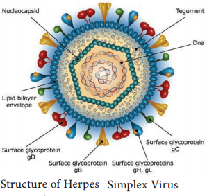
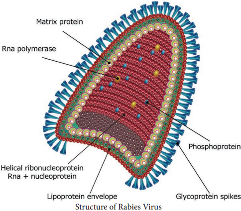
The rabies virus is bullet shaped, with one end rounded or conical and the other planar or concave. The lipoprotein envelope, carries knob like spikes, composed of glycoprotein G responsible for pathogenesis, virulence and immunity beneath the envelope is the matrix (M) protein layer which may be invaginated at the planar end. The membrane may project outwards forming a bleb. The genome is unsegmented linear RNA (Figure 10.6).
Rabies Virus Types and its Laboratory Diagnosis img 1

The rabies virus isolated from natural human or animal infection is termed ‘the street virus’. Rabies has been recognized from very ancient times as a disease transmitted to humans and animals by the bite of ‘mad dogs’. The name rabies comes from the Latin word rabidus, meaning ‘mad’, derived from the Sanskrit root rabhas, for frenzy.

Pathogenesis

Human infection is usually caused by the bite of rabid dogs or other animals. The virus present in the saliva of the animal is deposited in the wound (Figure 10.7). Rarely, infection can also occur following non-bite exposures such as licks or aerosols.
Rabies Virus Types and its Laboratory Diagnosis img 2

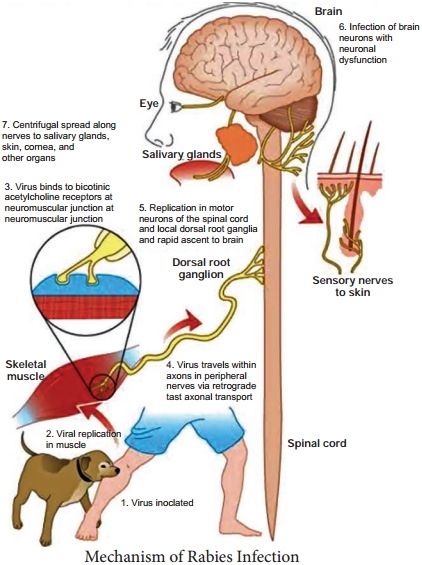
The virus appears to multiply in the muscles, connective tissue or nerves at the site of deposition for 48-72 hours. It penetrates the nerve endings and travels in the axoplasm towards the spinal cord and brain, at speed of about 3 mm per hour. The virus multiples and spreads centrifugally along the nerve trunks to
various parts of the body including the salivary glands. It multiplies in the salivary glands and is shed in the saliva.

The virus reaches every tissue in the body and dissemination may be interrupted at any stage by death. In humans the incubation period is usually from 1-3 months, short as 7 days or as long as three years. The incubation period is usually short in persons bitten on the face or head and long in those bitten on the legs. This may be related to the distance the virus has to travel to reach the brain. The incubation period is generally shorter in children than in adults.

The four stages of the disease are as follows, prodrome, acute encephalitic phase, coma and death. The onset is marked by symptoms such as fever, headache, malaise, fatigue and anorexia, anxiety, agitation, irritability, nervousness, insomnia or depression. The neurological phase begins with hyperactivity. Attempts to drink during such painful spasms of the pharynx and larynx produce choking or gagging that patients develop a dread of even the sight or sound of water (hydrophobia).

Animal Infection

In dogs, the incubation period is usually 3-6 weeks but it may range from 10 days to a year. The initial signs are an alert,troubled air and restlessness,snapping at imaginary objects, licking or gnawing at the site of the bite. After 2-3 days of this prodromal stage, the disease develops into either the furious or dumb types of rabies. In furious rabies, dog runs biting without provocation, the lower jaw droops and saliva drools from the mouth.

Paralysis convulsions and death follow. In dumb rabies, is the paralytic form, animals lies huddled, unable to feed. About 60% of rabid dogs shed the virus in saliva.
Rabid dogs usually die in 3-5days.

Laboratory Diagnosis

Human Rabies

The specimens tested are corneal smears and skin biopsy. Commonly used method for diagnosis is the demonstration of rabies virus antigens by immuno fluorescence. Direct immunofluorescence is done using antirabies serum tagged with fluorescein isothiocyanate.

Negri bodies in the brain, are demonstrated, Isolation of the virus by intracerebral inoculation in mice can be attempted from the brain, CSF, saliva and urine. The mice are examined for signs of illness, and their brains are examined after death.

Animal Rabies

The whole carcassof the animal suspended to have died of rabies may be sent to the laboratory. The brain may be removed sent for biological test and microscopy respectively. The portion of brain sent should include the hippocampus and cerebellum as negri bodies are most abundant. The following tests are done in the laboratory.

  1. Demonstration of rabies virus antigen by immuno fluorescence
  2. Demonstration of inclusion bodies – Negri bodies are seen as intracytoplasmic, round or oval purplish pink with characteristic basophilic inner granules. Negribodies vary in size from 3.27 Mm.

Antirabic Vaccines

Antirabic vaccines fall into two main categories neural and non-neural.

Neural Vaccines

Suspension’s of nervous tissues of animals infected with the fixed rabies virus. Following are the modified forms.

1. Semple Vaccine:

Vaccine developed by semple (1911). It is a 5% suspension of sheep brain infected with fixed virus and inactivated with phenol at 37°C leaving noresidual live virus.

2. Beta propiolactone (BPL) Vaccine:

Beta propiolactone is used as the inactivating agent instead of Phenol.

3. Infant Brain Vaccine:

The enceptalitogenic factor in brain tissue is a basic protein associated with myelin. Vaccines were developed using infant mouse, rat or rabbit brain. Infant brain vaccine is impractical in India.

Non-neural Vaccines

Non-neural vaccines includes

  1. Egg Vaccines
  2. Tissue Culture Vaccines
  3. Subunit Vaccine

Passive Immunisation

Human rabies immune globulin (HRIG) is free from the danger of sensitization but should be ensured free from HIV and hepatitis viruses.

Vaccines for Animals

Antirabies immunization in animals is to be done as pre-exposure prophylaxis concentrated cell culture vaccines – inactivated virus gives good protection after a single Intramuscular injection. Injections are given at 12 weeks of age and repeated at 1-3 years intervals.

Amines Class 12 Notes Chemistry Chapter 13

By going through these CBSE Class 12 Chemistry Notes Chapter 13 Amines, students can recall all the concepts quickly.

Amines Notes Class 12 Chemistry Chapter 13

Amines: Amines can be considered as derivatives of ammonia, obtained by replacement of H of NH3 by alkyl /aryl group.
Amines Class 12 Notes Chemistry 1
→ Structure of Amines: Like ammonia, the N atom of amines is trivalent and Carnes an unshared pair of electrons. N orbitals in amines are sp3 hybridized and the geometry of amines is pyramidal.
Amines Class 12 Notes Chemistry 2
(Pyramidal shape of trimethylamine)

→ Classification of Amines: Amines are classified as primary (1° secondary (2°) and tertiary (3°) depending upon the no. of hydrogen atoms replaced by alkyl/aryl groups in ammonia molecule.
Amines Class 12 Notes Chemistry 3
Amines are simple if R = R’ = R”
They are termed as mixed when R, R’, R” are different.

→ Nomenclature of Amines: In the IUPAC system ‘e’ of the alkane is replaced by ‘amine’ like Alkanamine. The simplest arylamine is aniline (C6H5—NH2) or benzene amine
Amines Class 12 Notes Chemistry 4
→ Preparation of Amines:
1. Reduction of Nitro Compounds
Amines Class 12 Notes Chemistry 5
2. Ammonolysis of Alkyl Halides
Amines Class 12 Notes Chemistry 6
The order of reactivity of halides is RI > RBr > RCI.
Amines Class 12 Notes Chemistry 7
3. Reduction of Nitriles
Amines Class 12 Notes Chemistry 8
4. Reduction of amides
Amines Class 12 Notes Chemistry 9
5. Gabriel Phthalimide Synthesis: This method is used to prepare pure primary amines. Aromatic amines cannot be prepared by this method.
Amines Class 12 Notes Chemistry 10
Hoffman Bromamide Degradation Reaction: Primary amines are obtained when an amide is treated with Br2 in an aqueous solution of NaOH.
Amines Class 12 Notes Chemistry 11
Physical Properties:

  1. Lower aliphatic amines are gases with a fishy odour. Primary amines with three or more C atoms are liquids and still higher ones are solids.
  2. Lower aliphatic amines are soluble in water because they can form H-bonds with water molecules. Higher amines are essentially insoluble in water.
  3. The order of boiling points of isomeric amines is Primary > Secondary > Tertiary. It is due to the presence of intermolecular H-bonding which is more in primary amines (due to the presence of two H- atoms attached to N than only one in secondary amines), less in 2° amines and absence in tertiary amines.
    Amines Class 12 Notes Chemistry 12

Chemical Reactions:
1. Basic character of Amines: Amines being basic in character (Lewis bases) react with acids to form salts.
Amines Class 12 Notes Chemistry 13
The basic character of amines can be better understood in terms of their PKb and kb values as explained below:
Amines Class 12 Notes Chemistry 14
Amines Class 12 Notes Chemistry 15
The larger the value of Kb or lower the value of PKb, the stronger is the base. PKb value of ammonia is 4.75. Aliphatic amines are stronger bases than ammonia due to the + I-effect of alkyl groups. The availability of lone pair of electrons on N increases. On the other hand, aromatic amines are weaker bases than ammonia due to the electron-withdrawing nature of the aryl group. The PKb values of few amines are given below:

Table: PKb values of Amines in Aqueous Phase:

Name of aminePKb
Methenamine3.38
N-Methylmethanamine3.27
N, N-Dimethylmethanamine4.22
Ethanamine3.29
N-Ehtylethamine3.25
Benzenamine9.38
Phenylmethanamine4.70
N-Methylaniline9.30
N, N-Dimethylaniline8.92

Besides the + I or – I effect, the interplay of other factors like solvation effect, steric hindrance etc. affect the basic strength of amines.

→ Structure-Basicity Relationship of Amines: The basicity of amines is related to their structure. The basic character of an amine depends upon the ease of formation of the cation by accepting a proton from the acid.

The more stable the cation is relative to the amine, the more basic is the amine.
(a) Alkamines versus Ammonia
Let us consider the reaction of an alkanamine and ammonia with a proton to compare their basicity.
Amines Class 12 Notes Chemistry 16
Due to the electron releasing nature of the alkyl group, it (R) pushes electrons towards nitrogen and thus makes the unshared electron pair more available for sharing with the proton. Moreover, the substituted ammonium ion formed from the amine gets stabilised due to dispersal of the positive charge by the +1 effect of the alkyl group. Hence, alkylamines are stronger bases than ammonia.

Thus the basic nature of aliphatic amines should increase with the increase in the number of alkyl groups. This trend is followed in the gaseous, phase. The order of basicity of amines in the gaseous phase follows the expected order: tertiary amine > secondary amine > primary amine > NH3.

The trend is not regular in the aqueous state as evident by their PKb values given in Table. In the aqueous phase, the substituted ammonium cations get stabilised not only by the electron releasing effect of the alkyl group (+1) but also by hydrogen bonds and solvation with water molecules. The greater the size of the ion, the lesser will be the solvation and the less stabilised the ion. The order of stability of ions are as follows:
Amines Class 12 Notes Chemistry 17
Decreasing order of extent of H-bonding in water and stabilization by solvation: Greater the stability of the substituted ammonium cation, the stronger base is the corresponding amine. Thus the order of basicity of aliphatic amines should be primary > secondary > tertiary, which is opposite to the inductive effect based order. Secondly, when the alkyl group is small like – CH3 group, there is no steric hindrance to H-bonding.

In case the alkyl group is bigger than the CH3 group, there will be some steric hindrance to H-bonding. Therefore, the change of nature of the alkyl group, e.g., from CH3 to – C2H5 results in a change in the order of basic strength. Thus, there is a subtle interplay of the inductive effect, solvation effect and steric hindrance of the alkyl group which decides the basic strength of alkylamines in the aqueous state.

The order of basic strength in the case of methyl-substituted amines and ethyl substituted amines in an aqueous solution is as follows:
(C2H5)2 NH > (C2H5)3N > C2H5NH2 > NH3
(CH3)2NH > CH3NH2 > (CH3)3N > NH3

(b) Arylamine Vs Ammonia: High value of PKb of aniline suggests that it is a much weaker base than aliphatic amines or even ammonia. It is due to the resonance shown by aniline.
Amines Class 12 Notes Chemistry 18
Aniline is a resonance hybrid of 5 canonical structures out of which there is no lone pair of electrons available for sharing on N in II, III and IV structures. It results in the unshared electron pair on N being in conjugation with the benzene ring making it less available for protonation.

On the other hand, aclidinium ion obtained by accepting a proton can have only two resonating structures (Kekule) as shown below:
Amines Class 12 Notes Chemistry 19
The greater the no, of resonating structures, the greater is the stability. Thus aniline is more resonance stabilized than anilinium ion. Hence the proton acceptability or the basic nature of aniline and other aromatic amines would be less than ammonia.

In the case of substituted aniline, electron-releasing groups like – OCH3 – CH3 increase the basic strength whereas electron-withdrawing groups like – NO2, – SO3H, – COOH, – X decrease it.
Thus (C2H5)2 NH > C2H5NH2 > NH3 > C6H5NH2

2. Alkylation: Amines undergo alkylation with alkyl halides.
Amines Class 12 Notes Chemistry 20
3. Acylation: Aliphatic and aromatic primary and secondary amines react with acid chlorides, anhydrides and esters by a nucleophilic substitution reaction. This reaction is known as acylation.

It is the replacement of H of – NH2 group
or
Amines Class 12 Notes Chemistry 21
group by the acyl group.
Amines Class 12 Notes Chemistry 22
Amines Class 12 Notes Chemistry 23
The reaction of amines with benzoyl chloride (CH5 COCl) is called Benzoylation.
Amines Class 12 Notes Chemistry 24
4. Carbylamine reaction: It is a test of primary amines-both aliphatic and aromatic.
Amines Class 12 Notes Chemistry 25
5. Reaction with Nitrous acid: All the three classes of amines react differently with nitrous acid, HNO2 (prepared in situ from HCl + NaNO2)
Amines Class 12 Notes Chemistry 26
Amines Class 12 Notes Chemistry 27
Secondary and tertiary amines react differently.

6. Reaction with aryl sulphonyl chloride: Benzensulphonyl chloride (C6H5SO2Cl) called Hinsberg’s reagent reacts with 1° and.2° amines.
Amines Class 12 Notes Chemistry 28
(c) Tertiary amines (R3N) do not react with benzene sulphonyl chloride. This property of 1°, 2° and 3° amines is made use of in their distinction and separation Now in place of benzene sulphonyl chloride,

p-toluenesulphonyl chloride is used.
Amines Class 12 Notes Chemistry 29
7. Electrophilic Substitution: NH2 group fused in the ring is a powerful activating group and is ortho and para directing.
(a) Bromination:
Amines Class 12 Notes Chemistry 30
Protection of the highly activating -NH2 group can be done by acetylation with acetic anhydride in which case only monobromo substituted aniline is the product.
Amines Class 12 Notes Chemistry 31
The lone pair of electrons on N of acetanilide interacts with oxygen atom due to resonance as shown below:
Amines Class 12 Notes Chemistry 32
Hence the lone pair of electrons on nitrogen is less readily available for donation to the benzene ring by resonance. Thus the activating effect of – NHCOCH3 is less than that of the – NH2 group.

(b) Nitration: Direct nitration of aniline yields tarry oxidation products in addition to nitro derivatives.
Amines Class 12 Notes Chemistry 33
However, by protecting the amino group by acetylation reaction with acetic anhydride the nitration can be controlled and the para nitro derivative obtained as the major product.
Amines Class 12 Notes Chemistry 34
(c) Sulphonation:
Amines Class 12 Notes Chemistry 35
Aniline does not undergo Friedel Crafts reaction (alkylation and acetylation) due to the salt formation with AlCl3– the Lewis acid-which is used as a catalyst. The N of -NH2 acquires a positive charge and hence acts as a strong deactivating group for further reaction.

Diazonium Salts are of the type Ar N2+ X where Ar stands for an aryl group and X can be Cl, Br, HSO4, BF4etc. N2+ group is called the diazonium group.

C6H5 N2+Cl is called benzene diazonium chloride and C6H5 N2+ HSO4is known as benzene diazonium hydrogen sulphate. The stability of the arene diazonium ion is explained on the basis of resonance.
Amines Class 12 Notes Chemistry 36
Method of Preparation:
Amines Class 12 Notes Chemistry 37
Physical Properties:
1. It is a colourless crystalline solid.
2. It is readily soluble in cold water and stable in it but reacts with water when warmed.
3. It decomposes easily in the dry state. Therefore it is used immediately after its preparation and is not stored.

Chemical Reactions A: Reactions involving displacement of Nitrogen -N2+ group is a very good leaving group. Therefore, it is substituted by other groups such as Cl, Br, I, CN and OH which displace nitrogen from the aromatic ring.
1. Replacement by halide or cyanide ion: Sandmeyer Reaction
Amines Class 12 Notes Chemistry 38
Alternatively, chlorine or bromine can also be introduced in the benzene ring by treating diazonium salt solution with corresponding halogen acid in the presence of copper powder. This is referred to as the Gatterman reaction.
Amines Class 12 Notes Chemistry 39
The yield in the Sandmeyer reaction is found to be better than the Gattermann reaction.

2. Replacement by Iodide Ion: Iodine is not easily introduced into the benzene ring directly, but, when the diazonium salt solution is treated with potassium iodide, iodobenzene is formed.
Ar N2+ Cl + KI → Arl + KCl + N2

3. Replacement by Fluoride Ion: When arene diazonium chloride is treated with fluoroboric acid, arene diazonium fluoroborate is precipitated which on heating decomposes to yield aryl fluoride.
Amines Class 12 Notes Chemistry 40
4. Replacement by H: Certain mild reducing agents like hypophosphorous acid (phosphinic acid) or ethanol reduce diazonium salts to arenes and themselves get oxidised to phosphorous acid and ethanol, respectively.
Ar N2+ Cl + H3PO2 + H2O → ArH + N2 + H3PO3 + HCl
Ar N2+ Cl + CH3CH2OH → ArH + N2 + CH3CHO + HCl

5. Replacement by hydroxyl group: If the temperature of the diazonium salt solution is allowed to rise up to 283 K, the salt gets hydrolysed to phenol.
Ar N2+ Cl + H2O → ArOH + N2 + HCl

6. Replacement by – NO2 group: When diazonium fluoroborate is heated with aqueous sodium nitrite solution in the presence of copper, the diazonium group is replaced by the NO2 group.
Amines Class 12 Notes Chemistry 41
(B) Reactions involving retention of diazo group: Coupling Reactions
(a) Reaction with Phenol in the presence of a weakly alkaline medium results in the formation of p-hydroxy azobenzene which is an orange dye.
Amines Class 12 Notes Chemistry 42
(b) Reaction with aniline: It reacts with aniline in a weakly acidic medium to form p-amino azobenzene (yellow dye)
Amines Class 12 Notes Chemistry 43
→ Importance of Diazonium salts in the synthesis of Aromatic Compounds: These salts are very good intermediates used to introduce – F, – Cl, – Br,-I, – CN, – OH, – NOz groups into the benzene ring. Ar-F or Ar-I cannot be prepared by direct halogenation. The cyanobenzene which cannot be prepared by the SN reaction of chlorobenzene can be easily obtained from diazonium salts. These compounds are useful for preparing several azo dyes by coupling reactions.

Hepatitis Viruses | Hepatitis Viruses Types, Laboratory Diagnosis

Learninsta presents the core concepts of Microbiology with high-quality research papers and topical review articles.

Hepatitis Viruses | Hepatitis Viruses Types, Laboratory Diagnosis

The term viral hepatitis refers to a primary infection of the liver, hepatitis viruses consists of types A, B, C, D, E and G. Except for type B which is a DNA virus all the others are RNA viruses. Two types of viral hepatitis had been recognised. Type one affects mainly children and young adults and transmitted by the fecal-oral route called as infective or infectious hepatitits or type A hepatitis.

Second type transmitted mainly by receiving serum inoculation or blood transfusion named as homologous serum jaundice, serum hepatitis transfusion hepatitis or type B hepatitis.

Type A Hepatitis (HAV)

HAV is a 27nm non enveloped RNA virus belonging to the picorna virus family. It is designated as ‘entero virus 72’, HAV is recognised as new genus ‘Hepatovirus’. It can be grown in human and simian cell cultures and is the only human hepatitis virus which can be cultivated in vitro.

HAV transmission is by the fecal oral route. Infection is by ingestion. The virus multiplies in the intestinal epithelium and reaches the liver by hematogenous spread. Once jaundice develops, it is rarely detectable in feces. The incubation period is 2-6 weeks. The clinical disease consists of two stages the prodromal and
the icteric stage. The onset may be acute with fever, malaise, anorexia, nausea, vomiting and liver tenderness.

These usually subside with the onset of jaundice. Recovery is slow, over a period of 4-6 weeks. The disease is milder in children. Type A hepatitis caused by contaminated food, water or milk. Over crowding and poor sanitation favour its spread.

Laboratory Diagnosis

Diagnosis of type A hepatitis may be made by demonstration of the virus or its antibody. Virus can be visualized by Immunelectron Microscopy (IEM) in fecal extracts during the late incubation period.

IgM anti-HAV antibody appears during the late incubation period disappears after 3-4 months. IgG peaks in 3-4 months and persists much longer for life. ELISA kits for detection of IgM and IgG antibodies are available.

A safe and effective formalin inactivated, alum conjugaged vaccine containing HAV grown in human diploid cell culture is used. Course consists of two intra muscular injections of the vaccine. Protection begins 4 weeks after injection and lasts for 10 to 20 years. No specific antiviral drug is available.

Type B Hepatitis (HBV)

HBV is a 42nm DNA virus with an outer envelope and an inner core 27nm in diameter. Enclosing the viral genome and a DNA polymerase. It belongs to the family Hepadna Viridae HBV is ‘Hepadna Virus type 1’. Australia antigen was found to be associated with serum hepatitis. It was the surface component of HBV, so named as hepatitis B surface antigen (HBsAg).

3 types of particles are visualized, most abundant form is a spherical particle, 22nm in diameter. The second type of particle is filamentous or tabular with a diameter of 22nm both are antigenically identical. Third type of particle are fewer in number, is a double walled spherical structure 42 nm in diameter. This particle is the complete hepatitis B virus, known as Dane particle.

The envelope proteins expressed on the surface contains hepatitis B surface antigen (HBsAg). HBsAg consists of two major polypeptides, one of which is glycosylated. The nucleocapsid or core contains hepatitis B core antigen (HBcAg A) (Figure 10.5). Third antigen called the hepatitis B e antigen (HBeAg)
is a soluble non particulate nucleocapsid protein.
Hepatitis Viruses img 1

The nucleocapsid encloses the viral genome consisting of two linear strands of DNA held in a circular configuration. One of the strands is incomplete (+ strand) DNA appears partially double stranded and partially single stranded. Associated with the + strand is a viral DNA polymerase (has both DNA dependent
DNA polymerase and RNA dependent reverse transcriptase functions).

This polymerase can repair the gap in the plus strand and render the genome fully double stranded. Natural infection occurs only in humans. The virus is maintained in carriers whose blood contains circulating virus for long periods. Carriers are of two categories, the highly infectious super carriers and the simple carriers. Former have high titre HBsAg along with HBsAg, DNA polymerase and HBV in ciruculation.

Simple carriers have low infectivity and low titre HBsAg in blood. HBV is a blood borne virus and the infection is transmitted by parenteral, sexual and perinatal models. The virus may also be present in other body fluids and excretions such as saliva, breast milk, semen, vaginal secretions, urine bile and feces of these semen and saliva are known to transmit the infection very commonly.

Transfusion of carrier blood once, the most widely known mode of infection has largely been eliminated by donor screening that is strictly enforced. Infection by direct contact with open skin lesions such as pyoderma, eczema, cuts and scratches is very common among young children in developing countries.

Certain groups and occupations carry a high risk of infection. These include medical and paramedical staff of blood banks, dialysis units, barbers, sex workers. The incubation period is long about 1-6 months. The onset is insidious and fever is not prominent.

Extra hepatic complications like arthralgia, urticaria and glomerulonephritis may occur. About 90-95% of adults with acute hepatitis B infection recover within 1-2months of onset and eliminate the virus from the body. They may be Asymptomatic carriers or may progress to recurrent or chronic liver disease.

Laboratory Diagnosis

Serology

Diagnosis of hepatitis B depends on the serological demonstration of the viral markers. HBsAg is the first marker to appear in blood after infection, being detectable. It remains in circulation throughout the symptomatic course of the disease (2-6months). Anti HBs is the protective antibody.

HBcAg is not demonstrable in circulation because it’s enclosed within the HBsAg coat but its antibody, anti HBc appears in serum a week or two after the appearance of HBsAg.

As anti HBc remains life long, it serves as a useful indicator of prior infection with HBV. HBeAg appears in blood concurrently with HBsAg, indicating the high infectivity. Molecular methods such as DNA: DNA hybridization and PCR at present used for HBV DNA testing are highly sensitive and quantitative.

Immunization

Both passive and active methods of immunization are available. Active immunization is more effective. The currently preferred vaccine is genetically engineered by cloning the S gene for HBV in Baker’s yeast. A special vaccine containing all antigenic components of HBsAg (Pre-S1, Pre-S2 and s) has been developed. No specific antiviral treatment is available for acute HBV infection.

Herpes Viruses Herpes Viruses | Herpes Viruses Structure, Classification, Clinical Features, Laboratory Diagnosis

Learninsta presents the core concepts of Microbiology with high-quality research papers and topical review articles.

Herpes Viruses | Herpes Viruses Structure, Classification, Clinical Features, Laboratory Diagnosis

The herpes virus family contains more than a hundred species of enveloped DNA viruses that affect humans and animals.

Structure

The herpes virus capsid is icosahedral, composed of 162 capsomers and enclosing the core containing the linear double stranded DNA genome. The nucleocapsid is surrounded by the lipid envelope derived from the host cell. The envelope carries surface spikes (Figure 10.4). Teguments are present in between the envelope and capsid. The enveloped virion measures about 200nm and the naked virion about 100 nm in diameter.
Herpes Viruses img 1

Classification

Herpes virus belongs to the family Herpesviridae.

i. Alpha herpes viruses

They have relatively short replicative cycle (12-18 hours) and a variable host range. They cause latent infection in sensory ganglia. Example: Herpes simplex virus and varicella zoster virus.

ii. Beta herpes viruses

They replicate slowly (more than 24 hours) and have a narrow host range, grow well in fibroblasts. They cause latent infection of salivary gland and other organs. Example: Cytomegalovirus.

iii. Gamma herpes viruses

They have a narrow host range and replicate in lymphoblastoid cells. They are specific for either B or T lymphocytes and causes latent infection in lymphoid tissue Example: Epstein – Barr Virus. Eight different types of herpes viruses are known whose primary hosts are humans. They have been designated as Human herpes virus type 1-8.

1. Herpes Simplex

The herpes simplex virus (HSV) occurs naturally only in humans, but it can produce experimental infection in laboratory animals. There are two types of the herpes simplex virus. HSV type 1 (Human herpes virus type 1) is isolated from lesions in and around the mouth and is transmitted by direct contact or droplet spread from carrier. HSV type 2 (Human herpes virus type 2 or HHV type 2) is responsible for the genital herpes infections transmitted venereally.

Pathogenesis

Herpes simplex is one of the most common viral infection in humans, the sources of infection are saliva, skin lesions or respiratory secretions. In type 2, transmission occurs by close contact and may be veneral in genital herpes. The virus enters through defects in the skin or mucous membranes and multiples
locally, with cell to cell spread.

The herpes lesions are thin walled, umbilicated vesicles, the roof of which breaks down, leaving tiny superficial ulcers. They heal without scarring.

Clinical features

The clinical manifestations depend on the site of infection, age and immune status of the host and the antigenic type of the virus. They are

  • Cutaneous infections
  • Mucosal infections
  • Ophthalmic infections
  • Nervous system infections
  • Visceral infections
  • Genital infections

Laboratory diagnosis

Microscopy

Smears are prepared from the lesions, from the vesicles and stained with 1% aqueous solution of toluidine blue ‘O’ for 15 seconds. Multinucleated giant cells with faceted nuclei with ground glass chromatin (Tzanck cells) are observed.

Virus isolation

Inoculation in mice and on chick embryo CAM is insensitive. Primary human embryonic kidney, human amnion cells are susceptible, but human diploid fibroblasts are preferred. Vesicle fluid, spinal fluid, saliva and swabs may be used. Cytopathic changes may appear as early as 24-48 hrs.

Serology

Antibodies develop within a few days of infection and rise in titre of antibodies may be demonstrated by ELISA, neutralization or complement fixation tests. Chemotherapy Indoxyuridine used topically in eye and skin infection, acyclovir and vidarabine are given for deep and systemic infections.

2. Varicella Zoster

In 1889, Von Bokay had suggested that varicella (Chicken pox) and herpes zoster are different manifestations of the same virus infection. The virus is therefore called Varicella zoster virus (VZV). The chicken pox follows primary infection in a non immune individual, while herpes zoster is a reaction of the latent virus when the immunity has fallen to infective levels.

VZV is similar to the herpes simplex virus in its morphology. It can be grown in cultures of human fibroblasts human amnion or HeLa cells. Chicken pox is one of the mildest and most common of child hood infections. The disease may, occur at any age.

3. Cytomegaloviruses

Cytomegaloviruses (CMV) formerly known as salivary gland viruses are a group of ubiquitous herpes viruses of humans and animals. They are characterized by enlargement of infected cells and intranuclear inclusions.

In 1926, cytomegalia presumed to be due to viral infection was reported in the salivary glands of guinea pigs and children and the viral agent was called the ‘salivary gland virus’.

4. Epstein – Barr Virus

A number of different viruses apparently ‘Passenger Viruses’ were isolated from cultured lymphoma cells. Epstein, Barr and Achong in 1964 observed a new type of herpes virus and named it has ‘EB Virus’ affecting B lymphocytes of only human and some sub human primate B cells have receptors (CD21 molecules)
for the virus.

The source of infection is usually the saliva of infected persons who shed the virus in oropharyngeal secretions. Intimate oral contact,as in kissing, appears to be the predominant mode of transmission. This accounts for infectious mononucleosis being called as ‘The kissing disease’.

5. Human Herpes Virus Types 6, 7, 8

A herpes virus, first isolated in 1986 from the peripheral blood of patients with lympho proliforative disease called as human B lymphotropic virus, renamed as HHV – 6. HHV – 7 was isolated in 1990 from peripheral CD4 cells of a healthy person appears to be widely distributed and transmitted through saliva.

In 1994, DNA sequences presumed to represent a new herpes virus from tissues of Kaposi’s sarcoma from AIDS patients was named as HHV8. Later Kaposi’s sarcoma was identified in persons not infected with HIV and referred to as Kaposi’s Sarcomaassociated Herpes Virus (KSHV).

Cultivation of Viruses Techniques

Learninsta presents the core concepts of Microbiology with high-quality research papers and topical review articles.

Cultivation of Viruses

Viruses areobligate intracellularparasites; they cannot be grown on any inanimate culture medium. Three methods are employed for the cultivation of viruses – inoculation into animals, embryonated eggs and tissue culture or cell culture.

i. Animal Inoculation

The earliest method for the cultivation of viruses causing human diseases was inoculation into human volunteers. Monkeys were used for the isolation of the polio virus by Landsteiner and Popper (1909). The embryonated hen’s egg was first used for cultivation of viruses by Good pasture (1931). The embryonated egg offers several sites for the cultivation of viruses. Non human primates provide the only method for virus cultivation. Mice are most widely employed animals in Virology.

ii. Embryonated Eggs

a. Chorioallantonic Membrane (CAM)

Inoculation on the chorioallantonic membrane produces visible lesions (pocks). Different viruses have different pock morphology. Example: variola or vaccinia

b. Allantonic Cavity

Inoculation on the allantonic cavity provides a rich yield of influenza and some paramyxo viruses.

c. Amniotic Sac

Inoculation into the amniotic sac is for the primary isolation of the influenza virus.

d. Yolk Sac

Inoculation into the yolk sac is for the cultivation of some viruses like Chlamydiae and Rickettsiae. Allantonic inoculation is employed for growing influenza virus for vaccine production (Figure 10.3).
Cultivation of Viruses img 1

iii. Tissue Culture

First tissue culture in Virology was maintained by Steinhardt and colleagues (1913) for the vaccinia virus in fragments of rabbit cornea. Bacterial contamination was the major limitation. Different types of culture used are:

a. Organ culture

Small bits of organs can be maintained, used for the isolation of some viruses.
Example: Corona virus (respiratory pathogen) cultured on tracheal ring organ culture.

b. Explant culture

Fragments of minced tissue are grown as ‘explants’. This is also known as tissue culture.
Example: Adeno virus cultured on Adenoid tissue explants.

iv. Cell Culture

Tissues are dissociated into the component cells by the action of enzymes (trypsin) or by mechanical process and are suspended in a growth medium (amino acids, vitamins, salts, glucose) supplemented with fetal calf serum of antibiotics and indicator (Phenol red).

This media is dispensed in bottles, tubes or petridishes. The cells adhere to the glass surface and on incubation divides to form a confluent monolayer sheet of cells covering the surface within about a week. The cell culture is classified into three types.

a. Primary cell cultures

In this culture, normal cells are taken from the body and cultured. They are capable of only limited growth in culture. Example: Monkey kidney, Human embryonic kidney, Chick embryo cell culture.

b. Diploid cell strains

These are cells of a single type that retain the original diploid chromosome number and serotype during serial sub cultivation for limited number of times. Example: Human fibroblast.

c. Continuous cell lines

These are single type, derived from cancer cells that are capable of continuous serial cultivation.
Example: Cells derived from cancers, such as Hela, Hep-2 and KB cell lines.