Immunology of Western Blot Techniques Principle and its Applications

Learninsta presents the core concepts of Microbiology with high-quality research papers and topical review articles.

Immunology of Western Blot Techniques

Macromolecules immobilized or fixed on nitrocellulose membrane i.e., blotted can be subjected to a variety of analytical techniques more easily. Southern blotting was the first blotting technique developed which made the analysis and recording of DNA easy.

Later the technique was extended for analysis of RNA and proteins and they have acquired the jargon terms Northern and Western Blotting respectively.

Western blotting is also known as immunoblotting because it uses antibodies to detect the protein. Western blotting is a quantitative test to determine the amount of protein in sample.

Principle

Western blotting technique is used for the identification of a particular protein from the mixture of a proteins. In this method, the proteins are first extracted from the sample. Extracted proteins are subjected to Poly Acryl – amide Gel Electrophoresis (PAGE).

Transfer of proteins from poly acryl amide to the nitrocellulose paper is achieved by applying electric field. When radio labelled specific antibody is added on such membrane it binds to the specific complementary protein. Finally the proteins on the membrane can be detected by staining or through ELISA technique.

Steps

Step I:
Extraction of Protein

The most common protein sample used for Western blotting is cell lysate. The protein from the cell is generally extracted by mechanical means or by adding chemicals which can lyse the cell. The extraction step is termed as tissue preparation.

Protease inhibitor is used to prevent the denaturing of proteins. Using spectroscopy the concentration of the protein sample is analysed and diluted in loading buffer containing glycerol. This will help the sample to sink in the well. Bromothymol blue is used as tracking dye and is used to monitor the movement of the sample.

Step II:
Gel electrophoresis

The protein sample is loaded in well of SDS-PAGE (Sodium dodecyl sulfatepoly-acryl amide gel electrophoresis). The proteins are separated on the basis of electric charge, isoelectric point, molecular weight, or combination of all these. Proteins are negatively charged, so they move toward positive (anode) pole as electric current is applied. Smaller proteins move faster than the larger proteins.

Step III:
Blotting

Blotting refers to the transfer of the protein from the gel to the nitrocellulose paper by capillary action. Electro blotting is done nowadays to speed up the process. In electro-blotting nitrocellulose membrane is sandwich between gel and cassette of filter paper and then electric current is passed through the gel causing transfer of protein to the membrane.

Step IV:
Blocking

The nitrocellulose membrane is nonspecifically saturated or masked by using casein or Bovine serum albumin (BSA) before adding the primary antibody. This blocking step is very important in western blotting as antibodies are also proteins and they are likely to bind to the nitrocellulose paper.

Step V:
Treatment with primary and secondary antibody

The primary antibody is specific to desired protein so it forms Ag-Ab complex. The secondary antibody is enzyme labelled and is against primary antibody (antiantibody) so it can bind with Ag-Ab complex. Alkaline phosphatase or Horseradish peroxidase (HRP) is labelled
with secondary antibody.

Step VI:
Treatment with suitable substrate

Finally, the reaction mixture is incubated with specific substrate. The enzyme convert the substrate to give visible coloured product, so band of colour can be visualized in the membrane (Figure 11.6).
Western Blot Techniques img 1

Application

  1. The size and concentration of protein in given sample is determined by western blotting.
  2. It is used in the detection of antibody against virus or bacteria in serum and helps in the disease diagnosis.
  3. Western blotting technique is the confirmatory test for HIV. It detects anti HIV antibody in patient’s serum.
  4. Useful to detect defective proteins.

Antigen Antibody Reactions and its types | Working principle, Applications

Learninsta presents the core concepts of Microbiology with high-quality research papers and topical review articles.

Antigen Antibody Reactions and its types | Working principle, Applications

The interaction between antigen and antibody is called antigen-antibody reactions. It is abbreviated as Ag-Ab reaction. This reaction is the basis of humoral immunity. The antigen and the antibody react to form immune complex.

Ag + Ab …………….. Ag – Ab complex The reaction between antigen and antibody is highly specific. It is compared to the lock and key system. The part of the antigen that combines with the antibody is called epitope or antigenic determinant. The part of antibody which combines with the antigen is called paratope or antigen determining site. Most of the antibodies have two binding sites and IgM has 5-10 binding sites.

Immunofluorescence

When antibodies are mixed with the fluorescent dyes such as fluorescein or rhodamine, they emit radiation. This phenomenon of emitting radiation by antibodies labelled with fluorescent dye is called immuno fluorescence. This reaction is well observed under fluorescent microscope. It is used to locate and identify antigens in tissues.

Types of Immunofluorescence

  • Direct method
  • Indirect method

Direct Method

In this method, the antibody labelled with fluorescent dye is directly applied on the tissue section. The labelled antibody binds with specific antigen. This can be observed under the fluorescent microscope.

Indirect Method

In this method, unlabelled antibodies are directly applied on the tissue sectionswhich bind with the specific antigens. Then the antibody labelled with the fluorescent dye is added to the tissue. Anti-antibody specifically binds with already added or linked unlabelled antibody (Figure 11.1).
Antigen Antibody Reactions img 1

ELISA (Enzyme Linked Immuno Sorbent Assay)

ELISA (Enzyme-Linked Immuno Sorbent Assay) is a plate-based assay technique designed for detecting and quantifying substances such as peptides, proteins, antibodies and hormones. It is also known as Enzyme Immuno Assay (EIA).

In 1971, after the descriptions of Peter Perlmann and Eva Engvall at Stockholm University in Sweden, ELISA has become the system of choice when assaying soluble antigens and antibodies. All assays for antibody production depend upon the measurement of interaction of elicited antibody with antigen.

Principle

The principle of ELISA is very simple. The test is generally conducted in micro titre plates. (Figure 11.2 Micro titre plate).
Antigen Antibody Reactions img 2

If the antigen is to be detected the antibody is fixed in the micro titre plate and vice versa. Test sample is added in the microtitre plate, if there is presence of Ag or Ab in the test sample, there will be Ag-Ab reactions (with immobilized Ab or Ag). Later enzyme labelled antibody is added in the reaction mixture, which will combine with either test antigen or Fc portion of test antibody.

The enzyme system consists of:

1. An enzyme:

Horse Radish Peroxidase(HRP), alkaline phosphatase which is labelled or linked, to a specific antibody.

2. A specific substrate:

  • O-Phenyl-diaminedihydrochloride for peroxidase
  • P nitrophenyl Phosphate – for Alkaline Phosphatase

Substrate is added after the antigenantibody reaction. The enzyme hydrolyses the substrate to give a yellow colour compound in case of alkaline phosphatase (Figure 11.3). The intensity of the colour is proportional to the amount of antibody or antigen present in the test sample, which can be quantified using ELISA reader
(Figure 11.4 ELISA reader)
Antigen Antibody Reactions img 3
Antigen Antibody Reactions img 4

Types

There are four kinds of ELISA assay tests. They are: Direct ELISA, Indirect ELISA, Sandwich ELISA and Competitive ELISA (Figure 11.5).
Antigen Antibody Reactions img 5

i. Direct ELISA

An antigen is immobilized in the well of an ELISA plate. The antigen is then detected by an antibody directly conjugated to an enzyme such as HRP. Direct ELISA detection is much faster than other ELISA techniques as fewer steps are required.

The assay is also less prone to error since fewer reagents and steps are needed, i.e. no potentially cross-reacting secondary antibody needed. Finally, the direct ELISA technique is typically used when the immune response to an antigen needs to be analyzed.

ii. Indirect ELISA

Indirect ELISA is used to detect antibody. A known antigen is coated on the micro titre plate. If the patient’s serum contains antibody specific to the antigen, the antibody will bind to the antigen.

After incubation the wells are washed and the enzyme labelled anti Human Gamma Globulin (HGG) is added to the well. AntiHGG can react with antigen antibody complex. The substrate for the enzyme is added finally which is hydrolysed by the enzyme which develops a colour.

iii. Sandwich ELISA

Sandwich ELISA is used to detect antigen. A known antibody is coated on the micro titre plate. A test antigen is added to each well and allowed to react with the bound antibody. If the patient’s serum contains antigen specific to the antibody, the antigen will bind to the antibody.

Specifically bound antigen and antibody will remain in the wells even after washing. The second antibody is added and allowed to react with bound antigen. Substrate is added to measure colour reaction.

iv. Competitive ELISA

It is used for the detection of antigens. Antibody is first incubated with a sample-containing antigen. The antigen and antibody complex is added to the antigen coated microtitre well. If more antigen present in the sample, the less free antibody will be available to bind to the antigen coated well.

Addition of an enzyme conjugated secondary antibody specific to the primary antibody can be used to determine the amount of primary antibody bound to the well. It is a quantitative test for the antigen detection.

Application

An ELISA test may be used to diagnose:

HIV, Lyme disease, pernicious anaemia, Rocky Mountain spotted fever, rotavirus, squamous cell carcinoma, syphilis, toxoplasmosis, varicella-zoster virus, which causes chickenpox and Zika virus.

Biomolecules Class 12 Notes Chemistry Chapter 14

By going through these CBSE Class 12 Chemistry Notes Chapter 14 Biomolecules, students can recall all the concepts quickly.

Biomolecules Notes Class 12 Chemistry Chapter 14

Carbohydrates: Most common examples of carbohydrates are glucose, fructose, cane sugar, starch etc. Most of them have a general formula Cx (H2O)y. Earlier they were considered hydrates of carbon. For example, glucose C6H12O6 fits into this general formula C6(H2O)6 But even acetic acid (CH3COOH) fits into this general formula C2(H2O) and it is not a carbohydrate. Similarly, rhamnose, C6H12O5 is a carbohydrate but does not fit into this definition.

Chemically, the carbohydrates may be defined as optically active polyhydroxy aldehydes or ketones or the compounds which produce such units on hydrolysis.

They are classified as:

  1. Sugars: They are sweet in taste and water-soluble, e.g. glucose, fructose, sucrose.
  2. Non-sugars: They are tasteless and water-insoluble, e.g., starch, cellulose. Carbohydrates are systemically classified as:

1. Monosaccharides: A carbohydrate that cannot be hydrolysed further to give simpler units of polyhydroxy aldehydes or ketones is called monosaccharides. Glucose (C6H1206) is an aldohexose and fructose (C6H1206) is a ketohexose.

2. Oligosaccharides: Carbohydrates that yield two to ten monosaccharides on hydrolysis are called oligosaccharides.
(a) Disaccharides: They hydrolyse to give two units of monosaccharides. They include sucrose, maltose, lactose.
Biomolecules Class 12 Notes Chemistry 1
(b) Trisaccharides: They yield three units of monosaccharides on hydrolysis, e.g. C18H32O16 (raffinose).
(c) Tetrasaccharides: Yields four units of monosaccharides on hydrolysis, e.g. stachyose C24H42O21

2. Polysaccharides: They yield a large number of monosaccharide units on hydrolysis: Common examples are starch, cellulose. They are not sweet in taste.

Reducing sugars are those which reduce Fehling’s solution and Tollen’s reagent. All monosaccharides whether aldoses and ketoses are reducing sugars.

Sugars that do not reduce Fehling solution or Tollen’s reagent are termed as non-reducing e.g., sucrose.

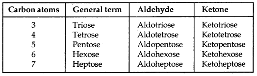
→ Monosaccharides: They contain three to seven carbon atoms. If they contain an aldehyde group (- CHO), they are termed aldoses. If they contain a keto group (C = O), they are termed ketoses.

Different Types of Monosaccharides:
Biomolecules Class 12 Notes Chemistry 2
1. Glucose:
Preparation:
(a) From Sucrose (Cane Sugar)
Biomolecules Class 12 Notes Chemistry 3
(b) From Starch:
Biomolecules Class 12 Notes Chemistry 4
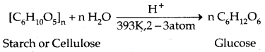
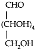
→ Structure of Glucose: It is an aldohexose and is also known as dextrose. Its structure (open chain) is
Biomolecules Class 12 Notes Chemistry 5
Evidence in favour of the above structure:

  1. Its molecular formula was determined to be C6H12O6.
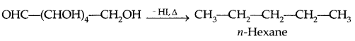
  2. On heating (prolonged) with HI, it formed an n-hexane suggesting that all the 6 carbon atoms are in a straight chain.
    Biomolecules Class 12 Notes Chemistry 6
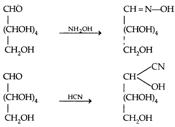
  3. It reacts with hydroxylamine to form an oxime and adds a molecule of hydrogen cyanide (HCN) to give cyanohydrin showing the presence of a carbonyl group in it,
    Biomolecules Class 12 Notes Chemistry 7
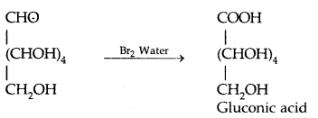
  4. Glucose is oxidised to gluconic acid by mild Oxidizing agent Br. water, confirming that a carbonyl group is an aldehyde group.
    Biomolecules Class 12 Notes Chemistry 8
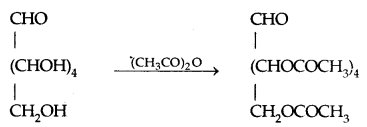
  5. Acetylation with acetic anhydride gives glucose pentaacetate which confirms the presence of five – OH groups attached to 5 different C atoms.
    Biomolecules Class 12 Notes Chemistry 9
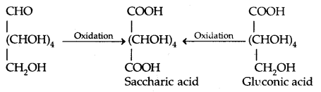
  6. on oxidation with nitric acid, glucose well as gluconic acid both yield a dicarboxylic acid, saccharic acid indicating the presence of -CH2OH group in it in addition to an aldehyde.
    Biomolecules Class 12 Notes Chemistry 10

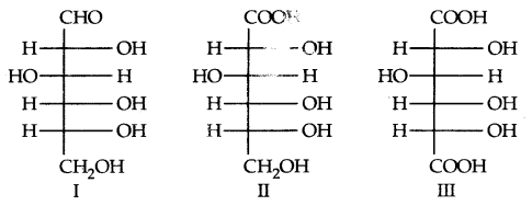
The exact spatial arrangement of different – OH groups was given by Fischer. Its exact configuration is correctly represented by I. Gluconic acid is II and Saccharic acid is III.
Biomolecules Class 12 Notes Chemistry 11
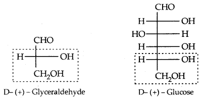
Glucose is correctly named as D (+) glucose. ‘D’ represents the configuration whereas (+) represents the dextro-rotatory nature of it. The meaning of D- and L- notations is given as follows:

[Note: It may be remembered that ‘D’ and ‘L’ notations have nothing to do with the optical activity of the compound.]

The letters ‘D’ or ‘L’ before the name of any compound indicate the relative configuration of a particular stereoisomer. This refers to their relationship with a particular isomer of glyceraldehyde. Glyceraldehyde contains one asymmetric carbon atom and exists in two enantiomeric forms as shown below.
Biomolecules Class 12 Notes Chemistry 12
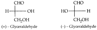
All those compounds which can be chemically correlated to (+) isomer of glyceraldehyde are said to have D-configuration whereas those which can be correlated to (-) isomer of glyceraldehyde are said to have L—configuration.

For assigning the configuration of monosaccharides, it is the lowest asymmetric carbon atom (as shown below) which is compared. As in (+) glucose, —OH on the lowest asymmetric carbon is on the right side which is comparable to (+) glyceraldehyde, so it is assigned D-configuration. For this comparison, the structure is written in a way that most oxidised carbon is at the top.
Biomolecules Class 12 Notes Chemistry 13
Cyclic Structure of Glucose

The structure (I) of glucose explained most of its properties but the following reactions and facts could not be explained by this structure.

  1. Despite having the aldehyde group, glucose does not give 2,4- DNP test, Schiff’s test and it does not form the hydrogen sulphite addition product with NaHSO3.
  2. The pentaacetate of glucose does not react with hydroxylamine indicating the absence of the free -CHO group.
  3. Glucose is found to exist in two different crystalline forms which are named a and b. The a-form of glucose (m.p. 419 K) is obtained by crystallization from a concentrated solution of glucose at 303 K while the (i-form (m.p. 423 K) is obtained by crystallisation from hot and saturated aqueous solution at 371 K,

This behaviour could not be explained by the open-chain structure (I) for glucose. It was proposed that one of the -OH groups may add to the -CHO group and form a cyclic hemiacetal structure. It was found that glucose forms a six-membered ring in which -OH at C-5 is involved in a ring formation. This explains the absence of -CHO group and also the existence of glucose in two forms as shown below. These two cyclic forms exist in equilibrium with an open-chain structure.
Biomolecules Class 12 Notes Chemistry 14
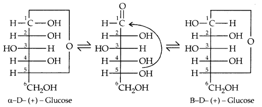
The two cyclic hemiacetal forms of glucose differ only in the configuration of the hydroxyl group at Cl, called anomeric carbon (the aldehyde carbon before cyclization). Such isomers, i.e., a-form and b-form, are called anomers.

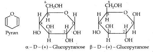
The six-membered cyclic structure of glucose is called the pyranose structure (α- or β-), in analogy with pyran. Pyran is a cyclic organic compound with one oxygen atom and five carbon atoms in the ring. The cyclic structure of glucose is more correctly represented by Haworth structure as given below:
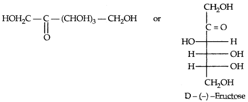
Biomolecules Class 12 Notes Chemistry 15
II. Fructose
Fructose is an important ketohexose. It is obtained along with glucose by the hydrolysis of disaccharide, sucrose. It has a ketonic group at C – 2. It belongs to D-series and is a laevorotatory compound. Therefore, it is written as D – (-) fructose. Its open-chain structures are given below:
Biomolecules Class 12 Notes Chemistry 16
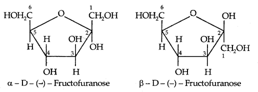
→ It differs from glucose only at C – 1 and C – 2. Its furanose form (cyclic) is:
Biomolecules Class 12 Notes Chemistry 17
→ The cyclic structures of two anomers of fructose as represented by Haworth are given below:
Biomolecules Class 12 Notes Chemistry 18
Disaccharides:
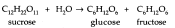
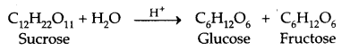
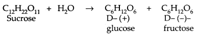
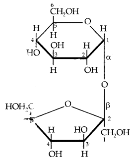
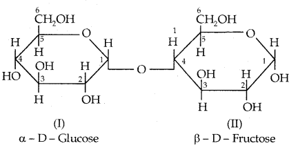
1. Sucrose: Sucrose on hydrolysis gives an equimolar mixture of D – (+) – glucose and D – (-) fructose.
Biomolecules Class 12 Notes Chemistry 19
Sucrose is a non-reducing sugar. Therefore, it has a glucoside linkage between C1 of α-glucose and C2 of β-fructose.
Biomolecules Class 12 Notes Chemistry 20
or
Biomolecules Class 12 Notes Chemistry 21
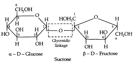
Sucrose is dextrorotatory but after hydrolysis gives dextrorotatory glucose and laevorotatory fructose. Since the laevorotation of fructose (- 92.4°) is more than the dextrorotation of glucose (+ 52.5°), the mixture is laevorotatory. Thus hydrolysis of sucrose brings about a change in the sign of rotation, from Dextro (+) to leave (-) and the product is named as invert sugar.

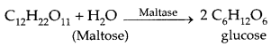
II. Maltose: Another disaccharide, maltose is composed of two α-D-glucose units in which C4 of one glucose (I) is linked to C4 of another glucose unit (II). Hie free aldehyde group can be produced at C1 of second glucose in solution and it shows reducing properties, so it is a reducing sugar.
Biomolecules Class 12 Notes Chemistry 22
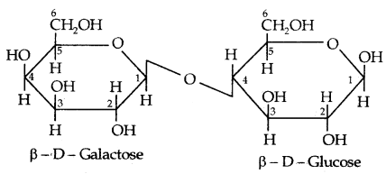
II. Lactose: It is more commonly known as milk sugar since this disaccharide is found in milk. It is composed of (β-D-galactose and β-D- glucose. The linkage is between C4 of galactose and C4 of glucose. Hence it is also a reducing sugar.
Biomolecules Class 12 Notes Chemistry 23
Polysaccharides: Polysaccharides contain a large number of monosaccharide units joined together by glycosidic linkages.
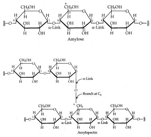
I. Starch: Starch is the main storage polysaccharide of plants. It is a polymer of a-glucose and consists of two components 15-20% of water-soluble Amylose and Amylopectin which is water-insoluble and constitutes about 80-85% of starch. Their structures have been given below:
Biomolecules Class 12 Notes Chemistry 24
II. Cellulose: Cellulose occurs exclusively in plants. It is a predominant constituent of the cell walls of plant cells. Cellulose is a straight-chain polysaccharide composed of only β-D-glucose units which are joined by the glycosidic linkage between C1 of one glucose unit and C4 of the next glucose unit.
Biomolecules Class 12 Notes Chemistry 25
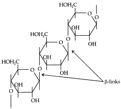
III. Glycogen: The carbohydrates are stored in the animal body as glycogen. It is also known as animal starch because its structure is similar to amylopectin and is more highly branched.

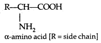
→ Proteins: Proteins are the most abundant biomolecules of the living system. Chief sources of proteins are milk, cheese, pulses, peanuts, fish and meat etc. They are required for the growth and maintenance of the body. All proteins are polymers of a-amino acids.

→ Amino acids: Amino acids contain an amino (- NH2) and carboxyl (- COOH) functional groups.
Biomolecules Class 12 Notes Chemistry 26
→ Classification of Amino acids: Amino acids are classified as acidic, basic or neutral depending upon the relative number of amino and carboxyl groups in their molecule. An equal number of amino and carboxyl groups makes it neutral; more amino than carboxyl groups makes it basic and more carboxyl groups as compared to amino groups makes it acidic.

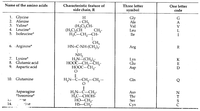
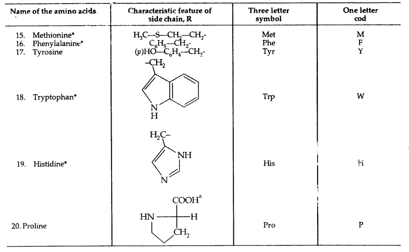
The amino acids, which can be synthesized in the body, are known as non-essential amino acids. On the other hand, which cannot be synthesized in the boxy and must be obtained through diet, are known as essential amino acids (marked with an asterisk in Table below).

Amino acids are usually colourless, crystalline solids. These are water-soluble, high melting solids and behave like salts rather than simple amines or carboxylic acids. This behaviour is due to the presence of both an acidic (carboxyl group) and a basic (amino group) group in the same molecule. In an aqueous solution, the carboxyl group can lose a proton and the amino group can accept a proton, giving rise to a dipolar ion known as a zwitterion. This is neutral but contains both positive and negative charges.
Biomolecules Class 12 Notes Chemistry 27
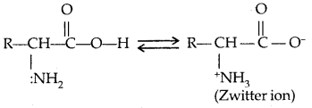
In zwitterionic form, amino acids show amphoteric behaviour as they react both with acids and bases.

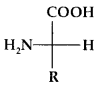
Except for glycine, all other naturally occurring a-amino acids are optically active. These exist both in D and L forms. Most naturally occurring amino acids have L-configuration. L-Amino acids are represented by writing the – NH2 group on the left hand.

Table: Natural Amino Acids,
Biomolecules Class 12 Notes Chemistry 28
Biomolecules Class 12 Notes Chemistry 29
Biomolecules Class 12 Notes Chemistry 30
→ Structures of Proteins: Proteins are the polymers of a-amino adds linked through peptide bond or peptide linkage.
Biomolecules Class 12 Notes Chemistry 31
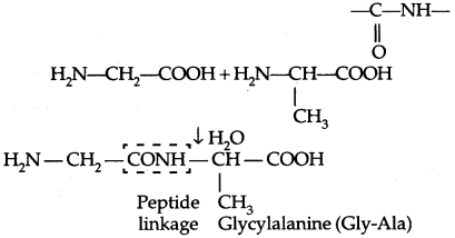
If a third amino acid combines with a dipeptide, the product is called a tripeptide. When the number of such amino acids is more than 10, then the products are called polypeptides. A polypeptide with more than 100 units of amino acid residues, having a molecular mass higher than 10,000 u is called a protein.

Proteins can be classified into two types:
(a) Fibrous proteins: When the polypeptide chains run parallel and held together by hydrogen and disulphide bonds, then a fibre-like structure is formed. Such proteins are generally insoluble in water.

(b) Globular proteins: This structure results when the chains of polypeptides coil around to give a spherical shape. These are usually soluble in water.

Insulin and albumins are common examples.
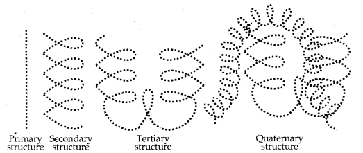
1. Primary structure of Proteins: Proteins may have one or more polypeptide chains. Each polypeptide is a protein that has amino acids linked with each other in a specific sequence and it is this sequence of amino acids that are said to be the primary structure of that protein.

2. Secondary structure of Proteins: The secondary structure of a protein refers to the shape in which a long polypeptide chain can exist. They are found to exist in two different types of structures, viz., a-helix and P-pleated sheet structure.

3. The tertiary structure of protein represents overall folding of the polypeptide chains i.e., further folding of the secondary structure. It gives rise to two major molecular shapes viz. fibrous and globular. The main forces which stabilise the 2° and 3° structures of proteins are hydrogen bonds, disulphide linkages, van der Waals and electrostatic forces of attraction.

4. Quaternary Structure of Proteins: Some of the proteins are composed of two or more polypeptide chains referred to as sub-units. The spatial arrangement of these subunits with respect to each other is known as a quaternary structure.

A diagrammatic representation of all these four structures is given in the figure below:
Biomolecules Class 12 Notes Chemistry 32
→ Denaturation of Proteins: When a protein in its native form is subjected to physical change like change in temperature or chemical change like change in pH, the hydrogen bonds are disturbed. The protein loses its biological activity. This is called denaturing of proteins, 2° and 3° structures are destroyed, but 1° structure remains intact. The coagulation of egg white on boiling is a common example.

→ Enzymes: The enzymes are biological catalysts produced by living cells that catalyse biochemical reactions. The enzymes differ from other types of catalysts in being highly specific and selective.
Biomolecules Class 12 Notes Chemistry 33
→ Mechanism of Enzyme Action: Enzymes, like catalysts, are needed only in small quantities and reduce the magnitude of activation energy of the activated complex. For example, the activation energy for acid hydrolysis of sucrose is 6.22 kJ mol-1 which is reduced to 2.15 kJ mol-1 when hydrolysed by the enzyme sucrase.

→ Vitamins: Certain organic compounds are required in small amounts in our diet but their deficiency in the body causes specific diseases. These compounds are called vitamins. In small quantities in the diet perform specific biological functions for normal maintenance of optimum growth and health of the organism.

Classification of Vitamins:

  1. Fat-soluble Vitamins: Vitamins like A, D, E and K are fat or oil-soluble, but insoluble in water. They are stored in the liver and adipose tissues.
  2. Water-soluble Vitamins: B group Vitamins and Vitamin C are soluble in water. They (except vitamin B12) cannot be stored in a body.

→ Nucleic acids: The particles in the nucleus of the cell, responsible for heredity, are called chromosomes which are made up of proteins and another type of biomolecules called nucleic acids. They are mainly of two types, deoxyribonucleic acid (DNA) and ribonucleic acid (RNA). Since nucleic acids are long-chain polymers of nucleotides, so they are also called polynucleotides.

→ Chemical composition of Nucleic acids: Complete hydrolysis of DNA (or RNA) yields a pentose sugar, phosphoric acid and nitrogen

Table: Vitamins, their sources and their deficiency diseases:
Biomolecules Class 12 Notes Chemistry 34
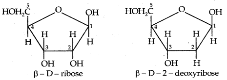
containing heterocyclic compounds called bases. In DNA molecules, the sugar part is β-D-2-deoxyribose whereas, in the RNA molecule, it is β-D- ribose.
Biomolecules Class 12 Notes Chemistry 35
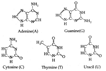
DNA contains four bases viz. adenine (A), guanine (G), cytosine (C) and thymine (T). RNA also contains four bases, the first three bases are A, G and C (as in DNA), but the fourth base is Uracil (U).
Biomolecules Class 12 Notes Chemistry 36
→ Structure of.Nucleic acids: A unit formed by the attachment of a base to the 1′ position of sugar is known as a nucleoside. In nucleosides, the sugar carbons are numbered as 1′, 2′, 3′ etc in order to distinguish these from the bases (Fig. (a) below). When nucleoside is linked to phosphoric acid at 5′-position of sugar moiety we get a nucleotide (Fig. (b) below)
Biomolecules Class 12 Notes Chemistry 37
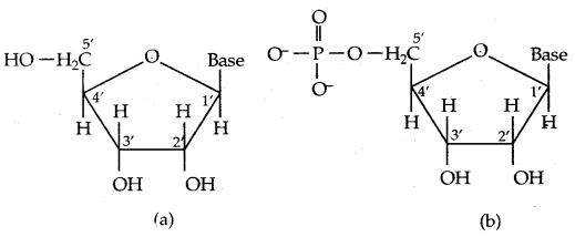
(a) Structure of a nucleoside
(b) Structure of a nucleotide.

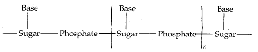
Nucleotides are joined together by phosphodiester linkage between 5′ and 3′ carbon atoms of the pentose sugar. The formation of a typical dinucleotide is:
Biomolecules Class 12 Notes Chemistry 38
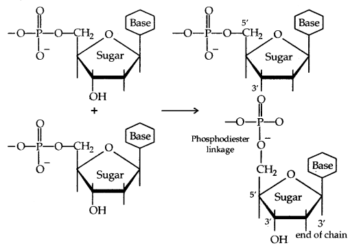
(Formation of a dinucleotide)

A simplified version of the nucleic acid chain is shown below:
Biomolecules Class 12 Notes Chemistry 39
RNA molecules are of three types and they perform different functions. They are named messenger RN A (m-RNA), ribosomal RNA (rRNA) transfer RNA (f-RNA).

DNA Fingerprinting is now used:

  • in forensic laboratories for the identification of criminals.
  • to determine the paternity of an individual.
  • to identify the dead bodies in an accident by comparing the DNAs of parents or children.
  • to identify racial groups to rewrite biological evolution.

Biological Functions of Nucleic Acids: DNA is the chemical basis of heredity and may be regarded as the reserve of genetic information. DNA is exclusively responsible for maintaining the identity of different species of organisms over millions of years. A DNA molecule is capable of self-duplication during cell division, and identical DNA strands are transferred to daughter cells.

Another important function of nucleic acids is the protein synthesis in the cell. Actually, the proteins are synthesised by various RNA molecules in the cell but the message is if the synthesis of a particular protein is present in DNA.

The first one is called Replication and the second one is called protein synthesis.

  1. Replication: The process by which a single DNA molecule produces two identical copies of itself is called cell division or replication. Replication of DNA is an enzyme catalysed process.
  2. Synthesis of Proteins: Another important function of DNA is the synthesis of proteins. In fact, DNA may be regarded as the instrument manual for the synthesis of all the proteins present in a cell.

The DNA directed synthesis of proteins occurs in the following two steps:

  1. Transcription,
  2. Translation

1. Transcription: It involves copying of DNA base sequence into an RNA molecule called the messenger RNA (m RNA).

2. Translation: The mRNA directs protein synthesis in the cytoplasm of the cell with the help of r RNA and t RNA. The process is called translation.

Overview of Arbo Virus and its Various Types

Learninsta presents the core concepts of Microbiology with high-quality research papers and topical review articles.

Overview of Arbo Virus and its Various Types

Arbo Viruses (arthropod – borne viruses) are viruses of vertebrates biologically transmitted by hematophagous insect vectors. They multiply in blood sucking insects and are transmitted by bite to vertebrate hosts. Arbo viruses are worldwide in distribution.

Arbo viruses have been named according to the disease caused (yellow fever), the place of isolation of the virus (kyasanur forest disease) or the local name for the disease (chikungunya). They are classified into Toga, Flavi, Bunya, Reo and Rhabdovirus families.

Arbo viruses have a very wide host range including many species of animals and birds. The most important arbo virus vectors are mosquitoes, followed by ticks. The virus enters the body through the bite of the insect vector.

After multiplication in the reticuloendothelial system, viremia of varying duration occurs, or the virus is transported to the target organs such as central nervous system in encephalitis, the liver in yellow fever and the capillary endothelium in hermorrhagic fever.

Clinical syndromes are fever with or without rash, encephalitis, hemorrhagic fever, systemic disease and yellow fever. Diagnosis may be established by virus isolation or serology.

Samples (Blood, CSF) are inoculated intra cerebrally into sucking mice. The animal develop fatal encephalitis. Viruses may be isolated in tissue cultures or in eggs. Isolates are identified by hemagglutination inhibition, complement fixation, gel precipitation, immunofluorescence and ELISA. Virus isolated from insect vectors and from reservoir animal.

Toga Viruses

Toga viruses are spherical enveloped viruses with a diameter of 50-70nm. Single stranded RNA genome. The virus replicates in the cyloplasm of the host cell and released by budding through host cell membranes. The name Toga Virus is derived from ‘toga’ meaning the Roman Mantle refers to the viral envelope.

The genus Alpha Virus was formerly classified as Group A arbo viruses which explains the name Alpha Virus. The genus Alpha Virus contains 32 species of which 13 infect humans. All are mosquito borne.

Chikungunya Virus

The virus was first isolated from human patients of Aedes aegypti mosquitoes (Figure 10.9) from Tanzania in 1952. The name Chikungunya is derived from the native word for the disease in which the patient lies ‘doubled up due to severe joint pains’. The virus first appeared in India in 1963 in Calcutta, Madras and
Other areas.
Overview of Arbo Virus and its Various Types img 1

The disease presents as a sudden onset of fever, Crippling joint pains, lymphadenopathy and conjunctivitis. A maculopapular rash in common. The fever is typically biphasic with a period of remission after 1-6 days of fever. The vector is Aedes aegypti. No animal reservoir has been identified. Antibody to the virus has been demonstrated in horses, cattle and other domestic animals.

Flavi viruses

The family flaviviridae contains only one genus flavivirus. They are smaller than alpha viruses, being 40nm in diameter. There are over 60 arthropod borne flava viruses classified as mosquito-borne and tick borne viruses. Examples of mosquito borne group known as encephalitis viruses they are St.

Louis encephalitis Virus, Ilheus virus, west nile virus, murray valley encephalitis virus and Japanese encephalitis. Tick borne viruses are classified in to tick borne encephalitis viruses and tick borne hemorrhagic fevers.

Dengue

The name dengue is derived from the ‘Swahili ki denga pepo’, meaning a sudden seizure by a demon. Dengue fever is similar to the illness caused by chikungunya. Four types of dengue virus exist: DEN1, DEN2, DEN3 and DEN4.

Dengue presents after an incubation period of 3-14 days as fever of sudden onset with headache, retrobulbarpain, conjunctival injection, pain in the back and limbs (break bone fever), lymphadenopathy and maculopapular rash. The fever is typically biphasic (saddle back) and lasts for 5-7 days.

Dengue may be more serious forms with hemorrhagic manifestations (dengue Hemorrhagic fever) or with shock (dengue shock syndrome). Dengue virus is transmitted from person to person by Aedes aegypti mosquitoes. The Incubation period is 8-10days. All four types of dengue virus are identified. Demonstration of circulating IgM antibody provides early diagnosis. IgM ELISA test offers reliable diagnosis. Difference between Dengue and Chikungunya is given in Table 10.2.

Difference between Dengue and Chikungunya
Overview of Arbo Virus and its Various Types img 3

Zika Virus

Zika virus is a mosquito-borne flavivirus that was identified in Uganda in 1947 in monkeys. Zika spreads by daytime-active Aedes mosquitoes, such as A. aegypti and A. albopictus. The infection is known as Zika fever or Zika virus disease. Zika is related to the dengue, yellow fever, Japanese encephalitis, and West Nile viruses.

Zika virus is enveloped and icosahedral and has a non segmented, single-stranded, positive-sense (+) RNA genome (Figure 10.10). A positive-sense RNA genome can be directly translated into viral proteins, the RNA genome encodes seven nonstructural proteins and three structural proteins. One of the structural proteins forms the envelope. The RNA genome forms a nucleocapsid along with copies of the 12-kDa capsid protein.
Overview of Arbo Virus and its Various Types img 2

Viral genome replication depends on the making of double-stranded RNA from the single-stranded, positive-sense RNA (ssRNA(+)) genome followed by transcription and replication to provide viral mRNAs and new ssRNA(+) genomes.

Pathogenesis and Clinical features

Zika virus replicates in the mosquito’s mid gut epithelial cells and then its salivary gland cells. After 5-10 days, the virus can be found in the mosquito’s saliva. If the mosquito’s saliva is inoculated into human skin, the virus can infect epidermal keratinocytes, skin fibroblasts in the skin and the Langerhans cells. The pathogenesis of the virus is hypothesized to continue with a spread to lymph nodes and the bloodstream.

Zika virus is primarily transmitted by the bite of an infected mosquito from the Aedes genus, mainly Aedes aegypti. The mosquitoes usually bite during the day, peaking during early morning and late afternoon or evening. This is the same mosquito that transmits dengue, chikungunya and yellow fever.

Zika virus is also transmitted from mother to fetus during pregnancy, through sexual contact, transfusion of blood and blood products, and organ transplantation.

The incubation period of Zika virus disease is estimated to be 3-14 days. The majority of people infected with Zika virus do not develop symptoms. Symptoms are generally mild including fever, rash, conjunctivitis, muscle and joint pain, malaise, and headache, and usually last for 2-7 days.

Zika fever (also known as Zika virus disease) is an illness caused by the Zika virus. Zika virus infection during pregnancy is a cause of microcephaly and other congenital abnormalities in the developing fetus and newborn. Zika infection in pregnancy also results in pregnancy complications such as fetal loss, stillbirth, and preterm birth.

Laboratory diagnosis

Virus can be demonstrated from the blood or other body fluids, such as urine or semen. Zika virus grow well in a variety of mammalian and insect cell lines. Zika virus is identified by NAAT – Nucleic acid Amplification test, Zika Antigen is detected by ELISA and PCR. Zika Antibody IgM is detected by MAC ELISA, IgG by ELISA and by PRNT plaque reduction neutralization test.

Prevention and Treatment

Protection against mosquito bites during the day and early evening is a key measure to prevent Zika virus infection. It is important to eliminate these mosquito breeding sites, Health authorities may also advise use of larvicides and insecticides to reduce mosquito populations and disease spread.

There is no treatment available for Zika virus infection or its associated diseases. No vaccine is yet available for the prevention or treatment of Zika virus infection. Development of a Zika vaccine remains an active area of research.

Human Immuno Deficiency Virus

Learninsta presents the core concepts of Microbiology with high-quality research papers and topical review articles.

Human Immuno Deficiency Virus

Human Immuno Deficiency Virus (HIV), the etiological agent of AIDS, belongs to the lentivirus subgroup of the family Retroviridae.

Structure

HIV is a spherical enveloped Virus, about 90-120 nm in size. The nucleo capsid has an outer icosahedral shell and an inner cone shaped core, enclosing the ribonucleo proteins. The genome is diploid, composed of two identical single stranded, positive sense RNA copies.

When the virus infects a cell, the Viral RNA is transcribed by the reverse transcriptase enzyme, first into single stranded DNA and then to double stranded DNA (provirus) which is integrated into the host cell chromosome. The virus coded envelope proteins are the projecting knob like spikes which binds to the CD4 receptors on susceptible host cells (Figure 10.8)
Human Immuno Deficiency Virus img 1

Viral Genes and Antigens

The genome of HIV contains the three structural genes (gag, pol and env) as well as other nonstructural and regulatory genes specific for the virus. These products of these genes, both structural and non structural act as antigens.

Genes coding for structural proteins

1. The gag gene → Determines the core and shell of the Virus. Precursor protein, p55 and it is cleaved into three proteins p15, p18 and p24. Major core antigen p24 can be detected in serum.

2. The env gene → Determines the synthesis of envelope glycoprotein gp160. Cleaved in to gp120 and gp41

3. The pol gene → Codes for the reverse transcriptase and other viral enzymes such as protease and endonucleases. It’s expressed as a precursor protein, which is cleaved into protein p31, p51 and p66.

Pathogenesis

Infection is transmitted when the Virus enters the blood or tissues of a person and comes into contact with a suitable host cell, principally the CD4 lymphocyte. The receptor for the virus is the CD4 antigen and therefore the virus may infect any cell bearing the CD4 antigen on the surface.

Specific binding of the virus to CD4 receptor is by the envelope glycoprotein gp120. Cell fusion is brought about by transmembrane gp41. After fusion with the host cell membrane, the HIV genome is uncoated and internalized into the cell.

Viral reverse transcriptase mediate transcription of its RNA into double stranded DNA, which is integrated into the genome of the infected cell through the action of the viral enzyme integrase, causing a latent infection.

The primary pathogenic mechanism in HIV infection is the damage caused to the CD4+T lymphocyte. The T4 cells decrease is numbers. Infected T4 cells do not release normal amounts of interleukin, gamma interferon and other lymphokines, this is damping effect on cell mediated immune response.

Clinical Features

AIDS is only the last stage in the wide spectrum in HIV infection.

1. Acute HIV infection

3-6 weeks of infection, persons experience low grade fever, malaise, headache, lymphadenopathy, with rash. Antibodies are usually negative at the onset of the illness but become positive during its course called ‘Sero conversion illness’.

2. Asymptomatic or latent infection

All HIV infected persons, whether or not they experience Sero conversion illness, pass through a phase of symptomless infection which may last up to several years. The infection progresses in course of time through various stages, CD4 lymphocytopenia, minor opportunistic infections, AIDS-related complex (ARC),
ultimately terminating to AIDS.

3. Persistent generalized
lymphadenopathy (PGL)

It is defined as the presence of enlarged lymph nodes at least 1cm, in diameter in two or more non contiguous extrainguinal, sites that persists for at least three months.

4. AIDS related complex (ARC)

This group includes patients with considerable immunodeficiency, suffering from various symptoms or minor opportunistic infections. eg. Oral candidiasis, Salmonellosis or Tuberculosis.

5. AIDS

End-stage disease, poor immune defence mechanism leading to the opportunistic infection and malignancies.

a. Commonest symptoms

Drycough, dyspnea and fever. Pheumonia may be viral (cmv) or fungal (Cryptococcus, Histoplasma).

b. Gastrointestinal system

The mouth is often involved with thrush, stomatitis, gingivitis, hairy leukoplakia. Dysphagia due to esophageal Candidiasis. Intestinal pathogen in AIDS is cryptosporidium. Other pathogens are Salmonellae, Mycobacteria, CMV or adeno viruses. ‘Gay bowel syndrome’ is common among the male homosexuals.

c. Central nervous system

The typical CNS opportunistic infections are toxoplasmosis and cryptococcosis. Lymphomas of the CNS are Common.

d. Malignancies

Kaposi’s Sarcoma was the lesion seen in male homosexuals. The tumours commonly seen are lymphomas, both the Hodgkin and non Hodgkin types.

e. Cutaneous

Herpes lesions, Candidiasis, Dermatitis, impetigo are common cutaneous lesions.

6. Dementia

Direct cytopathogenic damage in the CNS. It cross the blood-brain barrier and cause encepthalopathy leading to dementia.

7. Pediatric AIDS

Viral transmission may occur to the fetus in pregnancy. Many of the infected children may not survive for a year. Children may also acquire the infection from blood transfusion or blood products.

Laboratory Diagnosis

Lab diagnosis of HIV infection include tests for immuno deficiency in HIV infection.

A. Immunological tests

  • Total leukocyte and lymphocyte count to demonstrate leucopenia and a lymphocyte count usually below 2000/mm3.
  • Platelet count will show thrombocytopenia.
  • Raised IgG and IgA levels

B. Specific tests for HIV infection

1. Antigen detection

Single massive infection, as by blood transfusion, the virus antigens may be detectable in blood after about two weeks. The major core antigen p24 is the virus marker in blood.

2. Polymerase Chain reaction

It is the most senstitive and specific test.

3. Antibody detection

Demonstration of antibodies is the simplest and widely employed technique. It takes 2-8 weeks to months for antibodies to appear after infection, during this period, the individual may be highly infectious. This sero negative infective stage is known as the ‘window period’. Antibody can be detected by

  1. ELISA
  2. Western blot test.

Treatment

The treatment of AIDS include:

  • The treatment and prophylaxis of infections and tumours
  • General management
  • Immunorestrorative measures
  • Specific anti-HIV agents

Effective drugs are available, they are Zidovudine, Didanosine, Zalcitabine, Lamivudine and Protease inhibitors like Saquinavir, Ritonavir, Indinavir used as monotherapy or in various combination.