Learninsta presents the core concepts of Biology with high-quality research papers and topical review articles.
Selected Families of Angiosperms
Dicot Families
Family: Fabaceae (Pea family)
Systematic Position
General Characters
Distribution:
Fabaceae includes about 741 genera and more than 20, 200 species. The members are cosmopolitan in distribution but abundant in tropical and subtropical regions.
Habit:
All types of habits are represented in this family. Mostly herbs (Crotalaria), prostrate (Indigofera enneaphylla) erect (Crotalaria verrucosa), shrubs (Cajanus cajan), small trees (Sesbania), climbers (Clitoria), large tree (Pongamia, Dalbergia), woody climber (Mucuna), hydrophyte (Aeschynomene aspera) commonly called pith plant.
Root:
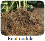
Tap root system, roots are nodulated, have tubercles containing nitrogen – fixing bacteria (Rhizobium leguminosarum)
Stem:
Aerial, herbaceous, woody (Dalbergia) twining or climbing (Clitoria).
Leaf:
Leaf simple or unifoliate (Desmodium gangeticum) bifoliate (Zornia diphylla,), Trifoliate (Lablab purpureus), alternate, stipulate, leaf base, pulvinate, reticulate venation terminal leaflet modifies into a tendril in Pisum sativum.
Inflorescence:
Raceme (Crotalaria verrucosa), panicle (Dalbergia latifolia) axillary solitary (Clitoria ternatea)
Flowers:
Bracteate, bracteolate, pedicellete, complete, bisexual, pentamerous, heterochlamydeous, zygomorphic hypogynous or sometimes perigynous.
Calyx:
Sepals 5, green, synsepalous, more or less united in a tube and persistant, valvate or imbricate, odd sepal is anterior in position.
Corolla:
Petals 5, apopetalous, unequal and papilionaceous, vexillary or descendingly imbricate aestivation, all petals have claw at the base. The outer most petal is large called standard petal or vexillum, Lateral 2 petals are lanceolate and curved. They are called wing petals or alae. Anterior two petals are partly fused and are called keel petals or carina which encloses the stamens and pistil.
Androecium:
Stamens 10, diadelphous, usually 9+1 (Clitoria ternatea). The odd stamen is posterior in position. In Aeschynomene aspera, the stamens are fused to form two bundles each containing five stamens (5)+(5). Stamens are monadelphous and dimorphic ie. 5 stamens have longer filaments and other 5 stamens have shorter filaments thus the stamens are found at two levels and the shape of anthers also varies in (Crotalaria verrucosa). (5 anthers are long and lanceolate, and the other 5 anthers are short and blunt). Anthers are dithecous, basifixed and dehiscing longitudinally
Gynoecium:
Monocarpellary, unilocular, ovary superior, with two alternating rows of ovules on marginal placentation. Style simple and bent, stigma flattened or feathery.
Fruit:
The characteristic fruit of Fabaceae is a legume (Pisum sativum), sometimes indehiscent and rarely a lomentum (Desmodium). In Arachis hypogea the fruit is geocarpic (fruits develops and matures under the soil). After fertilization the stipe of the ovary becomes meristematic and grows down into the soil. This ovary gets buried into the soil and develops into fruit.
Seed:
Endospermic or non-endospermic (Pisum sativum), mostly reniform.
Botanical Description of Clitoria Ternatea (Sangu Pushpam)
Habit:
Twining climber
Root:
Branched tap root system having nodules.
Stem:
Aerial, weak stem and a twiner
Leaf:
Imparipinnately compound, alternate, stipulate showing reticulate venation. Leaflets are stipellate. Petiolate and stipels are pulvinated.
Inflorescence:
Solitary and axillary
Flower:
Bracteate, bracteolate, bracteoles usually large, pedicellate, heterochlamydeous, complete, bisexual, pentamerous, zygomorphic and hypogynous.
Calyx:
Sepals 5, synsepalous, green showing valvate aestivation. Odd sepal is anterior in position.
Corolla:
Petals 5, white or blue apopetalous, irregular papilionaceous corolla showing descendingly imbricate aestivation.
Androecium:
Stamens 10, diadelphous (9)+1, nine stamens fused to form a bundle and the tenth stamen is free. Anthers are dithecous, basifixed, introse and dechiscing by longitudinal slits.
Gynoecium:
Monocarpellary, unilocular, with many ovules on mariginal placentation, ovary superior, style simple and incurved with feathery stigma.
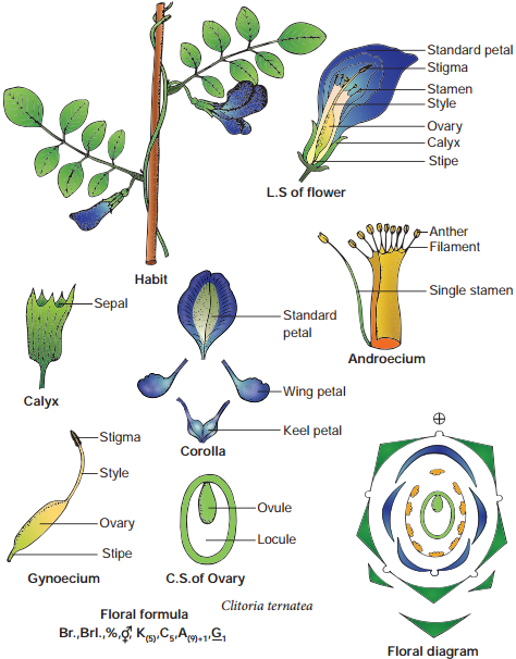
Fruit:
Legume
Seed:
Non-endospermous, reniform.
Floral Formula:

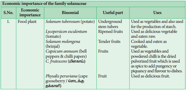
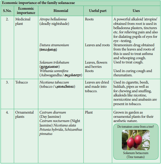
Economic Importance
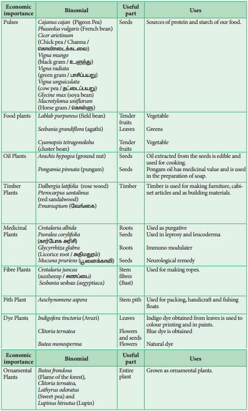
Family:
Solanaceae (Potato Family / Night shade family)
Systematic Position
General Characters
Distribution:
Family Solanaceae includes about 88 genera and about 2650 species, of these Solanum is the largest genus of the family with about 1500 species. Plants are worldwide in distribution but more abundant in South America.
Habit:
Mostly annual herbs, shrubs, small trees (Solanum violaceum) lianas with prickles (Solanum trilobatum)
Root:
Branched tap root system.
Stem:
Herbaceous or woody; erect or twining, or creeping; sometimes modified into tubers (Solanum tuberosum) it is covered with Spines (Solanum tuberosum)
Leaves:
Alternate, simple, rarely pinnately compound (Solanum tuberosum and Lycopersicon esculentum, exstipulate, opposite or sub-opposite in upper part, unicostate reticulate venation. Yellowish verbs present in Solanum tuberosum.
Inflorescence:
Generally axillary or terminal cymose (Solanum) or solitary flowers (Datura stramonium). Extra axillary scorpiod cyme called rhiphidium (Solanum americanum) solitary and axillary (Datura and Nicotiana) umbellate cyme (Withania somnifera).
Flowers:
Bracteate or ebracteate, pedicellate, bisexual, heterochlamydeous, pentamerous actinomorphic or weakly zygomorphic due to oblique position of ovary, hypogynous.
Calyx:
Sepals 5, Synsepalous, valvate persistent (Solanum americanum), often accrescent. (Physalis)
Corolla:
Petals 5, sympetalous, rotate, tubular (Solanum) or bell – shaped (Atropa) or infundibuliform (Petunia) usually alternate with sepals; rarely bilipped and zygomorphic (Schizanthus) usually valvate, sometimes convolute (Datura).
Androecium:
Stamens 5, epipetalous, filaments usually unequal in length, stamens only 2 in Schizanthus (others 3 are reduced to staminode), Anthers dithecous, dehisce longitudinally or poricidal.
Gynoecium:
Bicarpellary, syncarpous obliquely placed, ovary superior, bilocular but looks tetralocular due to the formation of false septa, numerous ovules in each locule on axile placentation.
Fruit:
A capsule or berry. (Datura & Petunia, Lycopersicon esculentum, Capsicum)
Seed:
Endospermous.
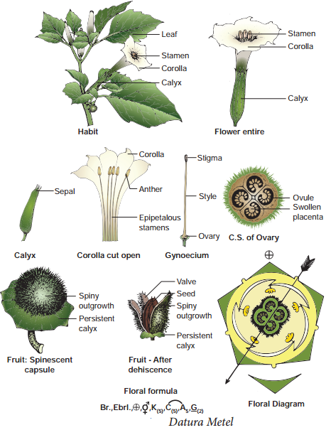
Botanical Description of Datura Metel
Habit:
Large, erect and stout herb.
Root:
Branched tap root system.
Stem:
Stem is hollow, green and herbaceous with strong odour.
Leaf:
Simple, alternate, petiolate, entire or deeply lobed, glabrous exstipulate showing unicostate reticulate venation.
Inflorescence:
Solitary and axillary cyme.
Flower:
Flowers are large, greenish white, bracteate, ebracteolate, pedicellate, complete, heterochlamydeous, pentamerous, regular, actinomorphic, bisexual and hypogynous.
Calyx:
Sepals 5, green synsepalous showing valvate aestivation. Calyx is mostly persistent, odd sepal is posterior in position.
Corolla:
petals 5, greenish white, sympetalous, plicate (folded like a fan) showing twisted aestivation, funnel shaped with wide mouth and 10 lobed.
Androecium:
Stamens 5, free from one another, epipetalous, alternipetalous and are inserted in the middle of the corolla tube. Anthers are basifixed, dithecous, with long filament, introse and longitudinally dehiscent.
Gynoecium:
Ovary bicarpellary, syncarpous superior ovary, basically bilocular but tetralocular due to the formation of false septum. Carpels are obliquely placed and ovules on swollen axile placentation. Style simple long and filiform, stigma two lobed.
Fruit:
Spinescent capsule opening by four apical valves with persistent calyx.
Seed:
Endospermous.
Floral Formula:

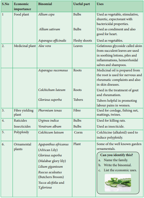
Economic Importance of the Family Liliaceae
Family: Liliaceae (Lily Family)
Systematic Position
General Characters
Distribution:
Liliaceae are fairly large family comprising about 15 genera and 550 species. Members of this family are widely distributed over most part of the world.
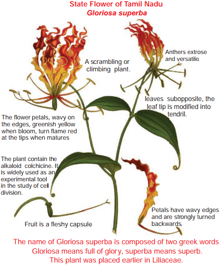
Habit:
Mostly perennial herbs persisting by means of a sympodial rhizome (Polygonatum), by a bulb (Lilium) corm (Colchicum), shrubby or tree like (Yucca and Dracaena) oody climbers, climbing with the help of stipular tendrils in Smilax. Trees in (Xanthorrhoea), succulents (Aloe).
Root:
Adventitious and firous, and typically contractile.
Stem:
Stems usually bulbous, rhizomatous in some, aerial, erect (Dracaena) or climbing (Smilax) in Ruscus the ultimate branches are modified into phylloclades, In Asparagus stem is modified into cladodes and the leaves are reduced to scales.
Leaf:
Leaves are radical (Lilium) or cauline (Dracaena), usually alternate, opposite (Gloriosa), sometimes fleshy and hollow, reduced to scales (Ruscus and Asparagus). The venation is parallel but in species of Smilax it is reticulate. Leaves are usually exstipulate, but in Smilax, two tendrils arise from the base of the leaf, which are considered modified stipules.
Inflorescence:
Flowers are usually borne in simple or branched racemes (Asphodelus) spikes in Aloe, huge terminal panicle in Yucca, solitary and axillary in Gloriosa, solitary and terminal in Tulipa.
Flowers:
Flowers are often showy, pedicellate, bracteate, ebracteolate, except Dianella and Lilium, bisexual, actinomorphic, trimerous, hypogynous, rarely unisexual (Smilax) and are dioecious, rarely tetramerous (Maianthemum), slightly zygomorphic (Lilium) and hypogynous.
Perianth:
Tepals 6 biseriate arranged in two whorls of 3 each, apotepalous or rarely syntepalous as in Aloe. Usually petaloid or sometimes sepaloid, odd tepal of the outer whorl is anterior in position, valvate or imbricate, tepals more than six in Paris quadrifolia.
Androecium:
Stamens 6, arranged in 2 whorls of 3 each, rarely stamens are 3 (Ruscus), 4 in Maianthemum, or up to 12, apostamenous, opposite to the tepals, sometimes epitepalous; fiaments distinct or connate, anthers dithecous, basified or versatile, extrose, or introse, dehiscing usually by vertical slit and sometimes by terminal pores; rarely synstamenous (Ruscus).
Gynoecium:
Tricarpallary, syncarpous, the odd carpel usually anterior, ovary superior, trilocular, with 2 rows of numerous ovules on axile placextation. Style simple, slender with simple stigma.
Fruit:
A loculicidal capsule
Seed:
Endospermous
Floral Formula:

Economic Importance of the Family Liliaceae