Biomolecules of Lipids:

Find free online Chemistry Topics covering a broad range of concepts from research institutes around the world.

Biomolecules of Lipids:

Lipids are organic molecules that are soluble in organic solvents such as chloroform and methanol and insoluble in water. The word lipid is derived from the Greek work ‘lipos’ meaning fat. They are the principal components of cell membranes. In addition, they also act as energy source for living systems. Fat provide 2-3 fold higher energy compared to carbohydrates/proteins.

Classification of Lipids:

Based on their structures lipids can be classified as simple lipids, compound lipids and derived lipids. Simple lipids can be further classified into fats, which are esters of long chain fatty acids with glycerol (triglycerides) and waxes which are the esters of fatty acids with long chain monohydric alcohols (Bees wax).

Compound lipids are the esters of simple fatty acid with glycerol which contain additional groups. Based on the groups attached, they are further classified into phospholipids, glycolipids and lipoproteins. Phospholipids contain a phospho-ester linkage while the glycolipids contain a sugar molecule attached. The lipoproteins are complexes of lipid with proteins.

Biological Importance of Lipids

  1. Lipids are the integral component of cell membrane. They are necessary of structural integrity of the cell.
  2. The main function of triglycerides in animals is as an energy reserve. They yield more energy than carbohydrates and proteins.
  3. They act as protective coating in aquatic organisms.
  4. Lipids of connective tissue give protection to internal organs.
  5. Lipids help in the absorption and transport of fat soluble vitamins.
  6. They are essential for activation of enzymes such as lipases.
  7. Lipids act as emulsifier in fat metabolism.

They include fats, waxes, oils, hormones, and certain components of membranes and function as energy-storage molecules and chemical messengers. Together with proteins and carbohydrates, lipids are one of the principal structural components of living cells.

Biological substances that are insoluble in water are classified as lipids. This characteristic physical property of lipids makes them very different from other biomolecules like carbohydrates, proteins, and nucleic acids. Some lipids are used to store energy.

The four main groups of lipids include:

  • Fatty acids (saturated and unsaturated)
  • Glycerides (glycerol-containing lipids)
  • Nonglyceride lipids (sphingolipids, steroids, waxes)
  • Complex lipids (lipoproteins, glycolipids)

Carbohydrates, nucleic acids, and proteins are often found as long polymers in nature. Lipids are not usually polymers and are smaller than the other three, so they are not considered macromolecules by some sources 1, 2 start superscript, 1, comma, 2, end superscript.

Fats and Oils. A fat molecule consists of two main components-glycerol and fatty acids. Glycerol is an organic compound (alcohol) with three carbons, five hydrogens, and three hydroxyl (OH) groups.

A lipid is any of various organic compounds that are insoluble in water. They include fats, waxes, oils, hormones, and certain components of membranes and function as energy-storage molecules and chemical messengers.

Biomolecules of Lipids img 1

Biomolecules of Proteins

Find free online Chemistry Topics covering a broad range of concepts from research institutes around the world.

Biomolecules of Proteins

Proteins are most abundant biomolecules in all living organisms. The term protein is derived from Greek word ‘Proteious’ meaning primary or holding first place. They are main functional units for the living things. They are involved in every function of the cell including respiration. Proteins are polymers of α-amino acids.

Amino Acids

Amino acids are compounds which contain an amino group and a carboxylic acid group. The protein molecules are made up a-amino acids which can be represented by the following general formula.

Biomolecules of Proteins img 1

There are 20 α-amino acids commonly found in the protein molecules. Each amino acid is given a trivial name, a three letter code and a one letter code. In writing the amino acid sequence of a protein, generally either one letter or three letter codes are used.

Classification of α-amino acids

The amino acids are classified based on the nature of their R groups commonly known as side chain. They can be classified as acidic, basic and neutral amino acids. They can also be classified as polar and non-polar (hydrophobic) amino acids.

Biomolecules of Proteins img 2

Amino acids can also be classified as essential and non-essential amino acids based on the ability to be synthesise by the human. The amino acids that can be synthesised by us are called non-essential amino acids (Gly, Ala, Glu, Asp, Gln, Asn, Ser, Cys, Tyr & Pro) and those needs to be obtained through diet are called essential amino acids (Phe, Val, Th, Trp, Ile, Met, His, Arg, Leu and Lys). These ten essential amino acids can be memorised by mnemonic called PVT TIM HALL.

Although the vast majority of plant and animal proteins are formed by these 20 α-amino acids, many other amino acids are also found in the cells. These amino acids are called as non-protein amino acids. Example: ornithine and citrulline (components of urea cycle where ammonia is converted into urea)

Properties of Amino Acid

Amino acids are colourless, water soluble crystalline solids. Since they have both carboxyl group and amino group their properties differ from regular amines and carboxylic acids. The carboxyl group can lose a proton and become negatively charged or the amino group can accept a proton to become positively charged depending upon the pH of the solution. At a specific pH the net charge of an amino acid is neutral and this pH is called isoelectric point. At a pH above the isoelectric point the amino acid will be negatively charged and positively charged at pH values below the isoelectric point.

In aqueous solution the proton from carboxyl group can be transferred to the amino group of an amino acid leaving these groups with opposite charges. Despite having both positive and negative charges this molecule is neutral and has amphoteric behaviour. These ions are called zwitter ions.

Biomolecules of Proteins img 3

Except glycine all other amino acids have at least one chiral carbon atom and hence are optically active. They exist in two forms namely D and L amino acids. However, L-amino acids are used predominantly by the living organism for synthesising proteins. Presence of D-amino acids has been observed rarely in certain organisms.

Peptide Bond Formation

The amino acids are linked covalently by peptide bonds. The carboxyl group of the first amino acid react with the amino group of the second amino acid to give an amide linkage between these amino acids. This amide linkage is called peptide bond.

The resulting compound is called a dipeptide. Addition an another amino acid to this dipeptide a second peptide bond results in tripeptide. This we can generate tetra peptide, penta peptide etc… When you have more number of amino acids linked this way you get a polypeptide. If the number of amino acids are less it is called as a polypeptide, if it has large number of amino acids (and preferably has a function) then it is called a protein.

Biomolecules of Proteins img 4

The amino end of the peptide is known as N-terminal or amino terminal while the carboxy end is called C-terminal or carboxy terminal. In general protein sequences are written from N-Terminal to C-Terminal. The atoms other than the side chains (R-groups) are called main chain or the back bone of the polypeptide.

Classification of Proteins

Proteins are classified based on their structure (overall shape) into two major types. They are fibrous proteins and globular proteins.

Fibrous Proteins

Fibrous proteins are linear molecules similar to fires. These are generally insoluble in water and are held together by disulphide bridges and weak intermolecular hydrogen bonds. The proteins are often used as structural proteins. Example: Keratin, Collagen etc…

Globular Proteins

Globular proteins have an overall spherical shape. The polypeptide chain is folded into a spherical shape. These proteins are usually soluble in water and have many functions including catalysis (enzyme). Example: myoglobin, insuline

Biomolecules of Proteins img 5

Structure of Proteins

Proteins are polymers of amino acids. Their three dimensional structure depends mainly on the sequence of amino acids (residues). The protein structure can be described at four hierarchal levels called primary, secondary, tertiary and quaternary structures as shown in the figure 14.16

1. Primary Structure of Proteins:

Proteins are polypeptide chains, made up of amino acids are connected through peptide bonds. The relative arrangement of the amino acids in the polypeptide chain is called the primary structure of the protein. Knowledge of this is essential as even small changes have potential to alter the overall structure and function of a protein.

Biomolecules of Proteins img 6

2. Secondary Structure of Proteins:

The amino acids in the polypeptide chain forms highly regular shapes (sub-structures) through the hydrogen bond between the carbonyl oxygen image 7 and the neighbouring amine hydrogen (-NH) of the main chain. α-Helix and β-strands or sheets are two most common sub-structures formed by proteins.

α-Helix

In the α-helix sub-structure, the amino acids are arranged in a right handed helical (spiral) structure and are stabilised by the hydrogen bond between the carbonyl oxygen of one amino acid (nth residue) with amino hydrogen of the fifth residue (n+4th residue). The side chains of the residues protrude outside of the helix. Each turn of an α-helix contains about 3.6 residues and is about 5.4 Å long. The amino acid proline produces a kink in the helical structure and often called as a helix breaker due to its rigid cyclic structure.

β-Strand

β-Strands are extended peptide chain rather than coiled. The hydrogen bonds occur between main chain carbonyl group one such strand and the amino group of the adjacent strand resulting in the formation of a sheet like structure. This arrangement is called β-sheets.

Biomolecules of Proteins img 7

3. Tertiary Structure:

The secondary structure elements (α-helix & β-sheets) further folds to form the three dimensional arrangement. This structure is called tertiary structure of the polypeptide (protein). Teritary structure of proteins are stabilised by the interactions between the side chains of the amino acids. These interactions include the disulphide bridges between cysteine residues, electrostatic, hydrophobic, hydrogen bonds and van der Waals interactions.

4. Quaternary Structure

Some proteins are made up of more than one polypeptide chains. For example, the oxygen transporting protein, haemoglobin contains four polypeptide chains while DNA polymerase enzyme that make copies of DNA, has ten polypeptide chains. In these proteins the individual polypeptide chains (subunits) interacts with each other to form the multimeric structure which are known as quaternary structure. The interactions that stabilises the tertiary structures also stabilises the quaternary structures.

Biomolecules of Proteins img 8

Denaturation of Proteins

Each protein has a unique three-dimensional structure formed by interactions such as disulphide bond, hydrogen bond, hydrophobic and electrostatic interactions. These interactions can be disturbed when the protein is exposed to a higher temperature, by adding certain chemicals such as urea, alteration of pH and ionic strength etc., It leads to the loss of the three-dimensional structure partially or completely. The process of a losing its higher order structure without losing the primary structure, is called denaturation. When a protein denatures, its biological function is also lost.

Since the primary structure is intact, this process can be reversed in certain proteins. This can happen spontaneously upon restoring the original conditions or with the help of special enzymes called cheperons (proteins that help proteins to fold correctly).

Example: Coagulation of egg white by action of heat.

Biomolecules of Proteins img 9

Importance of Proteins

Proteins are the functional units of living things and play vital role in all biological processes

  1. All biochemical reactions occur in the living systems are catalysed by the catalytic proteins called enzymes.
  2. Proteins such as keratin, collagen act as structural back bones.
  3. Proteins are used for transporting molecules (Haemoglobin), organelles (Kinesins) in the cell and control the movement of molecules in and out of the cells (Transporters).
  4. Antibodies help the body to fifth various diseases.
  5. Proteins are used as messengers to coordinate many functions. Insulin and glucagon control the glucose level in the blood.
  6. Proteins act as receptors that detect presence of certain signal molecules and activate the proper response.
  7. Proteins are also used to store metals such as iron (Ferritin) etc.

Enzymes:

There are many biochemical reactions that occur in our living cells. Digestion of food and harvesting the energy from them, and synthesis of necessary molecules required for various cellular functions are examples for such reactions. All these reactions are catalysed by special proteins called enzymes. These biocatalysts accelerate the reaction rate in the orders of 105 and also make them highly specific.

The high specif city is followed allowing many reactio ns to occur within the cell. For example, the Carbonic anhydrase enzyme catalyses the interconversion of carbonic acid to water and carbon dioxide. Sucrase enzyme catalyses the hydrolysis of sucrose to fructose and glucose. Lactase enzyme hydrolyses the lactose into its constituent monosaccharides, glucose and galactose.

Mechanism of Enzyme Action:

Enzymes are biocatalysts that catalyse a specific biochemical reaction. They generally activate the reaction by reducing the activation energy by stabilising the transition state. In a typical reaction enzyme (E) binds with the substrate (S) molecule reversibly to produce an enzyme-substrate complex (ES). During this stage the substrate is converted into product and the enzyme becomes free, and ready to bind to another substrate molecule. More detailed mechanism is discussed in the unit XI surface chemistry.

Biomolecules of Proteins img 10

Biomolecules of Carbohydrates:

Find free online Chemistry Topics covering a broad range of concepts from research institutes around the world.

Biomolecules of Carbohydrates:

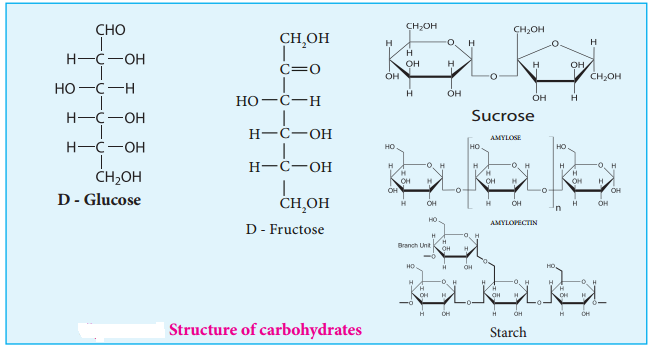
Carbohydrates are the most abundant organic compounds in every living organism. They are also known as saccharides (derived from Greek word ‘sakcharon’ which means sugar) as many of them are sweet. They are considered as hydrates of carbon, containing hydrogen and oxygen in the same ratio as in water. Chemically, they are defined as polyhydroxy aldehydes or ketones with a general formula Cn(H2O)n. Some common examples are glucose (monosaccharide), sucrose (disaccharide) and starch (polysaccharide).

Biomolecules of Carbohydrates img 1

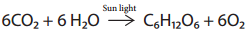
Carbohydrates are synthesised by green leaves during photo synthesis, a complex process in which sun light provides the energy to convert carbon dioxide and water into glucose and oxygen. Glucose is then converted into other carbohydrates and is consumed by animals.

Biomolecules of Carbohydrates img 2

Configuration of Carbohydrates:

Almost all carbohydrates are optically active as they have one or more chiral carbons. The number of optical isomers depends on the number of chiral carbons (2n isomers, where n is the total number of chiral carbons). We have already learnt in XI standard to represent an organic compound using Fischer projection formula. Fischer has devised a projection formula to relate the structure of a carbohydrate to one of the two enantiomeric forms of glyceraldehyde (Figure 14.2).

Biomolecules of Carbohydrates img 3

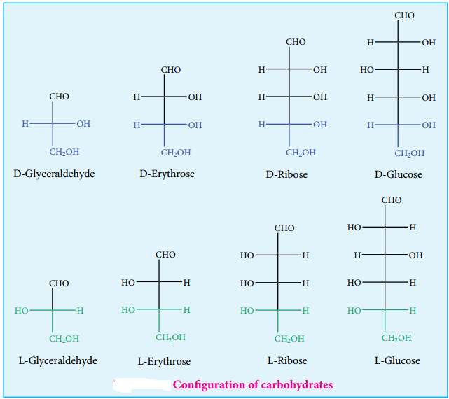
Based on these structures, carbohydrates are named as D or L. The carbohydrates are usually named with two prefixes namely D or L and followed by sign either (+) or (-). Carbohydrates are assigned the notation (D/L) by comparing the confiuration of the carbon that is attached to – CH2OH group with that of glyceraldehyde. For example D-glucose is so named because the H and OH on C5 carbon are in the same configuration as the H and OH on C2 carbon in D-Glyceraldehyde.

There + and – sign indicates the dextro rotatory and levo rotatory respectively. Dextro rotatory compounds rotate the plane of plane polarised light in clockwise direction while the levo rotatory compounds rotate in anticlockwise direction. The D or L isomers can either be dextro or levo rotatory compounds. Dextro rotatory compounds are represented as D-(+) or L-(+) and the levo rotatory compounds as D-(-) or L-(-)

Classification of Carbohydrates:

Carbohydrates can be classified into three major groups based on their product of hydrolysis, namely monosaccharides, oligosaccharides and polysaccharides.

Monosaccharides:

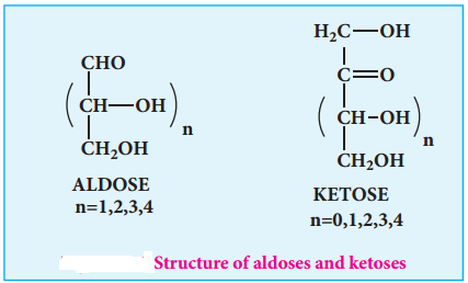
Monosaccharides are carbohydrates that cannot be hydrolysed further and are also called simple sugars. Monosaccharides have general formula Cn(H2O)n. While there are many monosaccharides known only about 20 of them occur in nature. Some common examples are glucose, fructose, ribose, erythrose.

Monosaccharides are further classified based on the functional group present (aldoses or ketoses) and the number of carbon present in the chain (trioses, tetroses, pentoses, hexoses etc…). If the carbonyl group is an aldehyde, the sugar is an aldose. If the carbonyl group is a ketone, the sugar is a ketose. The most common monosaccharides have three to eight carbon atoms.

Table 14.1 Different Types of Monosaccharides:

No.of carbon atoms in the chain

Functional group presentType of sugar

Example

3AldehydeAldotrioseGlyceraldehyde
3KetoneKetotrioseDihydroxy acetone
4AldehydeAldotetroseErythrose
4KetoneKetotetroseErythrulose
5AldehydeAldopentoseRibose
5KetoneKetopentoseRibulose
6AldehydeAldohexoseGlucose
6KetoneKetohexoseFructose

Glucose

Glucose is a simple sugar which serves as a major energy source for us. It is the most important and most abundant sugar. It is present in honey, sweet fruits such as grapes and mangoes etc… Human blood contains about 100 mg/dL of glucose, hence it is also known as blood sugar. In the combined form it is present in sucrose, starch, cellulose etc.,

Biomolecules of Carbohydrates img 4

Preparation of Glucose

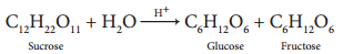
1. When sucrose (cane sugar) is boiled with dilute H2SO4 in alcoholic solution, it undergoes hydrolysis and
give glucose and fructose.

Biomolecules of Carbohydrates img 5

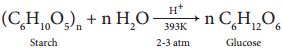
2. Glucose is produced commercially by the hydrolysis of starch with dilute HCl at high temperature under pressure.

Biomolecules of Carbohydrates img 6

Structure of Glucose

Glucose is an aldohexose. It is optically active with four asymmetric carbons. Its solution is dextrorotatory and hence it is also called as dextrose. The proposed structure of glucose is shown in the figure 14.4 which was derived based on the following evidences.

Biomolecules of Carbohydrates img 7

1. Elemental analysis and molecular weight determination show that the molecular formula of glucose is C6H12O6.

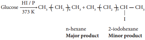
2. On reduction with concentrated HI and red phosphorus at 373K, glucose gives a mixture of n hexane and 2-iodohexane indicating that the six carbon atoms are bonded linearly.

Biomolecules of Carbohydrates img 8

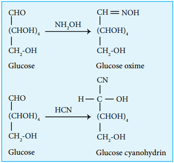
3. Glucose reacts with hydroxylamine to form oxime and with HCN to form cyanohydrin. These reactions indicate the presence of carbonyl group in glucose.

Biomolecules of Carbohydrates img 9

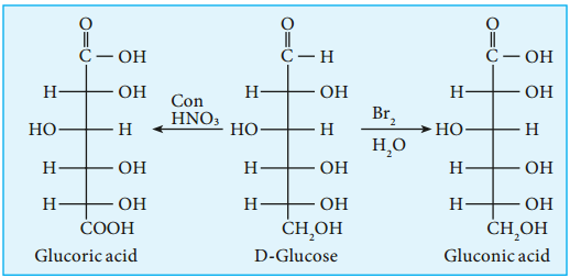
4. Glucose gets oxidized to gluconic acid with mild oxidizing agents like bromine water suggesting that the carbonyl group is an aldehyde group and it occupies one end of the carbon chain. When oxidised using strong oxidising agent such as conc. nitric acid gives glucaric acid (saccharic acid) suggesting the other end is occupied by a primary alcohol group.

Biomolecules of Carbohydrates img 10

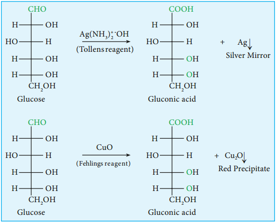
5. Glucose is oxidised to gluconic acid with ammonical silver nitrate (Tollen’s reagent) and alkaline copper sulphate (Fehling’s solution). Tollen’s reagent is reduced to metallic silver and Fehling’s solution to cuprous oxide which appears as red precipitate. These reactions further confirm the presence of an aldehyde group.

Biomolecules of Carbohydrates img 11

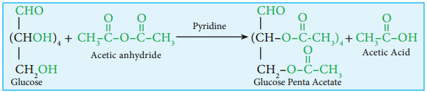
6. Glucose forms penta acetate with acetic anhydride suggesting the presence of fie alcohol groups.

Biomolecules of Carbohydrates img 12

7. Glucose is a stable compound and does not undergo dehydration easily. It indicates that not more than one hydroxyl group is bonded to a single carbon atom. Thus the five the hydroxyl groups are attached to five different carbon atoms and the sixth carbon is an aldehyde group.

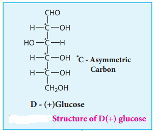
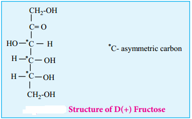
8. The exact spacial arrangement of -OH groups was given by Emil Fischer as shown in Figure 14.4. The glucose is referred to as D(+) glucose as it has D configuration and is dextrorotatory.

Biomolecules of Carbohydrates img 13

Cyclic Structure of Glucose

Fischer identified that the open chain penta hydroxyl aldehyde structure of glucose, that he proposed, did not completely explain its chemical behaviour. Unlike simple aldehydes, glucose did not form crystalline bisulphite compound with sodium bisulphite. Glucose does not give Schif ’s test and the penta acetate derivative of glucose was not oxidized by Tollen’s reagent or Fehling’s solution. Thus behaviour could not be explained by the open chain structure.

Biomolecules of Carbohydrates img 14

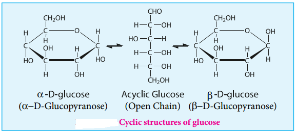
In addition, glucose is found to crystallise in two different forms depending upon the crystallisation conditions with different melting points (419 and 423 K). In order to explain these it was proposed that one of the hydroxyl group reacts with the aldehyde group to form a cyclic structure (hemiacetal form) as shown in figure 14.5. This also results in the conversion of the achiral aldehyde carbon into a chiral one leading to the possibility of two isomers.

These two isomers differ only in the configuration of C1 carbon. These isomers are called anomers. The two anomeric forms of glucose are called α and β-forms. This cyclic structure of glucose is similar to pyran, a cyclic compound with 5 carbon and one oxygen atom, and hence is called pyranose form.

The specific rotation of pure α- and β-(D) glucose are 112º & 18.7º respectively. However, when a pure form of any one of these sugars is dissolved in water, slow interconversion of α-D glucose and β-D glucose via open chain form occurs until equilibrium is established giving a constant specific rotation +53º. This phenomenon
is called mutarotation.

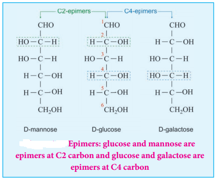
Epimers and Epimerisation:

Sugar differing in configuration at an asymmetric centre is known as epimers. The process by which one epimer is converted into other is called epimerisation and it requires the enzymes epimerase. Galactose is converted to glucose by this manner in our body.

Biomolecules of Carbohydrates img 15

Fructose

Fructose is another commonly known monosaccharide having the same molecular formula as glucose. It is levorotatory and a ketohexose. It is present abundantly in fruits andhence it is also called as fruit sugar.

Preparation

1. From Sucrose

Fructose is obtained from sucrose by heating with dilute H2SO4 or with the enzyme invertase

Biomolecules of Carbohydrates img 16

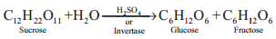
Fructose is separated by crystallisation. The mixture having equal amount of glucose and fructose is termed as invert sugar.
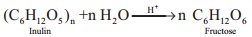
2. From Inulin

Fructose is prepared commercially by hydrolysis of Inulin (a polysaccharide) in an acidic medium.

Biomolecules of Carbohydrates img 17

Structure of Fructose:

Fructose is the sweetest of all known sugars. It is readily soluble in water. Fresh solution of fructose has a specific rotation -133° which changes to -92° at equilibrium due to mutarotation. Similar to glucose the structure of fructose is deduced from the following facts.

1. Elemental analysis and molecular weight determination of fructose show that it has the molecular formula C6H12O6

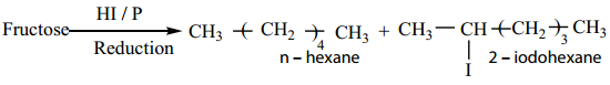
2. Fructose on reduction with HI and red phosphorus gives a mixture of n – hexane (major product) and 2 – iodohexane (minor product). This reaction indicates that the six carbon atoms in fructose are in a straight chain.

Biomolecules of Carbohydrates img 18

3. Fructose reacts with NH2OH and HCN. It shows the presence of a carbonyl groups in the fructose.

4. Fructose reacts with acetic anhydride in the presence of pyridine to form penta acetate. This reaction indicates the presence of five hydroxyl groups in a fructose molecule.

5. Fructose is not oxidized by bromine water. This rules out the possibility of absence of an aldehyde (-CHO) group.

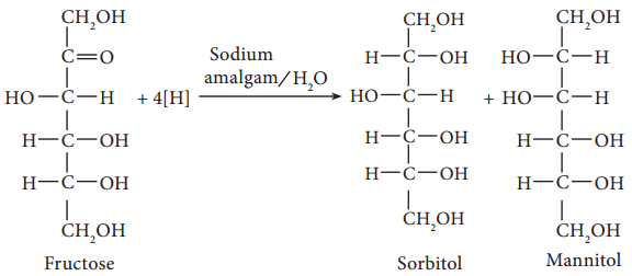
6. Partial reduction of fructose with sodium amalgam and water produces mixtures of sorbitol and mannitol which are epimers at the second carbon. New asymmetric carbon is formed at C-2. This confirms the presence of a keto group.

Biomolecules of Carbohydrates img 19

On oxidation with nitric acid, it gives glycollic acid and tartaric acids which contain smaller number of carbon atoms than in fructose.

Biomolecules of Carbohydrates img 20

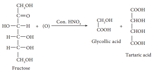
This shows that a keto group is present in C-2. It also shows that 1° alcoholic groups are present at C-1 and C-6. Based on these evidences, the following structure is proposed for fructose (Figure 14-7)

Biomolecules of Carbohydrates img 21

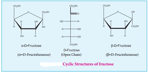
Cyclic Structure of Fructose

Like glucose, fructose also forms cyclic structure. Unlike glucose it forms a five membered ring similar to furan. Hence it is called furanose form. When fructose is a component of a saccharide as in sucrose, it usually occurs in furanose form.

Biomolecules of Carbohydrates img 22

Disaccharides

Disaccharides are sugars that yield two molecules of monosaccharides on hydrolysis. This reaction is usually catalysed by dilute acid or enzyme. Disaccharides have general formula Cn(H2O)n-1. In disaccharides two monosaccharide’s are linked by oxide linkage called ‘glycosidic linkage’, which is formed by the reaction of the anomeric carbon of one monosaccharide reacts with a hydroxyl group of another monosaccharide.

Example: Sucrose, Lactose, Maltose

Sucrose: Sucrose, commonly known as table sugar is the most abundant disaccharide. It is obtained mainly from the juice of sugar cane and sugar beets. Insects such as honey bees have the enzyme called invertases that catalyzes the hydrolysis of sucrose to a glucose and fructose mixture.

Biomolecules of Carbohydrates img 23

Honey in fact, is primarily a mixture of glucose, fructose and sucrose. On hydrolysis sucrose yields equal amount of glucose and fructose units.

Biomolecules of Carbohydrates img 24

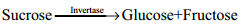
Sucrose (+66.6°) and glucose (+52.5°) are dextrorotatory compounds while fructose is levo rotatory (-92.4°). During hydrolysis of sucrose the optical rotation of the reaction mixture changes from dextro to levo. Hence, sucrose is also called as invert sugar.
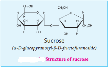
Structure:

In sucrose, C1 of α-D-glucose is joined to C2 of β-D-fructose. The glycosidic bond thus formed is called α-1,2 glycosidic bond. Since, both the carbonyl carbons (reducing groups) are involved in the glycosidic bonding, sucrose is a non-reducing sugar.
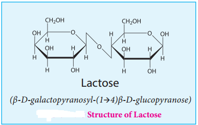
Lactose:

Lactose is a disaccharide found in milk of mammals and hence it is referred to as milk sugar. On hydrolysis, it yields galactose and glucose. Here, the β-D-galactose and β-D-glucose are linked by β-1,4 glycosidic bond as shown in the figure 14.10. The aldehyde carbon is not involved in the glycosidic bond hence, it retains its reducing property and is called a reducing sugar.

Biomolecules of Carbohydrates img 25

Maltose:

Maltose derives its name from malt from which it is extracted. It is commonly called as malt sugar. Malt from sprouting barley is the major source of maltose. Maltose is produced during digestion of starch by the enzyme α-amylase.

Biomolecules of Carbohydrates img 27

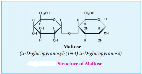
Maltose consists two molecules of α-D-glucose units linked by an α-1, 4 glycosidic bond between anomeric carbon of one unit and C-4 of the other unit. Since one of the glucose has the carbonyl group intact, it also acts as a reducing sugar.

Polysaccharides:

Polysaccharides consist of large number of monosaccharide units bonded together by glycosidic bonds and are the most common form of carbohydrates. Since, they do not have sweet taste polysaccharides are called as non-sugars. They form linear and branched chain molecules.

Polysaccharides are classified into two types, namely, homopolysaccharides and heteropolysaccharides depending upon the constituent monosaccharides. Homopolysaccharides are composed of only one type of monosaccharides while the heteropolysaccharides are composed of more than one. Example: starch, cellulose and glycogen (homopolysaccharides); hyaluronic acid and heparin (heteropolysaccharides).
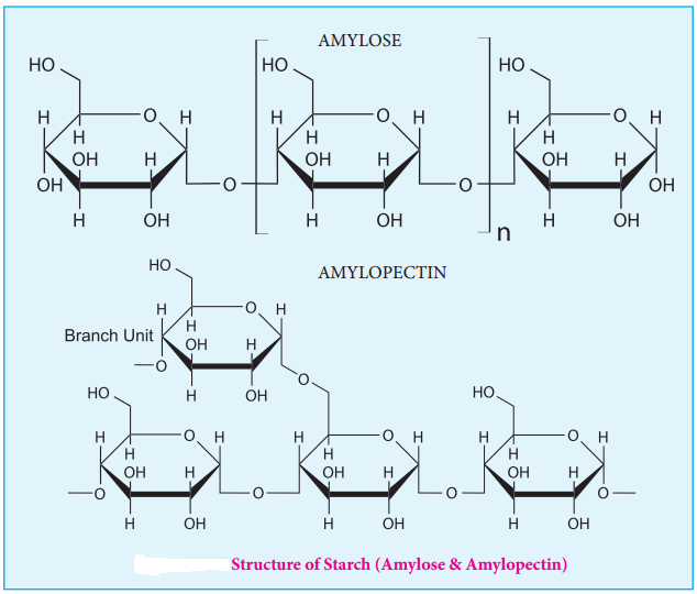
STARCH

Starch is used for energy storage in plants. Potatoes, corn, wheat and rice are the rich sources of starch. It is a polymer of glucose in which glucose molecules are lined by α(1, 4) glycosidic bonds. Starch can be separated into two fractions namely, water soluble amylose and water insoluble amylopectin. Starch contains about 20 % of amylose and about 80% of amylocpectin.

Amylose is composed of unbranched chains upto 4000 α-D-glucose molecules joined by α(1, 4)glycosidic bonds. Amylopetin contains chains upto 10000 α-D-glucose molecules linked by α(1, 4)glycosidic bonds. In addition, there is a branching from linear chain. At branch points, new chains of 24 to 30 glucose molecules are linked by α(1, 6)glycosidic bonds. With iodine solution amylose gives blue colour while amylopectin gives a purple colour.

Biomolecules of Carbohydrates img 28

Cellulose

Cellulose is the major constituent of plant cell walls. Cotton is almost pure cellulose. On hydrolysis cellulose yields D-glucose molecules. Cellulose is a straight chain polysaccharide. The glucose molecules are linked by β(1, 4)glycosidic bond.

Biomolecules of Carbohydrates img 29

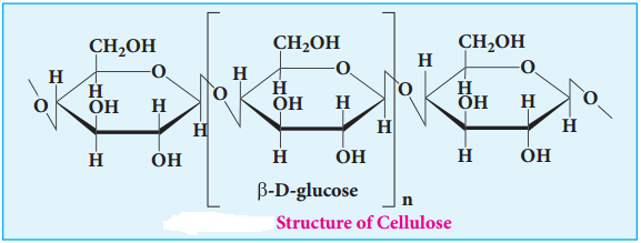
Cellulose is used extensively in the manufacturing paper, cellulose fires, rayon explosive, (Gun cotton – Nitrated ester of cellulose) and so on. Human cannot use cellulose as food because our digestive systems do not contain the necessary enzymes (glycosidases or cellulases) that can hydrolyse the cellulose.

Glycogen:

Glycogen is the storage polysaccharide of animals. It is present in the liver and muscles of animals. Glycogen is also called as animal starch. On hydrolysis it gives glucose molecules. Structurally, glycogen resembles amylopectin with more branching. In glycogen the branching occurs every 8-14 glucose units opposed to 24-30 units in amylopectin. The excessive glucose in the body is stored in the form of glycogen.

Importance of Carbohydrates

  1. Carbohydrates, widely distributed in plants and animals, act mainly as energy sources and structural polymers.
  2. Carbohydrate is stored in the body as glycogen and in plant as starch.
  3. Carbohydrates such as cellulose which is the primary components of plant cell wall, is used to make paper, furniture (wood) and cloths (cotton)
  4. Simple sugar glucose serves as an instant source of energy.
  5. Ribose sugars are one of the components of nucleic acids.
  6. Modified carbohydrates such as hyaluronate (glycosaminoglycans) act as shock absorber and lubricant.

Cyanides and Isocyanides

Find free online Chemistry Topics covering a broad range of concepts from research institutes around the world.

Cyanides and Isocyanides

These are the derivatives of hydrocyanic acid (HCN), and is known to exist in two tautomeric forms

Cyanides and Isocyanides img 1

Two types of alkyl derivatives can be obtained. Those derived by replacement of H – atom of hydrogen cyanide by the alkyl groups are known as alkyl cyanides (R-C≡N) and those obtained by the replacement of H – atom of hydrogen isocyanide are known as alkyl isocyanides

Cyanides and Isocyanides img 2

In IUPAC system, alkyl cyanides are named as “alkanenitriles” whereas aryl cyanides as “arenecarbonitrile”.

Table: Nomenclature of Cyanides

Cyanides and Isocyanides img 3

Methods of Preparation of Cyanides

1. From Alkyl Halides

When alkyl halides are treated in the solution NaCN (or) KCN, alkyl cyanides are obtained. In this reaction a new carbon – carbon bond is formed.

Example

Cyanides and Isocyanides img 4

Aryl cyanide cannot be prepared in this method because of their less reactivity towards nucleophilic substitution. Aryl cyanides are prepared using Sandmeyers reactions.

2. By Dehydration of Primary Amides and Aldoximes with P2O5

Cyanides and Isocyanides img 5

3. By dehydration of ammonium carboxylates with P2O5

Cyanides and Isocyanides img 6

This method suitable for large scale preparation of alkyl cyanides.

4. From Grignard Reagent

Methyl magnesium bromide on treatment with cyanogen chloride (Cl – CN) forms ethanenitrile.

Cyanides and Isocyanides img 7

Properties of Cyanides

Physical Properties

The lower members (up to C14) are colourless liquids with a strong characteristic sweet smell. The higher members are crystalline solids, They are moderately soluble in water but freely souble in organic solvents. They are poisonous. They have higher boiling points than analogous acetylenes due to their high dipole moments.

Chemical Properties

1. Hydrolysis

On boiling with alkali (or) a dilute mineral acid, the cyanides are hydrolysed to give carboxylic acids.

For Example

Cyanides and Isocyanides img 8

2. Reduction

On reduction with LiAlH4 2(or) Ni/H2, alkyl cyanides yields primary amines.

Cyanides and Isocyanides img 9

3. Condensation Reaction

(a) Thorpe Nitrile Condensation

Self condensation of two molecules of alkyl nitrile (containing α-H atom) in the presence of sodium to form iminonitrile.

Cyanides and Isocyanides img 10

(b) The nitriles containing α – hydrogen also undergo condensation with esters in the presence of sodamide in ether to form ketonitriles. This reaction is known as “Levine and Hauser” acetylation.

This reaction involves replacement of ethoxy (OC2H5) group by methylnitrile (- CH2CN) group and
is called as cyanomethylation reaction.

Cyanides and Isocyanides img 11

Alkyl Isocyanides (Carbylamines)

Nomenclature of Isocyanides

They are commonly named as Alkyl isocyanides. The IUPAC system names them as alkylcarbylamines

Table: Nomenclature of Alkylisocyanides

Cyanides and Isocyanides img a

Methods of Preparation of Isocyanides

1. From Primary Amines (Carbylamines Reaction)

Both aromatic as well as aliphatic amines on treatment with CHCl3 in the presence of KOH give carbylamines.

Cyanides and Isocyanides img 12

2. From Alkyl Halides

Ethyl bromide on heating with ethanolic solution of AgCN give ethyl isocyanide as major product and ethyl cyanide as minor product.

Cyanides and Isocyanides img 13

3. From N – alkyl formamide. By reaction with POCl3 in pyridine.

Cyanides and Isocyanides img 14

Properties of Isocyanides

Physical Properties

  • They are colourless, highly unpleasant smelling volatile liquids and are much more poisonous than the cyanides.
  • They are only slightly soluble in water but are soluble in organic solvents.
  • They are relatively less polar than alkyl cyanides. Ths, their melting point and boiling point are lower than cyanides.

Chemical Properties

1. Hydrolysis:

Alkyl isocyanides are not hydrolysed by alkalies. However they are hydrolysed with dilute mineral acids to give primary amines and formic acids.

Cyanides and Isocyanides img 15

2. Reduction:

When reduced catalytically (or) by nascent hydrogen, they give secondary amines.

Cyanides and Isocyanides img 16

3. Isomerisation:

When Alkyl isocyanides and heated at 250°C, they change into the more stable, isomeric cyanides

Cyanides and Isocyanides img 17

4. Addition Reaction:

Alkyl isocyanides add on halogen, sulphur, and oxygen to form the corresponding addition compounds.

Cyanides and Isocyanides img 18

Uses of Organic Nitrogen Compounds

Nitroalkanes

  1. Nitromethane is used as a fuel for cars
  2. Chloropicrin (CCl3NO2) is used as an insecticide
  3. Nitroethane is used as a fuel additive and precursor to explosive and they are good solvents for polymers, cellulose ester, synthetic rubber and dyes etc.,
  4. 4% solution of ethylnitrite in alcohol is known as sweet spirit of nitre and is used as diuretic.

Nitrobenzene

  1. Nitrobenzene is used to produce lubricating oils in motors and machinery.
  2. It is used in the manufacture of dyes, drugs, pesticides, synthetic rubber, aniline and explosives like TNT, TNB.

Cyanides and Isocyanides

1. Alkyl cyanides are important intermediates in the organic synthesis of larger number of compounds like acids, amides, esters, amines etc.

2. Nitriles are used in textile industry in the manufacture of nitrile rubber and also as a solvent particularly in perfume industry.

Organic Compounds of Diazonium Salts

Find free online Chemistry Topics covering a broad range of concepts from research institutes around the world.

Organic Compounds of Diazonium Salts

We have just learnt that aromatic amines on treatment with (NaNO2+HCl) gives diazonium salts. They are stable only for a short time and hence are used immediately after preparation.

Example

Diazonium Salts img 1

Resonance Structure

The stability of arene diazonium salt is due to the dispersal of the positive charge over the benzene ring.

Diazonium Salts img 2

Method of Preparation of Diazonium Salts

We have already learnt that benzene diazonium chloride is prepared by the reaction of aniline with nitrous acid (Which is produced by the reaction of NaNO2 and HCl) at 273 – 278K

Physical Properties

  • Benzene diazonium chloride is a colourless, crystalline solid.
  • These are readily soluble in water and stable in cold water. However it reacts with warm water.
  • Their aqueous solutions are neutral to litmus and conduct electricity due to the presence to ions.
  • Benzenediazonium tetrafloro borate is soluble in water and stable at room temperature.

Chemical Reactions

Benzene diazoniumchloride gives two types of chemical reactions

A. Replacement reactions involving loss of nitrogen

In these reactions diazonium group is replaced by nucleophiles such as X, CN, H, OH
etc.,

B. Reactions involving retention of diazogroup. Coupling reaction.

A. Replacement Reactions Involving loss of Nitrogen

1. Replacement by Hydrogen

Benzene diazonium chloride on reduction with mild reducing agents like hypophosphrous acid (phosphinic acid) or ethanol in the presence of cuprous ion gives benzene. This reaction proceeds through a free-radical chain mechanism.

Diazonium Salts img 3

2. Replacement by Chlorine, Bromine, Cyanide group

(a) Sandmeyer Reaction

On mixing freshly prepared solution of benzene diazonium chloride with cuprous halides (chlorides and bromides), aryl halides are obtained. This reaction is called Sandmeyer reaction. When diazonium salts are treated with cuprous cyanide, cyanobenzene is obtained.

Diazonium Salts img 4

(b) Gattermann Reaction

Conversion of benzene diazonium chloride into chloro/bromo arenes can also be effected using hydrochloric/hydrobromic acid and copper powder. This reaction is called Gattermann reaction.

Diazonium Salts img 5

The yield in Sandmeyer reaction is found to be better than the Gattermann reaction.

3. Replacement by Iodine

Aqueous solution of benzene diazonium chloride is warmed with KI to form iodobenzene

Diazonium Salts img 6

4. Replacement of Flourine (Baltz – schiemann reaction)

When benzene diazonium chloride is treated with floroboric acid, benezene diazonium tetra flouroborate is precipitated which on heating decomposes to give flourobenzene.

Diazonium Salts img 7

5. Replacement by Hydroxyl Group

Benzene diazonium chloride solution is added slowly to a large volume of boiling water to get phenol.

Diazonium Salts img 8

6. Replacement by Nitrogroup

When diazonium flouroborate is heated with aqueous sodium nitrite solution in the presence of copper, the diazonium group is replaced by – NO2 group.

Diazonium Salts img 9

7. Replacement by Aryl Group (Gomberg Reaction)

Benzene diazonium chloride reacts with benzene in the presence of sodium hydroxide to give biphenyl. This reaction in known as the Gomberg reaction.

Diazonium Salts img 10

8. Replacement by Carboxylic Acid Group

When diazonium flouroborate is heated with acetic acid, benzoic acid is obtained. This reaction is used to convert the of aliphatic carboxylic acid into aromatic carboxylic acid.

Diazonium Salts img 11

B. Reactions Involving Retention of Diazo Group

9. Reduction to Hydrazines

Certain reducing agents like SnCl2/HCl ; Zn dust/CH3COOH, sodium hydrosulphite, sodium sulphite etc. reduce benzene diazonium chloride to phenyl hydrazine.

Diazonium Salts img 12

10. Coupling Reactions

Benzene diazonium chloride reacts with electron rich aromatic compounds like phenol, aniline to form brightly coloured azo compounds. Coupling generally occurs at the para position. If para position is occupied then coupling occurs at the ortho position. Coupling tendency is enhanced if an electron donating group is present at the para – position to – N2Cl group. This is an electrophilic substitution.

Diazonium Salts img 13

Aryl flourides and iodides cannot be prepared by direct halogenation and the cyano group cannot be introduced by nucleophilic substitution of chlorine in chlorbenzene. For introducing such a halide group, cyano group -OH, NO2 etc.. benzenediazonium chloride is a very good intermediate Diazo compounds obtained from the coupling reactions of diazonium salts are coloured and are used as dyes.