Locomotion and Movement Class 11 Important Extra Questions Biology Chapter 20

Here we are providing Class 11 Biology Important Extra Questions and Answers Chapter 20 Locomotion and Movement. Important Questions for Class 11 Biology are the best resource for students which helps in Class 11 board exams.

Class 11 Biology Chapter 20 Important Extra Questions Locomotion and Movement

Locomotion and Movement Important Extra Questions Very Short Answer Type

Question 1.
What is a tendon?
Answer:
The dense connective tissue joins bone and skeletal muscle.

Question 2.
What are antagonistic muscles?
Answer:
The. pair of muscles which at a joint produce opposite movements.

Question 3.
What is tetanus?
Answer:
The continued state of muscular contraction is called tetanus.

Question 4.
What is threshold stimulus?
Answer:
The stimulus of minimum strength which is required to bring about muscular contraction is called the threshold stimulus.

Question 5.
What is a muscle twitch?
Answer:
The single contraction of muscle upon receiving the stimulus is called muscle twitch. (Contraction is followed by relaxation).

Question 6.
What is sarcomere?
Answer:
The functional unit of myofibril contracts and causes the shortening of muscle fibre.

Question 7.
How many bones are present in the human skeleton?
Answer:
The human skeleton contains 206 bones.

Question 8.
What are synovial joints?
Answer:
These are freely movable joints due to the presence of synovial fluid in the synovial cavity.

Question 9.
What is locomotion?
Answer:
The bodily movement in animals from one place to the other is called locomotion.

Question 10.
What is rigour mortis?
Answer:
Stiffening of muscle after death.

Question 11.
Name the proteins which help in muscle contraction.
Answer:
Myosin and actin.

Question 12.
What is the function of synovial fluid?
Answer:
Synovial fluid acts as a lubricant.

Question 13.
What is a pivot joint?
Answer:
The joint allows the turning or rotational movements, e.g., between atlas and axis vertebra.

Question 14.
Which of the movable joint makes the hip joint?
Answer:
Ball and socket joint.

Question 15.
Which muscle contracts to make your palm face upwards?
Answer:
Supinator.

Question 16.
How many bones are there in the human skull?
Answer:
29

Question 17.
Which type of movable joint is the knee joint?
Answer:
Hinge joint.

Question 18.
Name the band of the skeletal joint which permits movements in a single plane only.
Answer:
Hinge joint.

Question 19.
Differentiate between A-band and I-band.
Answer:

  • A-band is a dark band having myosin filaments.
  • I-band is a light band having thin filaments.

Question 20.
What is the total number of bones in our body?
Answer:
206.

Question 21.
Name the five different categories of vertebrae in your backbone.
Answer:

  1. Cervical,
  2. thoracic,
  3. lumber,
  4. sacral and
  5. coccygeal.

Question 20.
Where inside the bones are blood cells produced?
Answer:
The bone marrow of long bones.

Question 23.
Give one example of a ball and socket joint.
Answer:
Shoulder joint.

Locomotion and Movement Important Extra Questions Short Answer Type

Question 1.
List the mechanical function of the skeleton.
Answer:

  1. It provides a rigid framework of the body and definite shape to organs.
  2. It supports the weight of the body.
  3. It protects the internal organs.
  4. Its long bones function as a lever.
  5. Skeletal muscles with flexible connective tissue bands called tendons in association with endoskeleton and joints give locomotion and movements to different body parts.

Question 2.
List some biological function of the skeleton.
Answer:

  1. Provides attachment surface to muscles.
  2. Serves as storage depot of calcium and phosphate minerals.
  3. Act in erythropoiesis.
  4. Ear ossicles help in sound wave propagation.
  5. Redbone marrow present inside the marrow cavity of long bones such as femur, humerus and in interstices of spongy bones of vertebrae, sternum, scapula etc. help in the formation of RBCs, WBCs and platelets of the blood. This process is known as Haemopoiesis.

Question 3.
List different modes of locomotion and movement in hydra.
Answer:

  1. Contraction and expansion
  2. Bending and swaying
  3. Looping
  4. Somersaulting.
  5. Floating
  6. Gliding
  7. Swimming
  8. Walking.

Question 4.
What are the different molecules present in muscles?
Answer:

  1. Contractile proteins viz. actin, myosin and tropomyosin.
  2. Enzymes and other proteins like troponin.
  3. Carbohydrate as a substrate for energy.
  4. Energy carries viz. ATP, ADP, AMP and CP.
  5. Ions viz. Na+, K+, Mg++, Ca+, CI+.

Question 5.
Differentiate between isotonic and isometric contraction
Answer:

Isotonic contractionIsometric contraction
1. There is a change in the shape of muscles.1. There is no change in the shape of muscles
2. Muscles maintains tension.2. Muscles maintains length.
3. Muscles contracts and the load is lifted3. Muscles contracts against a load that can’t be lifted.

Question 6.
A red muscles fibre works for a prolonged period, whereas a white muscle fibre gets fatigued, why?
Answer:
Red muscle fibres contain oxygen storing pigment myoglobin and a large number of mitochondria, so they can have O2 supply for aerobic respiration and release of energy for a longer period.

White muscles fibres do not have myoglobin pigment. They face a short supply of O2 and much depends on anaerobic respiration, so they get fatigued soon.

Question 7.
What are the main types of joints present in the human body?
Answer:
The types of joints present in the body of man are:
1. Fixed or fibrous joints: There is no movement at all in articulating joints, because of the presence of tough, inextensible, white fibrous tissue, e.g., skull bones.

2. Slightly movable or cartilaginous joints: A limited movement is possible in articulating bones. A dense disc of white cartilage joins the articulating surfaces, e.g., vertebrae and public symphysis.
Locomotion and Movement Class 11 Important Extra Questions Biology 1
Locomotion and Movement Class 11 Important Extra Questions Biology 2
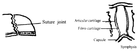
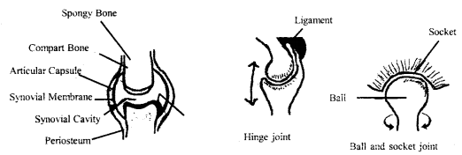
Different types of joints

3. Freely movable or synovial joints: Free movement is possible due to the presence of synovial fluid in the synovial cavity, between the articulating bones, e.g., hinge joint, ball and socket joint.

Question 8.
What are the advantages of the movement of body parts?
Answer:
The movement has the following advantages:

  1. With change in body posture and limb movement, equilibrium of the body is maintained.
  2. Limb movement causes locomotion.
  3. Food is captured by movement of tentacles, limbs, jaw, tongue etc. in different animals.
  4. Changes in environment surrounding can be sensed by the movement of the eyeball, pinna etc.
  5. Blood circulation is possible by heart movement.
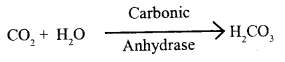
  6. Movement of the diaphragm causes inhales and exhale (breathing).

Question 9.
What are the advantages of locomotion?
Answer:
The bodily movements or locomotion has the following advantages:

  1. It enables the body to shift it entirely from one place to the other.
  2. It protects the organism from predation.
  3. It helps the animals to make the search for their food and other nutritional requirements.
  4. It helps the animal to seek a mate for reproduction.

Question 10.
Draw a labelled diagram of the joint found between the pelvic girdle and femur. Also, write the type of this joint.
Answer:
Type of the joint: The joint between pelvic and femur bones is a ball and socket synovial joint.
Locomotion and Movement Class 11 Important Extra Questions Biology 3
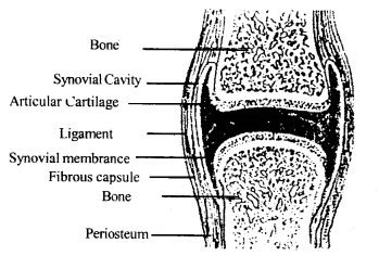
Synovial ball and socket joint between pelvic and femur

Question 11.
Why movement and locomotion are necessary among animals?
Answer:
Movement and locomotion are necessary among animals for the survival of them. It enables, them to procure food, search for shelter, find mates, protect themselves from predatory and perform many other life activities.

Question 12.
Elucidate the types of movements found among the animals.
Answer:
Movements among animals vary greatly. Movement involves three basic mechanisms.
These are:

  1. amoeboid,
  2. ciliary and
  3. muscular.

Amoeboid movement is typical of Amoeba. Amoeba moves with the help of pseudopodia. Amoeboid movement helps in the food capture and change of place as well.

The same method of movement is also employed by the leucocytes, like phagocytes and macrophages of the human lymphatic system for engulfment of antigen and migration of circulatory fluid. In protozoan ciliary movement is seen. Muscular movement is the basic mechanism used in the majority of vertebrates including humans. Most multicellular animals possess muscle fibres for the movement of different organs and at¬taining locomotion.

Question 13.
What is muscle? Write the names of different types of muscles?
Answer:
Locomotion in humans depends on the movements of muscle fibres (muscle cells). Muscles are made up of contractile fibres which in turn formed of myofibrils. In humans, muscles constitute nearly 40 – 50 per cent of the total body weight.

Muscles are broadly classified into three categories.

  1. Skeletal muscles: These are attached to the bones by tendons and help in the movement of the part of the skeleton. These muscles are under the control of the conscious mind and can be moved to the wall.
    Skeletal muscles are termed voluntary muscles.
  2. Cardiac muscles: These are also striated and occur exclusively on the heart.
  3. Smooth muscles: These are involuntary and non-striated muscles and are innervated by the autonomic nervous system.

Question 14.
How the skeletal muscle contracts?
Answer:
During contraction, the actin and myosin filaments slide past each other to reduce the length of the sarcomeres. The actin filaments move inwards towards the centre of the sarcomere. The heads of the myosin filaments operate as ‘hooks’, attaching to the F-actin they form cross-bridges, then change their relative configuration and pull the actin filaments.

As 3 result, the Z-lines limiting the sarcomeres are drawn closer together, but the length of A-bands remain unchanged. The I-bands reduce in length.

However, the net result is the shortening of the sarcomere. The actin filament slides out from the A-band resulting in the lengthening of the sarcomere.

Question 15.
What is arthritis? How is it caused?
Answer:
It is a disorder of bones in which fibrous tissues are attached with bones- and become ossified, making the joints immovable.

It is caused by the inflammation of the joints. It is of several types, e.g., rheumatoid arthritis, osteoarthritis ‘and gouty arthritis.

Question 16.
Write the names of the factors which are responsible for osteoporosis.
Answer:
Imbalances of hormones like thyrocalcitonin, parathyroid and sex hormones, deficiencies of calcium and vitamin D are the major Causativetors.

Question 17.
How do the joints help in the movement? Explain.
Answer:
A synovial or movable joint is a joint which allows the movement of collating bones such that they can move extensively upon each other. In joints, there is a space called a synovial cavity. This cavity remains filled in a fluid called synovial fluid.

The movement of an organ occurs due to the pulling of bones. Movement takes place along the joints which act as the fulcrum of the liver. In fact, the joints function as a lever. Due to the presence of a number of joints movement of the different body parts and the whole body is possible.

Question 18.
How calcium affects the process of muscle contraction?
Answer:
Muscle fibres are excitable. Normally, a nerve impulse arriving at the neuromuscular junction initiates a contractile response. A neurotransmitter released at the neuromuscular junction enters into the sarcomere through its membrane channel. The opening of the channel also results in the inflow of Na+ inside the sarcomere and generates an action potential in the muscle fibre.

The sarcoplasmic reticulum releases the stored Ca++, which binds with the specific sites present on the troponin component of the thin filament. As a result, the active sites present on the F-actin molecules are exposed. These sites are specific to the myosin head, which exhibits Mg++ dependent ATP as activity.

During relaxation of the muscle, the Ca++ is pumped back into the sarcoplasmic reticulum. As a result, the troponin component becomes free. The cross-bridge breaks and the thin filament occupies its normal position. The muscle relaxes.

Question 19.
Write the difference between movable and immovable joints.
Answer:

Movable jointsImmovable joints
1. The articulating surfaces are kept in close contact by a fibrous capsule and a slippery synovial fluid occurs in the space between the articulating surfaces of the bone.1. The articulating bones at this joint are firmly held together by dense bands of tough inextensible white fibrous tissue.
2. It permits considerable movement of the articulating bones.2. It does not permit any movement of the articulating bones.

Question 20.
Fill in the blanks:
Answer:

  1. Troponin is a part of Myosin filament.
  2. The Head of the myosin has AT passive activity.
  3. Humerus Radius and Ulna bones are found in the forearm.
  4. The acetabulum is present in the Pelvic girdle.
  5. The ball and socket joint is a Movable girdle.

Question 21.
Match column I with column II

Column IColumn- II
(a) Smooth muscle(i) Myoglobin
(b) Tropomyosin(ii) Third class lever
(c) Red muscle(iii) Thin filament
(d) Skull(iv) Sutures
(e) Forearm(v) Involuntary

Answer:

Column IColumn- II
(a) Smooth muscle(v) Involuntary
(b) Tropomyosin(iii) Thin filament
(c) Red muscle(i) Myoglobin
(d) Skull(iv) Sutures
(e) Forearm(ii) Third class lever

Question 22.
What is a joint? Write its type with an example.
Answer:
Joints are the place of articulation between two or more bones or between a bone and cartilage. Due to the presence of a number of joints, the movement of the different body parts and the whole body is possible.

There are three types of joints:

  1. Fixed or immovable joints: There is no space between the bones. They are attached very tightly with the help of white fibrous connective tissue.
  2. Slightly movable or cartilaginous: It is an articulation between the bones that allow a very little movement.
  3. Movable joints or synovial: It is a joint which allows the movement of articulating bones such that they can move extensively upon each other. In such joints a synovial vanity is present.

Question 23.
What is the role of the girdle in the skeleton?
Answer:
Girdle bones provide a connection between the axial skeleton and limbs. The two girdles are named pectoral and pelvic girdles. Each girdle is formed of two halves.

Locomotion and Movement Important Extra Questions Long Answer Type

Question 1.
(a) During muscular contraction what are the chemical changes that take place. Describe in a listed form.
Answer:
The main chemical events that happen during muscular contraction described by Albert Szent Gyorgi are
1. Acetylcholine is released from vesicles at the neuromuscular junction. It stimulates the muscle.

2. Hydrolysis of ATP in the presence of Ca++ and Mg++ Energy used up in muscle contraction.

3. ADP is charged again by taking phosphate from creatine phosphate (CP).
Locomotion and Movement Class 11 Important Extra Questions Biology 4
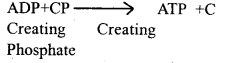
4. During relaxation, creatine is phosphorylated, energy being provided by anaerobic conversion of muscle glycogen into lactic acid.
Creatine + ATP— Creating-phosphate + ADP

5. Energy released by hydrolysis of ATP causes rotation of myosin heads and bring near the actin filaments, actomyosin complex is formed, eventually, sarcomere shortens.

6. Ca++ are actively transported to the sarcoplasmic reticulum, no more Ca++ available for ATP breakdown, no further energy available for further contraction of the sarcomere.

7. Part of the energy is utilized by breaking of cross-bridges and the muscle relaxes.

(b) What are the main groups of vertebrae in the vertebral column of man?
Answer:
There are 5 groups of vertebrae namely cervical, thoracic, lumbar, sacral and coccygeal vertebral.
(The vertebral formula is C7, T12, L5, C3-5 = 32 – 34).

Question 2.
(a) What purposes does movement of external body parts in relation to body axis serve in animals?
Answer:

  1. The movement of limbs, appendages, head and trunk serves to change the body posture to maintain equilibrium against gravity.
  2. Limb movements are prerequisites for carrying out locomotion.
  3. Prehension of food involves movement of tongue, jaws, snout, tentacles, limbs and appendages in different animals.
  4. Movement of eyeballs and pinna of ear help to collect information from the external environment.

(b) What are fibrous joint and cartilaginous joints and their biological function?
Answer:

  1. Fibrous joint: The articulating bones are firmly held together by the dense bands of tough, inextensible white fibrous tissues. They provide strength and support for the body or protection of delicate structures which cannot withstand any kind of deformation.
  2. Cartilaginous joints: In cartilaginous joints, a dense disc of white fibrocartilage joins the opposing surfaces of the articulating bones to each other. This allows a limited movement at the joints.

(c) Explain Antagonistic muscles.
Answer:
Antagonistic muscles: Antagonistic muscles are those which contract to produce opposite movements at the same joint. When a muscle contract to produce a movement, its antagonistic must relax to allow that movement to take place, e.g., the bicep is a FLEXER for the elbow joint and the tricep is it’s antagonistic and an EXTENSOR for that joint.

During flexion at the elbow, the biceps contract and the tricep relax, during extension at the same joint the tricep contracts and the biceps relaxes.

(d) Distinguish between muscles twitch and tetanus or explain muscle twitch and tetanus.
Answer:
A single isolated contraction caused by a single nerve impulse or electric shock is called a muscle twitch. Immediately after the brief twitch, the muscle fibres relax.

Tetanus is a continued state of concentration caused by many repeated stimuli. Much higher tension is developed in tetanus than in an isolated twitch. Almost all our daily activities are carried out by tetanic contractions of muscles.

Question 3.
How thick and thin filaments are arranged in a muscle fibre?
Locomotion and Movement Class 11 Important Extra Questions Biology 5
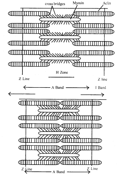
relationship between actin and myosin filaments in stretched and contracted states
Answer:
Each striated muscle contains thin actin and thick myosin filaments. These filaments are longitudinally arranged inside light I bands and dark A bands respectively. The actin and myosin filaments remain cross-linked with each other in the myofibril. Sarcomeres are the rows of functional unit in each myofibril, each extending from the dark Z- line of the next I band. Each sarcomere thus comprises of A band in the middle with 2 half I band on its two sides.

From each Z line, the actin filaments through half of the I band intermingles with the ends of myosin filaments in the A band. The myofibril is surrounded at each I band by the tubules and cisternae of sarcoplasmic reticulum and at each junction of A and I bands by a TI tubule communicating with the cell exterior, which is shown in the figure. The relationship between actin (thin filament) and myosin (thick filament).

Excretory Products and their Elimination Class 11 Important Extra Questions Biology Chapter 19

Here we are providing Class 11 Biology Important Extra Questions and Answers Chapter 19 Excretory Products and their Elimination. Important Questions for Class 11 Biology are the best resource for students which helps in Class 11 board exams.

Class 11 Biology Chapter 19 Important Extra Questions Excretory Products and their Elimination

Excretory Products and their Elimination Important Extra Questions Very Short Answer Type

Question 1.
What is a nephron?
Answer:
The functional unit of the kidney.

Question 2.
What is a flame cell?
Answer:
The excretory unit in planaria, tapeworm, and liver fluke.

Question 3.
What is micturition?
Answer:
It is the act of void of the urinary bladder, the activity under nervous and voluntary control.

Question 4.
What are ammonotelic animals?
Answer:
The animals which excrete nitrogenous wastes as ammonia are ed ammonotelic animals, e.g., certain fishes.

Question 5.
What is a green gland and in which animal it is found?
Answer:
It is an excretory structure found in prawns.

Question 6.
What is an antidiuretic hormone?
Answer:
It is the hormone that helps in the reabsorption of water in the nephron, also called vasopressin (Secreted by post pituitary gland).

Question 7.
Define excretion.
Answer:
Excretion is the process of elimination of metabolic wastes from the body.

Question 8.
What is the color rendering substance found in urine?
Answer:
Urochrome.

Question 9.
What are diuretics?
Answer:
The substances which increase the volume of water, to be excreted as urine, are called diuretics, e.g., tea, coffee, alcoholic beverages.

Question 10.
What is osmoregulation?
Answer:
It is the maintenance of water and osmotic concentration of blood.

Question 11.
Name the organ of the excretory system, which stores urine before its removal from the body.
Answer:
Urinary bladder.

Question 12.
In which part of the nephron does filtration occur?
Answer:
Glomerulus.

Question 13.
What happens to the useful substances that get filtered into the renal tubule?
Answer:
They are reabsorbed into the blood.

Question 14.
Point out the main excretory organ?
Answer:
Kidney.

Question 15.
Write down the products excreted by the following organs.
(a) Lung
Answer:
Lung: Carbon dioxide and water vapor

(b) Skin,
Answer:
Skin: Urea, water, and some salts

(c) Intestine.
Answer:
Intestine: Some salts like calcium and iron.

Question 16.
What is excreted by the kidney in urine?
Answer:
Urea.

Question 17.
In which part of the nephron does filtration occur?
Answer:
Glomemle.

Question 18.
Who filters the blood?
Answer:
The kidney filters the blood, which takes place between the glomerulus and Bowman’s capsule.

Question 19.
Why it is necessary to remove waste products by excretion?
Answer:
It is essential and necessary because all waste products are toxic and harmful.

Excretory Products and their Elimination Important Extra Questions Short Answer Type

Question 1.
Differentiate between sweat and sebum.
Answer:

SweatSebum
1. It is a liquid state excretion of tin.1. It is semisolid excretion
2. NaCl. urea, amino-acids are excreted.2. Waxes, fatty acids, and sterol are excreted.
3. Excreted in large amounts3. Excreted in small amounts
4. Also thermoregulatory role.4. No thermoregulatory role.

Question 2.
What consequences will follow with the failure of tubular reab¬sorption in nephrons?
Answer:
Nephrons are the structural and functional units of each kidney. With the failure of reabsorption in nephrons, much-needed substances like glucose, amino acids, water, salts, etc. will be excreted along with urine.

The biological functioning of organs and body will be impaired, ultimately death will occur.

Question 3.
How the net filtration pressure is obtained?
Answer:
The pressure of blood in afferent arterioles is (+ mm Hg 75). This is opposed by the osmotic pressure of plasma proteins by (-) 30 mm Hg and intertubular pressure of (-) 20 mm Hg. The net filtration pressure is (+) 25 mm Hg that acts in glomerular filtration as a driving force. About 172 liters of glomerular filtrate are produced in 24 hrs. which is nearly 4-1/ 2 times the total fluid in the human body.

Question 4.
List some important functions of kidneys?
Answer:
Kidneys play a vital role as follows:
(a) It removes nitrogenous wastes from the blood.
(b) It regulates fluid balance, between intake and fluid loss.
(c) It removes drugs, penicillin, poisons, etc. from blood.
(d) It maintains acid-base (pH) balance
(e) It regulates electrolyte balance.

Question 5.
Differentiate between ureter and urethra?
Answer:

UreterUrethra
1. It is a muscular tube.1. It is a membranous tube.
2. It is long.2. It is short.
3. It arises from the renal pelvis of the kidney.3. It arises from the urinary bladder.
4. It carries urine to the urinary bladder.4. It eliminates stored urine of the exterior.
5. No muscular splincter.5. Muscular splinter keeps urethra-closed except for micturition.

Question 6.
How does the excretion of uric acid take place in birds and reptiles?
Answer:
In birds and reptiles, uric acid is formed mostly in the liver, transported to the kidney through blood. It is separated by renal tubules and temporarily stored in cloacae. Water is absorbed by cloacal walls, needing only a minimum amount of water for excretion. In birds, urine is eliminated in a paste-like form along with feces.

Question 7.
Name and state in brief the processes involved in the formation of urine.
Answer:
The urine is formed by the combined processes as follows:
(a) Glomerular filtration: Metabolism wastes and other substances are filtered out by glomerulus due to the generation of net filtration pressure.
(b) Re-absorption: Water and other required substances are selectively reabsorbed from the filtrate, so that urine becomes concentrated.
(c) Tubular secretion: Tubules secrete certain ions (like K+ in exchange for Na+), urea, creatinine, uric acid, ammonia, etc. This process is of more significance in marine fishes and desert amphibians than mammals.

Question 8.
Differentiate between ureotelism and Uricotelism.
Answer:

UreotelismUricotelism
1. The process of elimination of main urea.1. The process of elimination of mainly uric acid.
2. Water moderately required for excretion.2. Much less water required for excretion.
3. Synthesis of urea requires less energy expenditure.3. Synthesis of uric acid needs more energy expenditure.

Question 9.
What is Polynephritis? What is uremia?
Answer:
It is a bacterial infection that causes inflammation of the renal pelvis, nephrons, and medullary tissues of the kidney. It affects the counter-current mechanism. Its main symptoms are frequent and painful urination, fever, and pain in the lumbar region.

A high concentration of urea, uric acid, creatinine, etc. in the blood due to some bacteria infection or some obstruction in the passage of the urinary system is called uremia.

Question 10.
Indicate whether the following statements are True or False
(a) Micturition is carried out by a reflex.
Answer:
True

(b) ADH helps in water elimination, making the urine hypotonic.
Answer:
False

(c) Protein-free fluid is filtered from blood plasma into the Bowman’s capsule
Answer:
True

(d) Henle’s loop plays an important role in concentrating the urine.
Answer:
True
(e) Glucose is actively reabsorbed in the proximal convoluted tubule.
Answer:
True

Question 11.
Match the items of Column I with these of Column II.

Column IColumn-II
(a) Ammonotelism(i) Birds
(b) Bowman’s Capsule(ii) Hypertonic   urine
(c) Micturition(iii) Counter-current system
(d) Uricotelism(iv) Bony fish
(e) Vasa recta(v) Urinary bladder
(f) Sebum(vi) Glucose
(g) ADH(vii) Glomerular Alteration
(h) Tubular reabsorption(viii) Skin

Answer:

Column IColumn-II
(a) Ammonotelism(iv) Bony fish
(b) Bowman’s Capsule(vii) Glomerular Alteration
(c) Micturition(v) Urinary bladder
(d) Uricotelism(i) Birds
(e) Vasa recta(iii) Counter-current system
(f) Sebum(viii) Skin
(g) ADH(ii) Hypertonic   urine
(h) Tubular reabsorption(vi) Glucose

Question 12
Fill in the blanks with appropriate words:
(a) During micturition, the urinary bladder, and the urethral sphincters contract, and relax
(b) Flame cells and malpighian tubules are found in and Bowman’s capsule and glomerulus respectively.
(c) Blood enters the glomerulus through the renal arteriole and leaves via the afferent arteriole.
(d) Two counter-current systems formed in the kidney are the Renal medulla and the renal cortex
(e) Sweat serves to eliminate mainly water and salt

Question 13.
Compare and contrast the osmoregulatory problems and adaptations of a marine bony fish with a freshwater bony fish.
Answer:
Osmoregulation in freshwater Marine bony fish, do not drink water to reduce the need to expel excess water. In this case, water uptake and salt loss are minimized by a specialized body covering. Freshwater animals have the ability to take up salts from the environment. The active transport of ions takes place against the concentration gradient, specialized cells called monocytes or chloride cells in the gill membrane of freshwater fish. These can import Na+ and CI from the surrounding water containing less than 1 mm NaCl when their plasma concentration of NaCl exceeds 100 mm.

Osmoregulation in marine environment Seawater has an osmolarity of about 1000m Osm L The osmoregulatory problems in marine water are opposite to those in a freshwater environment. Marine bony fish have the body fluids hypotonic to seawater and thereby, they tend to lose water from the body through permeable surfaces.

To compensate for the water loss, marine bony fish drink seawater, which results in a gain of excess salts. The monocytes or chloride cells of the gill membrane of marine bony fish help to eliminate excess monovalent ions from the body fluid to the seawater. Divalent cations are generally eliminated with feces.

Question 14.
State the importance of counter-current systems in renal functioning.
Answer:
Vasa rectal is responsible for the concentration of urine. The vase rectal is in the form of loops. Therefore, the blood flows in the opposite directions in two limbs of each vasa Fecta; the blood entering its descending limb comes into close contact with the outgoing blood in the ascending limb. This is called a Counter-Current System. The two limbs of the loops of Henle form another Counter-Current System.

Importance: The counter-current system significantly contributes to concentrating urine in mammals.

Question 15.
State the position and function of the juxtaglomerular apparatus?
Answer:
This is a specialized cellular apparatus located where the distal convoluted tubule passes close to the Bowman’s capsule between the afferent and efferent arterioles. JGA cells secrete substance like renin that modulates blood pressure and renal blood flow and thus, GFR is regulated.

Question 16.
Describe the hormonal feedback circuits in controlling renal functions.
Answer:
Two important hormonal control of the kidney function by negative feedback circuits can be identified:
1. Control by Antidiuretic Hormone ADH: ADH produced in the hypothalamus of the brain and released into the blood from the pituitary gland, enhances fluid retention by making the kidneys reabsorb more water. The release of ADH is triggered when osmoreceptors in the hypothalamus detect an increase in the osmolarity of the blood.

The osmoreceptors cells also promote thirst. Drinking reduces the osmolarity of the blood which inhibits the secretion of ADB, thereby completing the feedback circuit.

2. Control by Juxtaglomerular Apparatus (JGH): It operates a multihormonal Renin-Angiotensin-Aldosterone System (RAAS). JGA responds to decrease the blood pressure and release enzyme renin into the blood. In the blood, the enzyme initiates chemical reactions that convert a plasma protein called angiotensinogen to a peptide called angiotensin II which works as a hormone.

Angiotensin II increases blood pressure and stimulates the adrenal gland to release aldosterone, a hormone. This leads to an increase in blood volume and pressure completing the feed¬back circuit by supporting the release of renin.

Still another hormone, a peptide called Atrial Natriuretic Factor ANF), opposes the regulation by RAAS.

Thus, ADH, the RAAS, and ANF provide an elaborate system of checks and balance that regulate the kidney functioning to control body fluid, osmolarity, salt concentration, blood pressure, and blood volume.

Question 17.
State the normal and abnormal constituents of human urine.
Answer:
Urine is a pale yellow colored slightly acidic watery fluid.

  • Abnormal Urine: Various metabolic errors of kidney malfunctioning changes the composition of urine.
  • Proteinuria: Excess of protein level.
  • Albuminuria: The presence of albumin, usually occurs in nephritis.
  • Glycosuria: Presence of glucose in urea as in case of diabetes mellitus.
  • Ketonuria: Presence of abnormally high ketone bodies.
  • Hematuria: Presence of blood or blood cells in urine.
  • hemoglobinuria: Presence of hemoglobin in urine.
  • Uremia: Presence of excess urea.
  • Normal Urine: Normal urine is slightly heavier than water. It gives an aromatic odor due to the presence of volatile, bad-smelling organic substances, the ruined water, organic and inorganic materials are the main constituents of normal urine.

The other nitrogenous constituents of normal urine are ammonia, uric acid, hippuric acid, and creatinine.

Non-nitrogenous substances are vitamin C, oxalic acid, phenolic substances. In inorganic substances, sodium chloride is the principal mineral salt in the urine.

Question 18.
State the role of skin and lungs in excretion.
Answer:
Role of Skin: Human skin possesses glands for secreting sweat and sebum (from the sebaceous gland). Sweat contains NaCl, lactic acid, urea, amino acids, and glucose. The volume of sweat various negligible to 14 L a day. The principal function of sweat is the evaporative cooling of the body surface.

Sebum is a waxy protective secretion to keep the skin oily and this secretion eliminates some lipids, such as waxes, sterols, other hydrocarbons, and fatty acids. Integument in many animals is excreting ammonia into the surrounding by diffusion.

Role of lungs in excretion: Human lungs eliminate around 18L of CO2 per day and about 400 ml of water in normal resting conditions. Water loss via lungs is small in hot humid climates and large in cold dry climates. The rate of ventilation and ventilation pattern also affects the water loss through the lungs. Different volatile materials are also readily eliminated through the lungs.

Excretory Products and their Elimination Important Extra Questions Long Answer Type

Question 1.
Briefly state the mechanism of urine formation in the human kidney.
Answer:
Three main processes are involved in urine formation
1. Glomerular filtration: Kidneys filter the equivalent of blood volume every 4 – 5 minutes. Filtration slits are formed by the assemblages of fine cellular processes of podocytes (foot cells). The process of ultra-filtration depends upon two main factors, first the net hydrostatic pressure difference between the lumen of the capillary and the lumen of the Bowman’s capsule favor filtration.

The glomerular ultrafiltrate contains essentially all the constituents of the blood except for blood corpuscles and plasma proteins. Nearly 15% – 25% of the water and salutes are removed from the plasma that flows through the glomerulus. The glomerular filtration rate is about 125 ml min1 or about 180 L day-1 in human kidneys.

2. Two important intrinsic mechanisms provide autoregulation of glomerular filtration rate.
(a) Myogenic mechanism: Increase in blood pressure will tend to stretch the efferent arteriole, which would increase the blood flow to the glomerulus. The diameter of the arteriole is reduced, increasing the resistance to flow. This myogenic mechanism thus reduced variations inflow to the glomerulus in case of fluctuations in blood pressure.

(b) Juxtaglomerular apparatus (JGA): This specialized cellular apparatus is located where the distal convoluted tubule passes close to the Bowman’s capsule between the afferent and efferent arterioles. JGA cells secrete substances like renin that modulate blood pressure and renal blood flow and GFR are regulated.

Myogenic and juxtaglomerular mechanisms work together to autoregulate the GFR over a wide range of blood pressure. In addition to these extrinsic neural control also regulates the filtration rate.

3. Tubular re-absorption: The selective transport of substances across the epithelium of the excretory tubule from the ultrafiltrate to the interstitial fluid is called re-absorption. Nearly all the sugar, vitamins, organic substances (nutrients), and most of the water are reabsorbed.

4. Tubular secretion: It is a very selective process involving both passive and active transport. The filtrate travel through the nephron, substances that are transported across the epithelium from the surrounding interstitial fluid and join it. The net effect of renal secretion is the addition of plasma solutes to the filtrate within the tubule.

Question 2.
Explain the following:
(a) Skin functions as an accessory excretory organ.
Answer:
The skin retains some excretory role in many animals. Human skin possesses two glands for secreting fluid on its surface. These are; sweat from sweat glands and sebum from sebaceous glands.

(b) Mammals can eliminate hypotonic and hypertonic urine according to body needs.
Answer:
When the animal takes a large quantity of water the kidneys excrete a very high amount of hypotonic urine. At the same time when the animal takes a small number of water kidneys to excrete a very high amount of hypertonic urine.

At the same time when the animal takes a small number of water kidneys to excrete a small amount of hypertonic urine, as kidneys need to conserve water. In this way, the osmotic concentration of blood is maintained by the kidneys. This flexibility of kidney nephrons is highly observed in mammals.

Hypotonic urine removes excess water from the body in order to raise the osmotic concentration of the blood to normal. Excess of water in body fluids generally lowers the osmotic pressure of blood and increases the volume of blood. This increase in the volume of blood raises the blood pressure and hydrostatic pressure which increases the rate of ultrafiltration. In this way, a large amount of hypotonic urine is produced in order to bring the volume of fluids to normal.

(c) Micturition is a reflex process but is under some voluntary control.
Answer:
It is the process of passing out urine. Nephrons produce urine and drain. When enough urine collects in the bladder the distension of its walls raises enough pressure which generates a spontaneous nervous activity under the stimulation of the sympathetic and parasympathetic nervous system. This nervous stimulation causes the smooth muscles on the urinary bladder to rise too high to control.

Similarly, micturition can voluntarily be initiated even before enough urine has accumulated in the bladder. Backflow of the urine into the ureters from the urinary bladder is prevented because the terminal part of each ureter passes through the bladder and gets closed as soon as the contraction of the bladder occurs.

(d) Mammals are ureotelic, but birds are uricotelic.
Answer:
Mammals are ureotelic animals as they eliminate nitrogen mainly urea. It is very soluble in water and needs a considerable amount of water for its elimination. Mammals can thus form hypertonic urine which they excrete. While the birds cannot excrete urine as hypertonic since nitrogen occurs mainly in the form of uric acid. The uric acid is insoluble in water and does not require much water for its elimination.

Question 3.
Describe the functional anatomy of a human nephron.
Answer:
Nephrons are structural and functional units of each kidney to form the urine. Each nephron is fine; microscopic highly coiled tubular structure differentiated into malpighian body and the renal tubule. The malpighian body comprises a large double-walled cup-shaped structure the Bowman’s capsule present in the renal cortex. It is lined by thin, semipermeable epithelial cells, the podocytes. Bowman’s capsule receives the blood supply through a branch of the renal artery.

The afferent arteriole forms a fine capillary network in the form of glomerules with high hydrostatic pressure. The lumen between two layers of Bowman’s capsule is continuous with the lumen of the tubule. The Bowman’s capsule and the glomerulus together form a globular body, the Malpighian body or the renal capsules.

The capillaries forming the glomerulus at the exit of Bowman’s capsule unite to form a narrow efferent arteriole which breaks up into a peritubular network of capillaries with low hydrostatic pressure.

The renal tubule is a long highly coiled tubular structure differentiated into proximal convoluted tubule (PCT) Henle’s loop, distal convoluted tubule (DCT). The U-shaped loop-like structure, descending and ascending from the renal tubule is called Henle’s loop.

Collecting tubules of several nephrons open into a wider duct called the collecting duct. A number of collecting ducts unite with each other in the medulla to form the ducts of Bellini, which drains down the urine into the ureter from each kidney to be stored in the urinary bladder.

The efferent arteriole emerges out from the glomerules breaks up into a peritubular capillary network around the renal tubule in the cortex. These capillaries also form a thin-walled, straight capillary the vasa recta. The vasa recta help in retaining the reabsorbed ions and urea in medullary interstitial fluid to maintain high osmotic pressure in kidneys.

Glomerular filtrate undergoes tubular reabsorption and tubular secretion for the formation of urine. (See diagram opposite page)
Excretory Products and their Elimination Class 11 Important Extra Questions Biology 1
Uriniferous tubules Or nephron of the kidney

Question 4.
Describe the gross anatomical features of the human kidney with a suitable diagram.
Answer:
Kidney: Kidney is chocolate brown, bean-shaped, large-sized about 10 cm long and 5 – 7 cm broad, 3 – 4cm thick flattened, metamorphic. The weight of each kidney is 150 to 170 gm. They are situated against the back wall of the abdominal cavity, just below the diaphragm, between the 12th thoracic and 3rd lumbar vertebrae.

The outer margin is convex. The inner concave presents a longitudinal opening called the hilum. The renal artery and renal vein respectively enter and leave the kidney through its hilum.

The two kidneys are slightly asymmetrical in position because the right kidney is slightly at a lower level than the left. Kidneys are held in position by a mass of adipose tissue called Renal fat. These rest against the abdominal muscles. Each kidney is covered on the ventral side by the peritoneum and is thus retroperitoneal in nature.

Surrounding the kidneys and the renal fat is a sheath of fibro elastic tissue known as renal fascia or capsule. They protect the kidney. The renal fat forms a shock-absorbing cushion. The renal fascia fixes the kidney to the abdominal wall.
Excretory Products and their Elimination Class 11 Important Extra Questions Biology 2
Longitudinal section (Diagrammatic of Kidney)

Question 5.
(a)What is the role of the liver in excretion in mammals?
Answer:
Role of liver in excretion: The liver changes ammonia into urea which is less toxic than ammonia. Urea is eliminated from the body by the kidneys through urine.

The liver is the principal organ of excretion of cholesterol, bile pigments (bilirubin and biliverdin) some vitamins, drugs, and inactivated products of steroid hormones. The liver excretes these substances in the bile which carries them to the small intestine. Ultimately, these substances get eliminated along with feces.

(b) What are the diseases associated with the urinary system?
Answer:
Diseases associated with the urinary system:
1. Polynephritis: It is a bacterial infection, which causes inflammation of renal pelvic nephrons and medullary tissues of the kidney. It affects the counter-current mechanism. Its main symptoms are frequent and painful urination, fever, and pain in the lumbar region.

2. Uremia: It causes the presence of a high concentration of urea, uric acid, creatinine, etc, in the blood due to some bacterial infection or some obstruction in the passage of the urinary system. Urea poisons the cells. It is not passed in the urine and accumulates in the blood.

3. Renal stones: When uric acid precipitates and accumulates in the nephrons of kidneys in the form of renal stones or when calcium phosphates and oxalates accumulate in the nephrons of the kidneys in the form of renal stones. It causes blockage or frequent painful urination along with blood in the urine. Renal stone causes severe colic pain starting in the back and radiating down to the front of the thigh or vulva or testicle on that side.

4. Glomerulonephritis: It is characterized by the inflammation of Glomeruliduct, some injury to the kidney, abnormal allergic reaction, or by some streptococci bacteria infection. Proteins and red blood corpuscles become filtered into the glomerular filtrate. It may lead to kidney failure in severe infection.

5. Oedema: It is characterized by the increased volume of interstitial fluid mainly caused by retention of excess Na+ ions which in turn causes water retention. Blood pressure increases dining edema.

Question 6.
Write a short account on hemodialysis.
Answer:
In case of renal failure, an artificial kidney is used for removing excess urea from the blood of the patient by a process called hemodialysis. Blood is taken out from the artery of the patient, cooled to 0°C, mixed with an anticoagulant such as heparin, and then pumped into the apparatus called artificial kidney. In this apparatus, blood flows through channels
Excretory Products and their Elimination Class 11 Important Extra Questions Biology 3
Working of artificial kidneys for hemodialysis

bounded by cellophane membrane. The membrane is impermeable to macromolecules but permeable to small solutes. The membrane separates the blood flowing inside the channels from a dialyzing fluid flowing outside the membrane. The wastes like urea, uric acid, and creatinine diffuse from the blood to the dialyzing fluid across the cellophane membrane.

Thus the blood is considerably cleared of nitrogenous waste products without losing plasma proteins. Such a processor separation of macromolecules from small solute particles with the help of a permeable membrane is called dialysis. The blood coming out of the artificial kidney is warmed to body temperature, mixed with an Antiheparin to restore its normal coagulability, and returned to a vein of the patient.

Haemodialysis saves and prolongs the life of many uremic patients.

Body Fluids and Circulation Class 11 Important Extra Questions Biology Chapter 18

Here we are providing Class 11 Biology Important Extra Questions and Answers Chapter 18 Body Fluids and Circulation. Important Questions for Class 11 Biology are the best resource for students which helps in Class 11 board exams.

Class 11 Biology Chapter 18 Important Extra Questions Body Fluids and Circulation

Body Fluids and Circulation Important Extra Questions Very Short Answer Type

Question 1.
What is systole?
Answer:
The contraction phase of the cardiac chamber (s) is called systole.

Question 2.
What is diastole?
Answer:
The relaxation phase of the cardiac chamber is called diastole.

Question 3.
Where is SA-node located?
Answer:
It is located at the place of merge of sinus venous at the right wall of the right atrium.

Question 4.
In which animals we can find the sinus Venosus?
Answer:
Sinus venosus can be found in fishes, amphibians and reptiles.

Question 5.
What is the ‘lubb’ sound?
Answer:
It is the first sound produced by the heart due to the sharp closure of AV valves at the start of ventricular systole.

Question 6.
What is AV-node?
Answer:
It is a node of specialised fibres, located at the junction of the right atrium and right ventricle.

Question 7.
What is hemolymph?
Answer:
The blood of insects that lack haemoglobin is called haemolymph.

Question 8.
What is a sphygmomanometer?
Answer:
The instrument which measures blood pressure is called a sphygmomanometer.

Question 9.
What is the “dup” sound?
Answer:
It is the second sound produced by the heart due to the sharp closure of semilunar valves at the start of ventricular systole.

Question 10.
What is the role of AV-node?
Answer:
It collects the wave contraction generated by SA-node and passes down to a bundle of His and Purkinje fibres.

Question 11.
Where is RBCs (red blood cells) produced?
Answer:
RBCs are produced in the bone marrow.

Question 12.
Name different types of blood groups present in human beings.
Answer:
Types of blood group: O, A, B, AB group.

Question 13.
Name various parts of the circulation.
Answer:
Heart, blood, gases and same waste materials.

Question 14.
Name the blood vessel which carries impure blood to the heart.
Answer:
Vena cava.

Question 15.
The transport system in animals is called
Answer:
Circulatory system.

Question 16.
What is the difference between artery and vein?
Answer:

  1. Artery: Thin-walled
  2. Vein: thick-walled.

Question 17.
What are the different components of blood?
Answer:
Plasma, RBC, WBC, Sugar, salt, waste products.

Question 18.
What is the heartbeat rate/minute?
Answer:
72 beat/minute.

Question 19.
What are circulatory fluid and its component?
Answer:
Circulatory fluid is blood and its components are haemoglobin, RBC, WBC.

Question 20.
Give the function of blood platelets.
Answer:
They help in the clotting of blood.

Question 21.
Which blood group is known as a universal donor?
Answer:
‘O’ group is known as a universal donor.

Body Fluids and Circulation Important Extra Questions Short Answer Type

Question 1.
Why does the ventricle contract as a closed chamber in the early phase of its systole?
Answer:
In the early phases of systole the ventricle contracts as a closed chamber, so to increase the pressure in the atrium. Backflow of blood into atria is prevented by closure of AV-valves. In a closed chamber, the ventricles contract and increased pressure cause the opening of semi-lunar valves. The blood is passed into arteries with great force.

Question 2.
The blood vascular system is considered efficient than the water circulatory system in animals, why?
Answer:
The dissolved oxygen and nutrient present in water are in fewer amounts. Oxygen is supplied through oxygen carrier molecule (haemoglobin) present in plasma or cells, in higher animals. Oxygen and nutrients are supplied quickly and in fairly large amounts in animals with a blood vascular system, so considered comparatively efficient.

Question 3.
Mention, in brief, the important events that happen during the cardiac cycle.
Answer:
The cardiac cycle comprises the following three phases.
(a) Atrial systole
(b) Ventricular systole
(c) Joint diastole.

(a) Atrial systole: The atria contract from anterior to posterior and the blood is sent to respective ventricles. Time taken is 0.1 sec.
(b) Ventricular systole: The ventricles contract, deoxygenated blood is sent to lungs for oxygenation, while oxygenated blood received from lungs is sent to different parts of the body through the aortic arch. Time taken is 0.3 sec.
(c) Joint diastole: All the chambers are in systole, called joint diastole. Time taken is 0.4 sec.

Question 4
Describe in brief the types of valves present in the heart.
Answer:
Different valves present in the heart are described as under:
(a) Tricuspid valve: The valve present at the right atrioventricular aperture formed of three flaps.
(b) Bicuspid valves: The valve present at the left atrioventricular aperture, formed of two flaps, also called the mitral valve.
(c) Semi-lunar valve: Two sets of half-moon shaped (3) valves, present at openings of pulmonary aorta and aorta, present here to check backflow of blood.

Question 5.
What is hypertension? What are causative factors?
Answer:
It is a manifestation of an increase in blood pressure. A continuous or sustained rise in arterial blood pressure is known as hypertension.

The normal systolic and diastolic pressure of a healthy human is 120m and 80mm Hg respectively. Under various physiological conditions, a moderate level of fluctuation may occur and blood pressure increase. It is referred to as high blood pressure.

Question 6.
What is heart rhythm? Discuss.
Answer:
Heart muscles continuously generate impulses in a manner that causes rhythmic contraction and relaxation of the heart chambers. For the purpose of maintenance of the heart rhythm, a highly excitatory’ and conductive system is present, which includes sino-atrial node (SA) inter- nodal pathways, the atria-ventricular node (Avnode) AV bundle and the bundle of Purkinje fibres.

In normal conduction of impulse, the atrial contraction precedes that of the ventricle.

The automatic rhythmicity of the heart is its ability to contract spon¬taneously at a regular rate.

Question 7.
Define the terms:
(a) SA node,
Answer:
SA: It is a self-excitatory node that initiates excitatory waves at the highest rate. SA-node normally generates the cardiac impulse and working by knowing the rate of discharge of cardiac impulse. It can determine the rate of heartbeat. The SA-node works as the pacemaker of the heart.

(b) Systole,
Answer:
Systole: The pumping out of blood due to contraction of the heart is called systole.

(c) Diastole,
Answer:
Diastole: The relaxation of the heart is called diastole. During diastole, blood flows into the auricles to their ventricles through the open auriculoventricular valve.

(d) Pulmonary circulation.
Answer:
Pulmonary circulation: The circulatory system which is associated with the lungs through arteries and veins, is known as pulmonary circulation.

Question 8.
Fill in the blanks:
Answer:
(a) Eosinophil is a bilobed nucleus
(b) Pulmonary artery carries deoxygenated blood.
(c) Heart of cockroach is present in the pericardial sinus.
(d) Platelets helps in coagulation
(e) Haemoglobin is present in RBCs due to which its colour is red.

Question 9.
What is an artificial pacemaker? Explain.
Answer:
A pacemaker is a rhythmic centre that establishes a pace of activity. Sometimes the component of the impulse conduction system is disrupted, causing irregularity in the heart rhythm like heart failure. Such types of patients are provided with an artificial electronic device, which regularly sends a small amount of electrical charge for maintaining the rhythmicity of the heart.

The device is known as an artificial pacemaker. It is implanted subcu¬taneously in the upper thoracic region having a connection with the heart.

If patients having symptoms of ventricular escape, in which atrial impulse suddenly fails, the artificial pacemaker is connected to the right ventricle for controlling its rhythm. The artificial pacemaker consists of a pulse generator containing a cell to produce electric impulse.

Question 10.
How arteriosclerosis is different from atherosclerosis? Discuss.
Answer:
Arteriosclerosis: It is the hardening of the arteries due to deposition and thickening. In the case of arteriosclerosis, calcium salts precipitate with the cholesterol and plague. The affected artery loses the property of distension and its walls may rupture. The blood leaking from the ruptured walls may clot and block the pathway of blood flow. This may lead to a heart attack and even death.

Atherosclerosis: It is the deposition of lipids (cholesterol) on the wall lining the lumen of large and medium-sized arteries. This type of deposition is called atherosclerosis. Its formation starts with the deposition of cholesterol particles/crystals in the tunica intima and smooth muscles. This results in the reduction of the lumen size of the artery and the flow of the blood also reduce.

In extreme circumstances, these plaques may completely block the artery. The proliferation of smooth muscles occurs because these plaques provide a rough surface to the platelets causing the release of platelet, derived from growth factor (PDGF). Such plaques reduce the blood supply to the heart or may stop the supply due to complete blockage. This may result in a heart attack or stroke.

Question 11.
What is heart rhythm? Discuss.
Answer:
Heart muscles continuously generate impulses in a manner that causes rhythmic contraction and relaxation of the heart chambers. For the purpose of maintenance of the heart rhythm, a highly excitatory and conductive system is present, which includes sino-atrial node (SA) intermodal pathways, the atrioventricular node (Av node) AV bundle and the bundle of Purkinje fibres.

In normal conduction of impulse, the atrial contraction precedes that of the ventricle.

The automatic rhythmicity of the heart is its ability to contract spontaneously at a regular rate. ,

Question 12.
What is an electrocardiogram? Write about its significance.
Answer:
The passage of cardiac impulse through the heart spreads electric current into the tissues around the heart and a small portion spreads throughout the surface of the body.

These electrical changes can be recorded along the cardiac cycle. The recording of electrical potential generated by the spreading of cardiac impulse is called an electrocardiogram (ECG). The electrical activity of the heart can be graphically represented by ECG, which shows different waves.

Question 13.
Match column I with column II.

Column IColumn II
(a) Haemolymph(i) Coagulation
(b) RBC(ii) Immunity
(c) Antibody(iii) Cockroach
(d) Platelets(iv) Contraction
(e) Systole(v) Gas transport

Answer:

Column IColumn II
(a) Haemolymph(iii) Cockroach
(b) RBC(v) Gas transport
(c) Antibody(ii) Immunity
(d) Platelets(i) Coagulation
(e) Systole(iv) Contraction

Question 14.
What is blood? Describe its components.
Answer:
Blood is a complex connective tissue, which consists of two components:

  1. Plasma: It is the extracellular fluid of blood constituting about 55% of the blood volume. Plasma contains 91 – 92% of water, 7% of proteins, 0.9% of inorganic constituents, 0.1% of glucose and the rest includes various organic and inorganic substances. Proteins are the second-largest constituents of plasma.
  2. Blood corpuscles: Nearly 45% volume of blood consists of corpuscles or blood cells. Blood corpuscles are of three types: erythrocytes or red blood corpuscles (RBC), leucocytes or white blood corpuscles (WBC) and thrombocytes or blood platelets.

Question 15.
What is hypertension? What are different contributory fac¬tors?
Answer:
An abnormal rise in arterial blood pressure is called hypertension.

The contributory factors of this disease are:
(a) Hypercholesterolemia,
(b) Nervous strain,
(c) Renal disorder/impaired functioning.
(d) Arteriosclerosis.

Question 16.
What is the main advancement in avian and mammalian heart over the amphibians and reptiles heart?
Answer:
Avian and mammalian heart show complete double circulation and in them, oxygenated blood remains completely separate from deoxygenated blood. There are no accessory chambers in them. There is a single aorta in aves and mammals and double aorta in reptiles and amphibians which quickens the supply of O2 and food to release the energy and aids in the removal of wastes from the body.

Question 17.
What is the base efficiency of the heart?
Answer:
The efficiency of the heart on beating throughout life without fatigue is based upon the principle that it rests double the time it works i.e. contraction of the heart is followed immediately by relaxation but relaxation is not followed by the next contraction. The beating of the heart is 72 times per minute. It is indicated by pulse rate per minute. The amount of blood pumped out per minutes is called heart output.

Question 18.
What does ECG stand for? Define it.
Answer:
ECG stands for electrocardiograph. It is defined as a permanent record of electrical events (depolarization and ventricles) during a cardiac cycle made on graph paper with the help of an instalment called an electrocardiogram.

It shows 5 waves i.e., P, G, R, S, T. In this P, R, T are positive waves that lie above the baseline whereas Q and S are negative waves that lie below the baseline. The part of the baseline between two deflections is called an interval.

Question 19.
What are the advantages of physical exercise?
Answer:
Any physical exercise initiates an impulse from the nervous system which causes the release of adrenaline hormone from adrenal glands into the blood.

Advantages of physical exercise:

  1. Exercise increases heartbeat which reduces the workload of the heart. It increases blood circulation so that every tissue gets a supply of nutrients and oxygen.
  2. It maintains normal blood pressure by lowering the chances of developing atherosclerosis- a disease, which causes narrowing of arteries.
  3. Exercise increases the level of high-density lipoproteins (HDLS) which help in reducing the harmful cholesterol from arteries.
  4. Exercise prevents heart attack with the development of additional or collateral blood vessels which provide alternative routes for blood supply to the muscles of the heart.
  5. Exercise adds more number RBCs
  6. Haemoglobin of blood increases which helps in supplying more O, to the tissues.

Question 20.
What is blood pressure? What do you mean by diastolic and systolic pressure?
Answer:
It is the pressure against the walls of blood vessels produced by the discharge of blood from the heart. It is high in arteries and Ipw in veins. It rises and falls in arteries due to heartbeat.

The temporary rise in B.P. during contraction of the heart is called systolic pressure. The temporary fall in the B.P during relaxation of the heart is called diastolic pressure.

Question 21.
What is a lymph node?
Answer:
The lymphatic vessels at intervals bear small lymph nodes which act as the filters of poisonous and foreign substances such as dust, debris, bacteria and other injurious substances. A lymph node has an inner border called the hilum. These are the masses of dense connective tissue, rich in phagocytic white corpuscles and macrophages.

Lymph nodes also produce lymphocytes, monocyte and antibodies into the lymph which are carried to the blood. These lymph nodes help in detecting and destroying cancer cells at the initial stages.

Question 22.
What is systemic circulation?
Answer:
The system of blood vessels that ensures the supply of oxygenated blood from the left ventricle to all the body organs and the return of deoxygenated blood to the right atrium is called systematic circulation.

Question 23.
Describe the importance of pulmonary circulation.
Answer:
The circulatory system associated with the lungs through arteries and veins forms the pathway of the circulatory system.

The flow of blood from the heart to the lungs and back to the heart is called pulmonary circulation. The regular oxygenated blood is returned to the heart and the pulmonary artery supplies deoxygenated blood from the right ventricle to the lungs. It is the pathway of the blood through arteries from t aorta arising from the heart and returning to the right atrium.

Question 24.
What is an electrocardiogram? Write about its significance.
Answer:
The passage of cardiac impulse .through the heart spreads electric current into the tissues around the heart and a small portion spreads throughout the surface of the body.

These electrical changes can be recorded along the cardiac cycle. The recording of electrical potential generated by the spreading of cardiac impulse is called an electrocardiogram (ECG). The electrical activity of the heart can be graphically represented by ECG, which shows different waves.

Question 25.
Write true or false:
(a) Atrio-ventricular node is the natural pacemaker of the heart.
Answer:
False

(b) Human heart has inter-auricular foramen.
Answer:
True

(c) Heart of the cockroach is segmentally arranged.
Answer:
True

(d) Right atriventricular valve is a semilunar valve
Answer:
True

(e) Normal systolic and diastolic pressure of humans is 120 and 60 mm Hg, respectively.
Answer:
False

Body Fluids and Circulation Important Extra Questions Long Answer Type

Question 1.
(a) Why is the AV bundle essential for the conduction of cardiac muscles? Explain.
Answer:
The sinoatrial node (pacemaker) of the heart spreads the cardiac impulse over the two atria to bring about their systole. It, however, cannot spread along the common cardiac muscle fibres from the atria to the ventricles. It is because, in the mammalian heart, there is no continuity between the cardiac muscle fibres of the atria and those of the ventricles.

Although the fibres of each individual chamber exist in a functional syncytium (not separatable). It is because a bond of specialized cardiac muscle fibres exists on the interatrial septum called the ATRIOVENTICULAR BUNDLE (AV bundle).

AV bundle forms the only muscular continuity between atrial and ventricular muscles. The AV bundle descends from the AV node along the interatrial septum and the interventricular septum. It branches into right and left bundle branches as it enters the ventricle.

From each AV bundle branch Purkinje which fibres spread out and connect with the common ventricular muscle fibre. Thus, the cardiac impulse spreads over the atria to reach the AV node; the AV bundle is necessary for the conductions of impulse through its two branches and the Purkinje fibres to reach the ventricular muscle fibres causing contraction of ventricles.

(b) Make a graphic representation of double circulation in the mammalian heart.
Answer:
Body Fluids and Circulation Class 11 Important Extra Questions Biology 1
Representation of double circulation in animals.

Question 2.
Define portal system. How is the hepatic portal system useful to our body?
Answer:
Portal system: A portal system is the circulatory system in which blood collected from one set of organs or tissues is conveyed to another organ through capillaries before entering the heart.

For example, in the hepatic portal system, blood collected from the alimentary -canal is first conveyed to the liver by the hepatic portal vein. After passing through the capillaries of the liver, it passes directly to the posterior vena cave by the hepatic vein.
Body Fluids and Circulation Class 11 Important Extra Questions Biology 2
Hepatic portal circulation

Use of hepatic portal system:
(i) Veins coming from the various parts of the alimentary canal carry deoxygenated and food-laden blood. Through the portal vein, it reaches the network of portal vein capillaries in the liver. The excess of food is filtered and stored in the liver as glycogen. Thus, heavily food loaded blood is not allowed to go to the heart, which may have to work more in pumping the blood.

(ii) Liver also consumes drugs and toxins present in the blood coming from the intestine. So, that heart can be saved from their harmful effects.

Question 3.
What is the lymphatic system? Discuss its importance.
Answer:
The lymphatic system consists of vessels and lymph organs, like lymph nodes, bone marrows, spleen and thymus. The fluid present in the lymphatic system is called lymph. This fluid has a composition similar to that of plasma except it is low in protein.

Fine channels present in the tissue are called lymph vessels, these are similar to veins. Besides these, a number of lymph nodes are present. The lymphatic vessels are distributed in the limbs, abdomen, thorax and neck.

The lymphatic system provides an accessory route for the flow of interstitial fluid into the blood. The lymphocytes present in the lymphatic system play important role in the defence against foreign agents or microbes.
Body Fluids and Circulation Class 11 Important Extra Questions Biology 3
Shows the various lymphatic organs in the human body

Question 4.
Describe the structure of the human heart.
Answer:
The human heart consists of four chambers: Two upper thin layered (Atrium) and two lower thick layered ventricles. The atria are situated at the broader end while the ventricles are situated at the lower conical end. Externally a transverse groove is present between the atria and ventricles, called the anterior interventricular sulcus and posterior intraventricular sulcus.

The right and left atria receive blood from different body parts. The right atrium receives deoxygenated blood from all parts of the body except the lungs, through the superior and inferior vena cava. Pulmonary veins bring oxygenated blood to the left atrium horn of the lungs.
Body Fluids and Circulation Class 11 Important Extra Questions Biology 4
Extma1 features of the human heart

The right and left atria pump their blood into the right and left ventricles. From the right ventricle, the pulmonary trunk arises which bifurcates into right and left pulmonary arteries, which supply deoxygenated blood to the lungs. The left ventricle gives rise to an ascending aorta from ‘which the oxygenated blood is supplied to the coronary- arteries and the systemic circulation of the body occurs.

Question 5.
Write the differences between
(a) Blood and haemolymph
Answer:

BloodHaemolymph
I. It is composed of blood plasma RBCs, WBCs and platelets.1. It is composed of watery fluid, lymphocytes. few monocytes, but no RBCs.
2. It is red in colour2. It is a watery fluid.
3. Blood contains more plasma proteins, calcium and phosphorus ions.3. Lymph contains fewer proteins, less calcium and phosphorus ions
4. Flow of blood is fairly rapid.4. Haemolymph flow is very slow.
5. Blood is pumped out from the heart, flow through the arteries, capillaries and veins.5. Haemolymph flow begins from the tissue spaces, goes to lymph capillaries.
6. Glucose concentration is comparatively low.6. Glucose Concentration is comparatively high.

(b) Open and closed system of circulation
Answer:
Open type of circulatory System and Closed system of circulation:
Open type of circulatory System: It mainly occurs in arthropods and molluscs, such as insects, prawns, spiders, oysters, etc. In these colourless blood coming through blood vessels, flows through open spaces and channels called lacunae and sinuses.

Lacunae and sinuses together called haemocoel, which carries colourless blood called haemolymph. The oxygen-carrying pigments are generally dissolved in the plasma of blood. Deoxygenated blood from these goes to the gills for oxygenation after oxygenation blood is returned to the sinus surrounding the heart.

Closed type of circulatory system: It mainly occurs in annelids and chordates. Heart and blood vessels together constitute the closed circulatory system. The heart is a pumping organ that is provided with valves. It pumps the blood into arteries, which earn’ the blood to different organs to the body.

Question 6.
Describe the circulatory system of cockroach.
Answer:
The circulatory system is of open type. It consists of the heart and dorsal blood vessel, sinuses and haemolymph. The haemocoel is divided into three chambers or sinuses dorsal, middle and ventral. The dorsal chamber is called the pericardial sinus, the middle is the perivisceral sinus whereas the ventral is the perineural sinus. The dorsal and ventral diaphragms bear a number of pores through which haemolymph flows.

The heart of a cockroach is an elongated tubular structure, closed behind and open in front. It has thirteen funnel-shaped and segmentally arranged chambers i.e. three in the thoracic segment and ten in the abdominal segment. Valves, ensuring the unidirectional flow of blood, guard the passage of each heart chamber.

Blood flows from the posterior to the anterior end and is discharged into the tissue space of the head. Laterally each heart chamber bears a pair of apertures called Ostia which communicates with the pericardial sinus.

In each segment, a pair of triangular alary muscles is present on either side of the heart.
The blood of the cockroach is not responsible for the transportation of respiratory gases but serves for
(1) the transportation of nutrients.
(2) maintains hydrostatic pressure and
(3) acts as a reservoir of water.

The haemolymph of cockroach circulates due to contraction and relaxation of the heart and ciliary muscles,
Body Fluids and Circulation Class 11 Important Extra Questions Biology 5
Open type of circulatory system in cockroach

Breathing and Exchange of Gases Class 11 Important Extra Questions Biology Chapter 17

Here we are providing Class 11 Biology Important Extra Questions and Answers Chapter 17 Breathing and Exchange of Gases. Important Questions for Class 11 Biology are the best resource for students which helps in Class 11 board exams.

Class 11 Biology Chapter 17 Important Extra Questions Breathing and Exchange of Gases

Breathing and Exchange of Gases Important Extra Questions Very Short Answer Type

Question 1.
Name the site of the exchange of gases in man.
Answer:
Alveoli in the lungs.

Question 2.
Name the respiratory organs in insects.
Answer:
Tracheae and spiracles.

Question 3.
Name the respiratory pigment of the blood of mammals.
Answer:
Haemoglobin.

Question 4.
What makes the exchange of gases in gills and lungs possible?
Answer:
Thin-walled blood capillaries.

Question 5.
Why are erythrocytes unable to carry out cellular oxidation?
Answer:
They lack mitochondria.

Question 6.
What is the main source of energy?
Answer:
Carbohydrates lipids and proteins.

Question 7.
Why energy obtained from ATP molecules is called biologically useful energy?
Answer:
Because it drives the life processes.

Question 8.
How does the exchange of gases across a respiratory membrane occur?
Answer:
By diffusion and circulatory system.

Question 9.
What holds the energy in the body?
Answer:
Molecules of food hold energy in their chemical bonds.

Question 10.
What is external respiration?
Answer:
It is the intake of oxygen by the blood from air or water to the respiratory organs and the elimination of CO2.

Question 11.
Name the parts of the human respiratory system in a sequence starting from the nose.
Answer:
External nostrils, nasal cavity, internal nostrils, pharynx, trachea, bronchi, lungs.

Question 12.
What is glottis?
Answer:
The opening of the pharynx into the trachea is called the glottis.

Question 13.
What is breathing?
Answer:
Breathing is one phase of respiration. During breathing, we get oxygen from the atmosphere inside our body.

Question 14.
How breathing is different from circulation give a reason?
Answer:
Breathing is a phase of respiration. It is a physical process.

Question 15.
Which gases take part in breathing?
Answer:
Carbon dioxide, oxygen, nitrogen and mixture.

Question 16.
What is glomerulus?
Answer:
The glomerulus is a lift of blood capillaries in a cup-shaped Bowman’s capsule.

Question 17.
What is tidal volume?
Answer:
It is the volume of air breathed in and out normally.

Question 18.
Why gaseous exchange continues in the lungs even after expiration?
Answer:
Because of the presence of residual volume.

Question 19.
Where does the exchange of gases occur in our body?
Answer:
In lung alveoli and tissue cells.

Question 20.
What is vital capacity in regard to breathing?
Answer:
Vital capacity is the volume of air that can be inspired and expired with maximum efforts.

Question 21.
Name excretory organs of earthworm.
Answer:
Nephridia.

Breathing and Exchange of Gases Important Extra Questions Short Answer Type

Question 1.
Define partial pressure of a gas.
Answer:
It is the pressure exerted in a mixture of gases and is equal to the total pressure of the mixture of gases divided by the percentage of that gas in a mixture. The atmospheric air pressure at sea level is 760mm of Hg. Oxygen forms 35% of the air. The partial pressure of oxygen is 760 × 35/ 100 = 266 mmHg.

Question 2.
How would you differentiate between tidal volume and residual volume?
Answer:

  1. Tidal volume is the amount of air inhaled or exhaled in one complete breathing. It is about 500 ml.
  2. Residual volume is the volume of air that remained in the lungs after the maximum effort of exhalation. It is about 1500ml.

Question 3.
What is the need for a circulatory system in a bigger animal?
Answer:
In larger animals the deeper cells cannot obtain oxygen directly from the atmosphere simply through the process of diffusion or eliminate CO2. In such a case, the respiratory system transports the respiratory gases from the respiratory’ surface to the deep-lying tissues.

Question 4.
Why does one experience difficulty at a high altitude?
Answer:
At high altitude, the pressure of air falls and the person cannot get enough oxygen in the lungs for diffusion in the blood. Due to insufficient O2, the person has difficulty breathing at high altitude. The person feels difficulty such as breathlessness, headache, dizziness, irritability, nausea, vomiting, mental fatigue and a blush (///) on the skin, nails and lips.

Question 5.
What are the conditions essential for effective respiration?
Answer:
Conditions essential for effective respiration:

  1. The respiratory surface should be thin and permeable to O2 and CO2
  2. The rich supply of blood to the respiratory surface.
  3. Passage for bringing oxygen to the respiratory surface and removing CO2 through the same passage.
  4. The respiratory surface should be moist.
  5. Presence of a circulatory system.
  6. Presence of a respiratory pigment to carry out the respiratory gases (CO2 and O2)

Question 6.
What is a specialized respiratory surface and what are its advantages?
Answer:
A specialized respiratory surface is thin, moist and highly vascular. It remains in contact with the environment outside the body and tissues inside the body. Diffusion of gases takes place from the respiratory surface between the body and outside the environment. The epidermal capillaries release carbon dioxide and take up oxygen dissolved in the film of surface moisture.

Question 7.
What is respiration?
Answer:
A process of physiochemical change by which environmental oxygen is taken in to oxidise the stored food to release CO2 water and energy; the energy released is used for doing various life activities whereas CO2 being foul gas is thrown out from the body. The main source of energy are carbohydrates, lipids and proteins. Respiratory mediums are air and water.

Question 8.
Define the
(a) Inspiratory reserve volume (IRV)
Answer:
Inspiratory reserve volume: It is the amount of air that can be inhaled forcibly after a normal inspiration. It is about 200 – 250 ml.

(b) Expiratory reserve volume (ERV)
Answer:
Expiratory reserve volume: It is the volume of ah, which can be exhaled forcibly after a normal expiration. It is about 1000 – 1500 ml.

(c) Vital capacity of lungs (VC)
Answer:
Vital capacity of lungs: It is the amount of air that one can exhale with maximum effort. It is about 3500-4500 ml.

(d) Residual volume. (RV)
Answer:
Residual volume: The amount of air left in the lungs after forcible expiration is called residual volume. It is about 1500ml.

Question 9.
What is Bronchial Asthma? How it is caused? What are the symptoms of this disease?
Answer:
It is characterised by the spasm of the smooth muscles present in the walls of the bronchiole. It is generally caused due to the hypersensitivity of the bronchiole to the foreign substances present in the air passing through it.

The symptoms of this disease are coughing, difficulty in breathing mainly due to expiration, the mucous membrane starts secreting an excess amount of mucous.

Question 10.
What is the preventive measure of the disease Bronchitis?
Answer:
Bronchitis is caused by cigarette smoking and exposure to air pollutants like carbon monoxide. It can be prevented and cured by avoiding exposure to the cause i.e. smoke, chemicals and pollutants. The underlying infection of the disease is treated with suitable antibiotics Bronchodilator drugs (for widening the constriction of bronchial passage by relaxing the smooth muscles) provide symptomatic relief.

Question 11.
How respiration fulfil the energy requirement of an organism?
Answer:
Respiration is a catabolic process. A catabolic biochemical process of exchange of gases by which atmospheric oxygen is taken in to oxidise the stored food to liberate energy, CO2 and water. Energy set free is used for doing life activities. For oxidation usually, oxygen is used. The energy is released in steps from the continuous breakdown of foodstuffs and is stored in the high energy bonds of ATP molecules. This energy obtained from ATP molecules is termed biologically useful energy as it allows the working of all life processes.

Question 12.
What is chloride shift? Write its significance during respiration.
Answer:
The chloride ions (CI) inside RBC combine with potassium ion (K+) to form potassium chloride (KCL), whereas hydrogen carbonate ions (HCO3) in the plasma combine with Na’ to form sodium hydrogen carbonate (NaHCO3) Nearly 70% of carbon dioxide is transported from tissues to the lungs in this form.

In response to chloride ions (CI) diffuse from plasma into erythrocytes to maintain the ionic balance. This is called the chloride shift.

Significance: It maintains electrochemical neutrality during respiration.

Question 13.
Write true or false.
(a) Inspiratory reserve volume is the volume of air, which can be inspired in addition to the normal inspiration.
Answer:
False

(b) Vital capacity is a measure of maximum inspiration.
Answer:
true

(c) During the gaseous exchange the gases diffuse from high partial pressure to low partial pressure.
Answer:
true

(d) Carbon dioxide cannot be transported with haemoglobin.
Answer:
true

(e) Earthworm respires through parapodia.
Answer:
false.

Question 14.
What is the role of the carbonic anhydrase enzyme in the transport of gases during respiration?
Answer:
Carbon dioxide produced by the tissues diffuses passively into the bloodstream and passes into the red blood corpuscles where it reacts with water to form carbonic acid (H2CO3). This reaction is catalysed by the enzyme, carbonic anhydrase found in the erythrocytes and takes less than one second to complete the process. Immediately after its formation, carbonic acid dissociates into hydrogen (H+) and bicarbonate (HCO3) ions. The majority of bicarbonate ions (HCO3 ) formed within the erythrocytes diffuse out into the plasma along a concentration gradient. These combine with haemoglobin to form the haemoglobin acid (H.Hb).
Breathing and Exchange of Gases Class 11 Important Extra Questions Biology 1

Question 15.
What is partial pressure? How does it help in gaseous exchange during respiration?
Answer:
During inspiration and expiration, gases move freely by the process of diffusion. Diffusion of any molecule takes place from high to low concentration. The process of diffusion is directly proportional to the pressure caused by the gas alone. The pressure exerted by an individual gas is called partial pressure. It is represented as PO2, PCO2, and PN2, for oxygen, carbon dioxide and nitrogen respectively.

The inspired air ultimately reaches the alveoli of the lung, which in turn receives the blood supply of the pulmonary circulation. At this stage the oxygen of the inspired air is taken in by the blood and carbon dioxide is released into the alveoli for expiration.

In this way, the gases exchange takes place due to partial pressure.

Question 16.
How does haemoglobin help in the transport of oxygen from the lung to tissues?
Answer:
Blood is the medium for the transport of oxygen from the respiratory organ to the different tissues and carbon dioxide from tissues to the respiratory organs. 97% of the oxygen is transported from the lungs to the tissues in combination with haemoglobin (Hb + O2 — HbO2), oxyhaemoglobin and 3% is transported in dissolved condition by the plasma.

Under high partial pressure oxygen easily binds with haemoglobin in the pulmonary capillaries. When this oxygenated blood reaches the different tissues, the partial pressure of oxygen declines and the bonds holding oxygen to haemoglobin become unstable. As a result, oxygen is released from the capillaries.

Question 17.
Write the names of the respiratory organs present in human.
Answer:
The human respiratory system consists of external nares or nostrils, nasal cavity, nasopharynx, larynx, trachea, bronchi, bronchiole and lungs.

Question 18.
How skin of the earthworm helps in respiration?
Answer:
Earthworms exchange O2 and CO2 between their looped epidermal blood capillaries and their body surface have a moist film. The epidermal capillaries release carbon dioxide; take up the oxygen dissolved in the film of surface moisture.

Question 19.
Fill in the blanks.
Answer:
(a) 15ml of oxygen is transported per decilitre of blood,
(b) Total lung capacity is 3400 to 4800 ml.
(c) There are 10 pairs of spiracles in the cockroach.
(d) Lung is enclosed by a pleural membrane
(e) Streptococcus bacteria causes pneumonia.

Question 20.
Explain breathing disorders in brief.
Answer:

  1. Asthma is caused by an allergic reaction. There is difficulty in breathing.
  2. Pneumonia is caused by bacterial infection. There are fever, pain and severe cough.
  3. Tuberculosis is an infectious bacterial disease of the lungs and in serious cases, blood may come out while coughing.

Question 21.
With the help of arrow marks show1 the sequence of airflow up to lungs.
Answer:
Air → Nostrils → Nasal cavity → Pharynx → Larynx → Bronchi Bronchioles → Lung, alveoli

Question 22.
In what form O2 is carried in blood? What happens to it when blood reaches the tissue?
Answer:
O2 is carried in combination with the haemoglobin of RBCs and forms oxyhaemoglobin.

In tissues, there is the dissociation of oxyhaemoglobin and release of Or It diffuses into the tissue cells where it is used in oxidation.

Breathing and Exchange of Gases Important Extra Questions Long Answer Type

Question 1.
Explain gas transport in the blood.
Answer:
It may be explained in two steps.
(a) Transport of O2 from lungs to tissues.
(b) Transport of CO2 from tissues to lungs.

A. Oxygen Transport

  1. O2 is transported in the blood via haemoglobin.
  2. O2 diffuses into RBC and combines with haemoglobin to form oxyhaemoglobin.
  3. Oxyhaemoglobin breaks into haemoglobin and oxygen at the tissues, where there are high PCO2 and PO2.
  4. In the lungs, oxyhaemoglobin is formed due to high PO2 and low PCO2.

B. CO2 Transport: CO2 is transported in 3 ways with blood.

  1. 70% of CO2 in RBC reacts with H2O to form H2CO3
    Breathing and Exchange of Gases Class 11 Important Extra Questions Biology 2
  2. The rest 30% CO2 combining with Hb to form carbon haemoglobin. (HCO3 carried by RBC and plasma)
  3. Some CO2 dissolves in plasma on reaching the lungs.
    HCO3 + H + H2CO3
    H2CO3 CO2 + 2H2O
    And this CO2 is expelled out through the lungs.

Question 2.
Name and explain the respiratory organs of the following,
(i) Insect
Answer:
Insect: The integument of insects is thick and highly impermeable to minimise the loss of water through the environment. The exchange of gases cannot take place through the skin covering of these insects. These insects have a highly developed complex system called the tracheal. This mode of respiration is called tracheal respiration.

(ii) Neries
Answer:
Neries: Parapodia is the respiratory oxygen in neries. In this organism respiratory occurs through the skin covering the parapodia (Locomotory organs), which is again very thin, moist, permeable and highly vascular.

(iii) Prawn
Answer:
Prawn: Gills, in the animals like prawns, certain molluscs, fishes, tadpoles, the process of gaseous exchange occur by special respiratory organs called gills. These are richly supplied with blood and readily absorb oxygen found dissolved in water and release CO2 back into the water.

(iv) Birds
Answer:
Birds: (lungs). In birds and mammals, the skin is impermeable. These have a high metabolic rate and their oxygen requirement is very high. Birds have spongy lungs to have a more extensive respiratory surface. These lungs always remain in the body to keep the respiratory surface moist, which is necessary for the exchange of respiratory gases.

(v) Fishes
Answer:
Oxygen and carbon dioxide dissolves in water, and most fishes exchange dissolved oxygen and carbon dioxide in water by means of the gills.

(iv) Earthworm.
Answer:
Earthworms do not have lungs. They breathe through their skin. Oxygen and carbon dioxide pass through the earthworm’s skin by diffusion

Question 3.
Define the following terms:
(a) Anaerobic respiration,
Answer:
Anaerobic respiration: It is a process that does not involve the use of molecular oxygen. Food is not completely oxidised to CO2 and water. Less energy is present in anaerobic respiration.

(b) Breathing,
Answer:
Breathing: It is a physical process, which brings in fresh air to the respiratory surface and removes foul impure airs from the outside. It occurs outside the cells and is thus an extracellular process.

(c) Vital capacity,
Answer:
Vital capacity: It is defined as an important measure of pulmonary capacity. It is the maximum amount of air a person can expel from the lungs after first filling the lungs to their maximum extent.

Vital capacity is the sum total of inspiration reserve volume, tidal volume and expiratory reserve volume.
(1 + 1 + VC = IRV = TV/ERV)

(d) Tidal volume,
Answer:
Tidal volume: It is defined as the volume of air normally inspired or expired in one breath without doing any effort. It is about 500 ml in an adult person. It represents the volume of air, which is renewed in the respiratory system during every breathing.

(e) Respiratory centre.
Answer:
Respiratory centre: A number of groups of neurons located bilaterally in the medulla oblongata control the respiratory. These are called respiratory centres. These centres are named the dorsal respiratory group. Ventral respiratory group and pneumatic centre.

Question 4.
Write the role of the diaphragm and its Costals muscles in the breathing process.
Answer:
During breathing, when the lungs contract their volumes decrease resulting in the increase of air pressure in the lungs. Hence, the air is exhaled from the lungs. These two processes are called inspiration and expiration. During normal breathing, the downward and upward movement of the diaphragm takes place. When the diaphragm, contracts, the lower surface of the lung is pulled downward consequently the volume of the lungs increases.

This causes the inhalation of air or inspiration. When the diaphragm relaxes, lungs are compressed and air exhaled, expiration takes place. The demand for extra oxygen is fulfilled by the expansion of the rib cage, during exercise when the rate of breathing increases.

During expiration, high pressure is generated in the lungs and air moves out. The upward movement of the rib cage is caused mainly by the external intercostals muscles present between the ribs along with the assistance of few other adjacent muscles.

Similarly, the downward movement of the rib cage is facilitated by the internal intercostals, external oblique and internal oblique muscles, position of the diaphragm, ribs and sternum during breathing as shown in the diagram
Breathing and Exchange of Gases Class 11 Important Extra Questions Biology 3
Position of diaphragm, ribs and sternum during breathing

Digestion and Absorption Class 11 Important Extra Questions Biology Chapter 16

Here we are providing Class 11 Biology Important Extra Questions and Answers Chapter 16 Digestion and Absorption. Important Questions for Class 11 Biology are the best resource for students which helps in Class 11 board exams.

Class 11 Biology Chapter 16 Important Extra Questions Digestion and Absorption

Digestion and Absorption Important Extra Questions Very Short Answer Type

Question 1.
How does pepsinogen become active in the stomach?
Answer:
Due to the presence of HCI.

Question 2.
What is pancreatic amylase?
Answer:
The pancreatic juice contains a starch-digesting enzyme called pancreatic amylase.

Question 3.
Name any animal starch.
Answer:
Glycogen.

Question 4.
Name the milk-coagulating enzyme.
Answer:
Chymotrypsin.

Question 5.
Which enzyme is necessary for the digestion of fat?
Answer:
Pancreatic lipase.

Question 6.
Which part of the ruminant stomach secretes gastric juice?
Answer:
Abomasum of the ruminant stomach secretes gastric juice.

Question 7.
Name the water-soluble vitamins.
Answer:
Vitamin B-complex and vitamin C.

Question 8.
State the function of ascorbic acid.
Answer:
It is necessary for the proper development of teeth and gums.

Question 9.
Which is the largest gland in our body?
Answer:
Liver.

Question 10.
Name the cobalt-containing vitamin.
Answer:
Vitamin Bp or Cobalamine.

Question 11.
What are the finger-like projections present in small intestines called?
Answer:
Villi.

Question 12.
Name the enzyme that converts proteins into protons in the stomach.
Answer:
Pepsin.

Question 13.
Name the glands associated with digestion.
Answer:
Salivary glands, liver and pancreas.

Question 14.
Name the acid that takes part in the digestion process.
Answer:
HCI (Hydrochloric acid).

Question 15.
Write four types of teeth present in the human jaw.
Answer:

  1. Incisor,
  2. Canines,
  3. Pre-molar
  4. Molar.

Question 16.
Write two main functions of saliva.
Answer:

  1. Saliva cleans the mouth cavity.
  2. It makes the food soft.

Question 17.
Name the different parts of the larger intestine.
Answer:

  1. Calcium,
  2. Colon,
  3. Rectum.

Question 18.
Name the secretion of liver and pancreatic juice.
Answer:
Bile and pancreatic juice.

Question 19.
Name the largest gland of the human body.
Answer:
Liver.

Question 20.
What is digestion?
Answer:
Digestion is the process of changing food into a simpler form so that it can be used by the body.

Digestion and Absorption Important Extra Questions Short Answer Type

Question 1.
What is the passive absorption of food?
Answer:
It is the absorption of nutrients from higher concentration to lower concentration without the expenditure of energy. This requires the nutrients to be in higher concentration in the intestinal lumen than inside the cell. The diffusion of molecules would continue as long as the concentration difference persists.

The diffusion is a slow process. Water is absorbed by osmosis from the interstitial lumen to the intestinal cells and then to blood as long as the solute concentration is higher in the blood than in the intestinal content.

Question 2.
What do you mean by incomplete and complete type of digestive tracts?
Answer:
The incomplete digestive tract has only one opening for intake of food and elimination of indigestible matter e.g. coelenterates planaria, liver fluke etc. Complete digestive tracts have a separate opening for intake of food and elimination of indigestible matter e.g. in man, frog, rabbit, etc.

Question 3.
What are wisdom teeth? Write the dental formula of permanent and milky teeth.
Answer:

  • The last molar grown at maturity age in both upper and lower jaws on both sides are called wisdom teeth
  • The dental formula of permanent teeth = 2, 1, 2, 3/2, 1, 2, 3 = 32; milk teeth = 2, 1, 2, 0/2, 1, 2, 0 = 20

Question 4.
What is the role of
(a) Enterogastrone
Answer:
Enterogastrone slows gastric contractions and stops the secretion of gastric juice.

(b) Cholecystokinin
Answer:
Cholecystokinin of CCK causes the release of bile from the gall bladder and the release of enzymes in pancreatic juice.

(c) Secretion
Answer:
Secretion causes the release of sodium bicarbonate in the pancreatic juice.

(d) Duocrinn
Answer:
Duocrinin causes the release of mucus from Brunner’s glands into the intestinal juice.

(e) Enterocrinin.
Answer:
Enterocrinin brings the release of enzymes from crypts of Leiberkuhn into intestinal juice.

Question 5.
Name some of the symbiotic bacteria residing in a healthy human colon. What is the role of this bacteria?
Answer:
Escherichia coli and streptococcus Faecalis, this synthesise vitamins B and K and convert bile pigment into brown pigments to impart colour to the farces; prevent the establishment of pathogenic microorganisms in the intestine.

Bile pigment contains water, mucin, lecithin, cholesterol, bile salts and bile pigments.

Question 6.
Define chemotrophs and heterotrophs?
Answer:
Organisms such as nitrifying bacteria e.g. Nitrosomonas and nitrobacteria, which capture the energy released during oxidation of inorganic chemical substances and prepare organic food with its help are called chemotrophs.

Organisms such as animal, fungi, some protestants (Trypanosoma) and any kind of bacteria which cannot utilize sun energy but use chemical bond energy in the form of organic molecules or food synthesized by other organisms in building up their own organic molecules are called Heterotro

Question 7.
How does the gastrovascular cavity in the cnidarian help in digestion?
Answer:
More organized animals, like cnidari&n (e.g. Hydra, Aurelia) have developed sac-like coelenteron or gastro Oscular cavity, which is lined by various types of endoderm cells. Gland, cells of the endoderm secrete their enzymes into the cavity and digest the food extracellularly. This kind of digestion outside cells within a cavity is extracellularly digestion. However in cnidarian, as soon as the food is, reduced to small fragments, the nutritive cells ingest them and complete the process of digestion intracellularly.

Question 8.
Crop, mid-gut-hepatic caeca, gizzard, buccal cavity, afraid gut- arrange these parts of the alimentary canal of a cockroach in proper sequence in relation to digestion.
Answer:
Buccal cavity, crop, gizzard, mid-gut, hepatic caeca, hindgut, are the parts of the alimentary canal of a cockroach.

Question 9.
Write down the human dental formula. What is meant by diphyodont?
Answer:

  • The dental formula (arrangement of teeth) of human is represented as.
  • 1 2/2, C1/1 Pm2/2, M3/3. Thus, human teeth are dissimilar or heterodont.
  • Human teeth are diphyodont because milk teeth are replaced by permanent teeth in the adult.

Question 10.
What is the function of the oesophageal sphincter?
Answer:
A ring of muscle, the oesophageal sphincter, controls the opening of the oesophagus into the stomach.

Question 11.
What are microvilli? State their function.
Answer:
The finger-like projections produced by mucose, called villi. The cells that line the surface of the villi produce numerous microscopic .bristle like projections called the microvilli or brush border.

These cells mainly secrete mucus that acts as a lubricant and protects the epithelial surface from excoriation and digestion.

Question 12.
Name the cell which secretes mucus. What are the functions of mucus?
Answer:
Mucus is secreted by mucous or goblet cells.

Mucus acts as a lubricant and protects the epithelial surface from excoriation.

Question 13.
What are crypts of Leiberkuhn?
Answer:
The surface area of the gastrointestinal tract is lined by invaginations of the epithelium (pits) into the submucosa. These pits of the intestine are called the crypts of Leiberkuhn. These crypts contain specialized secretory cells.

Question 14.
Name three accessory digestive organs in a human.
Answer:

  1. Tongue: It is located in the buccal cavity. Tongues are provided with gustatory receptors called taste buds.
  2. Salivary glands: Humans have three pairs of salivary glands-parotid glands in the cheek, submandibular and sublingual, opening into the floor of the mouth.
  3. Liver: It is situated in the right upper part of the abdomen. In an adult, the liver weighs about 1.5kg.

Question 15.
What is peristalsis how does it help in digestion?
Answer:
When food passes down into the oesophagus, a travelling wave of constrictions, which pushes the luminal contents downward is called peristalsis. Peristalsis is produced by the involuntary contraction of circular muscles. When a peristaltic wave reaches the end of the oesophagus the sphincter opens allowing the passage of bolus food to the stomach.

Question 16.
State the anatomical location of the pancreas.
Answer:
It is a soft, lobulated and elongated gland. It is the second-largest gland in the human body. It is located in the loop of the duodenum and extends up to the spleen behind the stomach. It consists of head, body and tail parts. It is a Heterocrine gland i.e. partly exocrine and partly endocrine.

The exocrine part consists of a large number of rounded glandular lobules. The endocrine part consists of a group of cells called islets of Langerhans formed of symbol cells. The alpha cells secrete glucagon hormone and symbol cells insulin hormone.

Digestion and Absorption Important Extra Questions Long Answer Type

Question 1.
What are the accessory digestive organs of a human digestive system?
Answer:
The human digestive system has many accessory organs, the tongue, which is located in the buccal cavity is a muscular organ with bony attachments with the floor of the buccal cavity. Tongues are provided with gustatory receptors called taste buds.

The accessory digestive glands include the salivary glands, the liver and the gall bladder and the pancreas. Humans have three pairs of salivary glands. Parotid glands in the cheek, submandibular and sublingual, opening into the floor of the mouth. The liver is situated in the right upper part of the abdomen. The gall bladder is a small end elongated muscular sac situated below the liver.

The pancreas is an elongated gland, situated near the junction of the stomach and the duodenum. Both the liver and pancreas act as endocrine and exocrine glands, whereas the gall bladder acts as a storing organ. The duct system of these organs is shown below diagram.

Question 2.
Discuss the five steps involved in the process of nutrition.
Digestion and Absorption Class 11 Important Extra Questions Biology 1
Digestive System
Answer:
Nutrition: Sum total of certain processes that enable a cell to utilize nutrients is called nutrition.

The entire process of nutrition includes the following steps: ingestion digestion, absorption, assimilation and egestion.
(a) Ingestion and Digestion: The process of taking in food through the mouth is called ingestion. The digestion of food starts from the mouth and ends in the intestines.
1. Mouth: The food is ingested through the mouth, carbohydrates, such as starch, are broken down or digested to form sugar. The saliva contains the enzyme salivary amylase that helps in the digestion of starch into sugar. The saliva also helps in lubricating the food and making it easier for swallowing. The tongue helps in rolling and pushing food into the oesophagus.
Digestion and Absorption Class 11 Important Extra Questions Biology 2
2. Oesophagus: TSie oesophagus or the food pipe helps in pushing the food into the stomach. The expansion and contraction of muscles of the oesophagus are called peristalsis or peristaltic movement.

3. Stomach: The stomach is a highly muscular organ. The gastric glands present in its walls secrete gastric juices and help in the digestion of food. These juices contain hydrochloric acid (HCI) and enzymes like pepsin. HCI created an acidic medium for the activation of enzymes and kills bacteria. These enzymes break down the proteins into smaller fragments called peptones. The muscles of the stomach help in churning the food so that it is properly mixed with the digestive juices.
Digestion and Absorption Class 11 Important Extra Questions Biology 3

4. Small intestine: The food moves from the stomach to the duodenum. Here emulsification of fat takes place with the help of the bile juice secreted by the liver. The bile juice is stored in the gall bladder. The pancreas secretes trypsin, amylase and lipase which are poured into the duodenum.

The food moves to the ileum, which is the lower part of the small intestine. The inner surface of the ileum contains thin finger-like projections called villi. Villi are responsible for the absorption of digested food. Blood then carries the absorbed food to a different part of the body and undigested food is pushed into the large intestine.
Digestion and Absorption Class 11 Important Extra Questions Biology 4
Digestion and Absorption Class 11 Important Extra Questions Biology 5
5. Large intestine: This part of the body absorbs water from the undigested food and solid waste is lubricated to form the faeces. The faeces pass on to the lower part of the large intestine, called the rectum, and thrown out of the body through the anus.

Question 3.
Where does the digestion of starch, proteins and fats take place and what is the role played by the associated glands?
Answer:
Starch: Digestion of starch takes place in the mouth. Carbohydrates are broken down or digested to form sugar. The saliva contains an enzyme salivary amylase that helps in the digestion of starch into sugar.
Digestion and Absorption Class 11 Important Extra Questions Biology 6
Proteins: The gastric glands present in the stomach secretes gastric juice which contains (HCI) hydrochloric acid and enzymes like pepsin. These enzymes breakdown the proteins into smaller fragments called peptones. Pepsin
Digestion and Absorption Class 11 Important Extra Questions Biology 3
Fat: Fat is digested in the duodenum (small intestine) with the help of the bile juice secreted by the liver. The bile juice is stored in the gall bladder. The pancreas secretes trypsin, amylase and lipase which are poured into the duodenum.
Digestion and Absorption Class 11 Important Extra Questions Biology 7