Fluorescence Microscope – Definition, Principle, Parts, Uses

Learninsta presents the core concepts of Microbiology with high-quality research papers and topical review articles.

Fluorescence Microscope – Definition, Principle, Parts, Uses

Fluorescence microscope is a very powerful analytical tool that combines the magnifying properties of light microscope with visualization of fluorescence.

Fluorescence microscope is a type of light microscope which instead of utilizing visible light to illuminate specimens, uses a higher intensity (lower wavelength) light source that excites a fluorescent molecule called a fluorophore (also known as fluorochrome).

Fluorescence is a phenomenon that takes place when the substances (fluorophore) absorbs light at a given wavelength and emits light at a higher wavelength. Thus, fluorescence microscopy combines the magnifying properties of the light microscope with fluorescence technology.
Fluorescence Microscope - Definition, Principle, Parts, Uses img 1

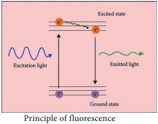
The fluorophore absorbs photons leading to electrons moving to a higher energy state (excited state). When the electrons return to the ground state by losing energy, the fluorophore emits light of a longer wavelength (Figure 2.5). Three of the most common fluorophores used are Diamidino – phenylindole (DAPI) (emits blue), Fluorescein isothiocyanate (FITC) (emits green), and Texas Red (emits red).

Principle

Light source such as Xenon or Mercury Arc Lamp which provides light in a wide range of wavelength, from ultraviolet to the infrared is directed through an exciter filter (selects the excitation wavelength). This light is reflected toward the sample by a special mirror called a dichroic mirror, which is designed to reflect light only at the excitation wavelength.

The reflected light passes through the objective where it is focused onto the fluorescent specimen. The emissions from the specimen are in turn, passed back up through the objective where magnification of the image occurs and through the dichroic mirror.

This light is filtered by the barrier filter, which selects for the emission wavelength and filters out contaminating light from the arc lamp or other sources that are reflected off from the microscope components. Finally, the filtered fluorescent emission is sent to a detector where the image can be digitized.

Components of Fluorescence

Microscope:

The main components of the fluorescent microscope resemble the traditional light microscope. However, the two main difference are the type of light source used and the use of the specialized filter
elements (Figure).

Light source:-
Fluorescence microscopy requires a very powerful light source such as a Xenon or Mercury Arc Lamp. The light emitted from the Mercury Arc Lamp 10 – 100 times brighter than most incandescent lamps and provides light in a wide range of wavelengths from ultra-violet to the infrared. Lasers or high-power LEDs were mostly used for complex fluorescence microscopy techniques.

Filter elements:-
A typical fluorescence microscope consists of three filters: excitation, emission and the dichroic beam splitter.

Excitation filters:
It is placed within the illumination path of a fluorescence microscope. Its purpose is to filter out all
Fluorescence Microscope - Definition, Principle, Parts, Uses img 2

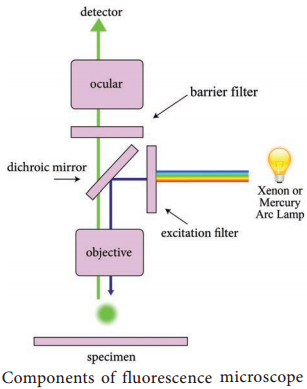
wavelength of the light source, except for the excitation range of the fluorophore in the sample or specimen of interest.

Emission filters:-
The emission filter is placed within the imaging path of a fluorescence microscope. Its purpose is to filter out the entire excitation range and to transmit the emission range of the fluorophore in the specimen.

Dichroic filter or beam splitter:-
The dichroic filter or beam splitter is placed in between the excitation filter and emission filter, at 45° angle. Its purpose is to reflect the excitation wavelength towards the fluorophore in the specimen, and to transmit the emission wavelength towards the detector.

Working Mechanism

The specimen to be observed are stained or labeled with a fluorescent dye and then illuminated with high intensity ultra violet light from mercury arc lamp. The light passes through the exciter filter that allows only blue light to pass through. Then the blue light reaches dichroic mirror and reflected downward to the specimen.

The specimen labeled with fluorescent dye absorbs blue light (shorter wavelength) and emits green light. The emitted green light goes upward and passes through dichroic mirror, reflects back blue light and allows only green light to pass the objective lens then it reaches barrier filter which allows only green light. The filtered fluorescent emission is sent to a detector where the image can be digitized Figure.
Fluorescence Microscope - Definition, Principle, Parts, Uses img 3

Application

  • Fluorescence microscope has become one of the most powerful techniques in biomedical research and clinical pathology.
  • Fluorescence microscope allows the use of multicolour staining, labeling of structures within cells, and the measurement of the physiological state of a cell.
  • Fluorescence microscope helps in observing texture and structure of coal.
  • To study porosity in ceramics, using a fluorescent dye.
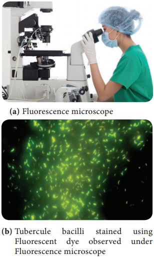
  • To identify the Mycobacterium tuberculosis.

Phase Contrast Microscope – Definition, Principle, Parts, Uses

Learninsta presents the core concepts of Microbiology with high-quality research papers and topical review articles.

Phase Contrast Microscope – Definition, Principle, Parts, Uses

Frits Zernike a Dutch Physicist invented the Phase Contrast Microscope and was awarded Nobel Prize in 1953. It is the microscope which allows the observation of living cell. This microscopy uses special optical components to exploit fine differences in the refractive indices of water and cytoplasmic components of living cells to produce contrast.
Phase Contrast Microscope img 1

Principle

The phase contrast microscopy is based on the principle that small phase changes in the light rays, induced by differences in the thickness and refractive index of the different parts of an object, can be transformed into differences in brightness or light intensity. The phase changes are not detectable to human eye whereas the brightness or light intensity can be easily detected.

Optical Components of Phase Contrast Microscope (PCM)

The phase contrast microscope is similar to an ordinary compound microscope in its optical components. It possesses a light source, condenser system, objective lens system and ocular lens system (Figure 2.1).

A phase contrast microscope differs from bright field microscope in having,

(i) Sub-stage annular diaphragm (phase condenser):-

An annular aperture in the diaphragm is placed in the focal plane of the sub-stage which controls the illumination of the object. This is located below the condenser of the
microscope. This annular diaphragm helps to create a narrow, hollow cone of light to illuminate the object.

(ii) Phase – plate (diffraction plate or phase retardation plate):-

This plate is located at the back focal plane of the objective lenses. The phase plate has two portions, in which one is coated with light retarding material (Magnesium fluoride) and the other portion devoid of light retarding material but can absorb light. This plate helps to reduce the phase of the incident light (Figure 2.2).
Phase Contrast Microscope img 2

Working Mechanism of Phase Contrast Microscopy

The unstained cells cannot create contrast under the normal microscope. However, when the light passes through an unstained cell, it encounters regions in the cell with different refractive indexes and thickness. When light rays pass through an area of high refractive index, it deviates from its normal path and such light
rays experience phase change or phase retardation (deviation). Light rays pass through the area of less refractive index remain non-deviated (no phase change). Figure 2.3 shows the light path in phase contrast microscope.
Phase Contrast Microscope img 3

The difference in the phases between the retarded (deviated) and un-retarded (non-deviated) light rays is about ¼ of original wave length (i.e., λ/4). Human eyes cannot detect these minute changes in the phase of light. The phase contrast microscope has special devices such as annular diaphragm and phase plate, which convert these minute phase changes into brightness (amplitude) changes, so that a contrast difference can be created in the final image. This contrast difference can be easily detected by human eyes.

In phase contrast microscope, to get contrast, the diffracted waves have to be separated from the direct waves. This separation is achieved by the sub-stage annular diaphragm.

The annular diaphragm illuminates the specimen with a hollow cone of light. Some rays (direct rays) pass through the thinner region of the specimen and do not undergo any deviation and they directly enter into the objective lens. The light rays passing through the denser region of the specimen get regarded and they
run with a delayed phase than the nondeviated rays.

Both the deviated and non deviated light has to pass through the phase plate kept on the back focal plane
of the objective to form the final image. The difference in phase (Wavelength) gives the contrast for clear visibility of the object. Figure 2.4 Microscopic image comparing phase and bright field microscopy.
Phase Contrast Microscope img 4

Applications (Uses)

  • Phase contrast microscope enables the visualization of unstained living cells.
  • It makes highly transparent objects more visible.
  • It is used to examine various intracellular components of living cells at relatively high resolution.
  • It helps in studying cellular events such as cell division.
  • It is used to visualize all types of cellular movements such as chromosomal and flagellar movements.

Basic Equipments and Microbiological Techniques

Learninsta presents the core concepts of Microbiology with high-quality research papers and topical review articles.

Developments in Microbiology – Equipments

Confocal Microscopy

Confocal microscopy offers several advantages over conventional optical microscopy, including shallow depth of field, elimination of out-of-focus glare, and the ability to collect serial optical sections from thick specimens. In the biomedical sciences, a major application of confocal microscopy involves imaging either fixed or living cells and tissues that have usually been labeled with one or more fluorescent probes.

Confocal microscopy, most frequently confocal laser scanning microscopy (CLSM) or laser confocal scanning microscopy (LCSM), is an optical imaging technique for increasing optical resolution and contrast of a micrograph by means of using a spatial pinhole to block out of-focus light in image formation.

Capturing multiple two-dimensional images at different depths in a sample enables the reconstruction of three-dimensional structures (a process known as optical sectioning) within an object.

This technique is used extensively in the scientific and industrial communities and typical applications are in life sciences, semiconductor inspection and materials science. Light travels through the sample under a conventional microscope as far into the specimen as it can penetrate, while a confocal microscope only focuses a smaller beam of light at one narrow depth level at a time. The CLSM achieves a controlled and highly limited depth of focus.

DNA Sequencing System

Sequencing means finding the order of nucleotides on a piece of DNA. Nucleotide order determines amino acid order, and by extension, protein structure and function (proteomics). An alteration in a DNA sequence can lead to an altered or non functional protein, and hence to a genetic disorder. DNA sequence is important to detect the type of mutations in genetic diseases and offer hope for the eventual development of treatment DNA.

Methods of sequencing

1. Sanger dideoxy (primer extension/chain-termination) method:-
Most popular protocol for sequencing, very adaptable, scalable to large sequencing projects.

2. Maxam-Gilbert chemical cleavage method:-

DNA is labelled and then chemically cleaved in a sequence dependent manner. This method is not easily scaled and is rather tedious.

It provides an important tool for determining the thousands of nucleotide variations associated with specific genetic diseases, like Huntington’s, which may help to better understand these diseases and advance treatment.

Nanoparticles Production Using Microbes

Learninsta presents the core concepts of Microbiology with high-quality research papers and topical review articles.

Nanoparticles Production Using Microbes

Particles with one or more dimensions of the order of 100 nm or less. There are a large number of physical, chemical, biological, and hybrid methods available to synthesize different types of nanoparticles. Although physical and chemical methods are more popular in the synthesis of nanoparticles, the use of toxic chemicals greatly limits their biomedical applications, in particular in clinical fields.

Therefore, development of reliable, nontoxic, and eco-friendly methods for synthesis of nanoparticles is of utmost importance to expand their biomedical applications. One of the options to achieve this goal is to use microorganisms to synthesize nanoparticles.

Nanoparticles are biosynthesized when the microorganisms grab target ions from their environment and then turn the metal ions into the element metal through enzymes generated by the cell activities. It can be classified into intra-cellular and extracellular synthesis according to the location where nanoparticles are formed.

The intracellular method consists of transporting ions into the microbial cell to form nanoparticles in the presence of enzymes. The extracellular synthesis of nanoparticles involves trapping the metal ions on the surface of the cells and reducing ions in the presence of enzymes.

The biosynthesized nanoparticles have been used in a variety of applications including drug carriers for targeted delivery, cancer treatment, gene therapy and DNA analysis, antibacterial agents, biosensors, enhancing reaction rates, separation science, and magnetic resonance imaging (MRI).

Many microorganisms can produce inorganic nanoparticles through either intracellular or extracellular routes. This section describes the production of various nanoparticles via biological methods following the categories of metallic nanoparticles including gold, silver, alloy and other metal nanoparticles, oxide nanoparticles consisting of magnetic and nonmagnetic oxide nanoparticles, sulfide nanoparticles, and other miscellaneous nanoparticles (Figure 1.4).
Nanoparticles Production Using Microbes img 1

Molecular Biology and Genetic Engineering

Learninsta presents the core concepts of Microbiology with high-quality research papers and topical review articles.

Molecular Biology and Genetic Engineering

Molecular biology – is the study of the structure, function & makeup of the molecular building blocks of life. It focuses on the interactions between the various system of a cell, including the interrelationship of DNA, RNA & Protein synthesis &how these interaction are regulated. Bioscience, Molecular biology closely interrelate with the fields of Biochemistry, Genetics & Cell biology.

Molecular biology is a specialised branch of biochemistry, the study of the chemistry of molecules which are specifically connected to living processes. Importance to molecular biology are the nucleicacids (DNA and RNA) and the proteins which are constructed using the genetic instructions encoded in those molecules.

Other biomolecules, such as carbohydrates and lipids may also be studied for the interactions they have with nucleic acids and proteins. Molecular biology is often separated from the field of cell biology, which concentrates on cellular structures (organelles and the like), molecular pathways within cells and cell life cycles.

Genetic Engineering

Genetic Engineering is the act of modifying the genetic makeup of an organism. Modification can be generated by methods such as gene therapy, nuclear transplantation, transfection of synthetic chromosome or viral insertion.

The manipulation of genetic make up of living cells by inserting desired genes through a DNA vector, is the genetic engineering. The gene is a small piece of DNA that encodes for a specific protein. The gene is inserted into a vector DNA so that a new combination of vector DNA is formed.

The DNA formed by joining DNA segments of two different organisms is called recombinant DNA. The organism whose genetic make up is manipulated using recombinant DNA technique, is called genetically manipulated organism (GMO). Genetic engineering has many application in agriculture, animal science, industry and medicines (Figure 1.3).

Biology Lesson Class 12 MCQ Questions with Answers Chapter 1.3 Molecular Biology and Genetic Engineering img 1

Genetically Modified Organism (GMO)

Organism genome has been engineered in the laboratory in order to favour the expression of desired physiological traits or the production of desired biological products. In conventional livestock production, crop farming, and even pet breeding, it has long been the practice to breed select individuals of a species in order to produce offspring that have desirable traits.

In genetic modification, however, recombinant genetic technologies are employed to produce organisms whose genomes have been precisely altered at the molecular level, usually by the inclusion of genes from unrelated species of organisms that code for traits that would not be obtained easily through conventional selective breeding.

GMOs are produced through using scientific methods that include recombinant DNA technology and reproductive cloning. In reproductive cloning, a nucleus is extracted from a cell of the individual to be cloned and is inserted into the enucleated cytoplasm of a host egg.

The process results in the generation of an offspring that is genetically identical to the donor individual. The first animal produced by means of this cloning technique with a nucleus from an adult donor cell (as opposed to a donor embryo) was a sheep named Dolly, born in 1996.

Since then a number of other animals, including pigs, horses, and dogs, have been generated by reproductive cloning technology. Recombinant DNA technology, on the other hand, involves the insertion of one or more individual genes from an organism of one species into the DNA (deoxyribonucleic acid) of another.

Whole-genome replacement, involving the transplantation of one bacterial genome into the “cell body,” or cytoplasm, of another microorganism, has been reported, although this technology is still limited to basic scientific applications.