Major Groups of Antimicrobial Chemical Agents

Learninsta presents the core concepts of Microbiology with high-quality research papers and topical review articles.

Major Groups of Antimicrobial Chemical Agents

A large number of chemical agents are in common use. Some of the more common groups are listed below.

1. Phenol and Phenolics

Phenol was the first widely used chemical antiseptic and disinfectant. In 1867, Joseph Lister employed carbolic spray to reduce the risk of infection in surgical theatres. Phenol derivatives called phenolics contain altered molecules of phenol useful as antiseptics and disinfectants. The phenolics damage cell membranes and inactivate enzymes of microorganisms, while denaturing the proteins.

Phenolics includes cresols, such as Lysol, as well as several bisphenols, such as hexachlorophene. Today phenol and phenolics such as cresol, xylenol, and orthophenyl phenol are used as disinfectants in laboratories and hospitals.

The commercial disinfectant Lysol is made of mixture of phenolics. Phenolics are tuberculocidal, effective in the presence of organic material, and remain active on surfaces long after application. However, they have a disagreeable odour and can cause skin irritation.

Hexachlorophene is one of the most popular antiseptics because it persists on the skin once applied and reduces skin bacteria for a long period. It is mainly used in soaps and creams. It is an ingredient of various dermatological preparation used for skin disorders.

2. Alcohols

Alcohols are among the most widely used disinfectant and antiseptic. They are bactericidal and fungicidal but not sporicidal. Alcohols can destroy the lipid component of enveloped viruses. The two most popular alcoholic germicides are ethanol and isopropanol. They act by denaturing proteins and dissolving membrane lipids. The recommended optimum concentration of ethanol is 70%, but concentration between 60% and 95% are employed to kill germs as well. Thermometers and small instruments are disinfected by immersing in alcohol for 10 to 20 minutes.

3. Halogens

Halogen compounds are broad spectrum compounds that are considered low toxicity, low cost and easy to use. Among the halogens, iodine and chlorine are important antimicrobial agents. Small quantities of drinking water can be disinfected with halazone tablets.

a. Iodine:-

Iodine compound are broad spectrum and considered effective for a variety of bacteria, mycobacteria, fungi and viruses. The alcoholic tincture of iodine is highly active against gram positive organisms and so is used as a skin antiseptic. It stains the skin. Iodine combines with microbial protein and inhibits their function.

b. Chloride:-

Chloride is also used as a gas to maintain a low microbial count in drinking water. Chlorine together with ammonia called chloramines are used to sanitize glasswall and eating utensils. Sodium hypochlorite (NaOCl) is one of the most widely used chlorine containing disinfectants. Low concentrations (2-500ppm) are active against vegetative bacteria, fungi and most viruses.

Rapid sporicidal action can be obtained around 2500ppm, however this concentration is very corrosive so should be limited in its use. High concentrations are also irritating to the mucous membranes, eyes and skin. Chlorine compounds are rapidly inactivated by light and some metals so fresh solutions should always be used. Hypochlorites should never be mixed with acids or ammonia as this will result in the release of toxic chlorine gas.

c. Iodophores:-

The combinations of iodine and organic molecules are called Iodophores. They include wescodine, betadine and previdone. These iodophore contains surface active agents. They cause less irritation to the skin than free Iodine and do not stain. They are used for cleaning wounds and as a general purpose laboratory disinfectant for discarded jars.

4. Heavy Metals

For many years the ions of heavy metals such as mercury, silver, arsenic, zinc, and copper were used as germicides. More recently these have been superseded by other less toxic and more effective germicides. Many heavy metals are more bacteriostatic than bactericidal. There are a few exceptions. 1% solution of Silver nitrate is often applied to the eyes of infants to prevent ophthalmic gonorrhea. Silver sulfadiazine is used on burns. Copper sulfate is an effective algicide used in lake and swimming pools to retard the growth of algae.

Heavy metals combine with sulfhydryl (SH) groups of proteins and inactivate them. High concentration of metallic salts, particularly those of mercury, silver and copper coagulate cellular proteins that results in damage or death of the microbial cell. The most toxic heavy metals are the mercury, silver, and copper.

5. Quaternary Ammonium Compounds (Quats)

The most widely used surface active agents are the cationic detergents, especially the quaternary ammonium compounds (quats).

Quaternary Ammonium compounds are strongly bactericidal against Gram positive bacteria and less active against gram negative bacteria. These include agents such as cetrimide, bromide and benzalkonium chloride. Their antibacterial activity is antagonized by soaps and certain organisms like Pseudomonas.

They are useful for washing cutlery in catering industry and for cleaning wounds in hospitals. Savlon, a popular antiseptic, is a mixture of cetrimide and chlorohexidine and is active against Gram negative bacteria. They are used as skin disinfectants and as a preservative of ophthalmic solution.

The combined properties of germicidal activity and low toxicity, high solubility in water, stability in solution and non-corrosiveness have resulted in many applications of quaterneries as disinfectants and sanitizing agents.

Quats are also fungicidal, amoebicidal, and virucidal against enveloped viruses. They do not kill endospores or mycobacteria.

6. Aldehydes

Aldehydes are highly effective, broad spectrum disinfectant. The most which typically achieve its anitimicrobial action by denaturing proteins and disrupting nucleic acids. Commonly used aldehydes are formaldehyde and glutaraldehyde. Formaldehyde is usually dissolved in water or alcohol before use. Formaldehyde is used as a surface disinfectat and a fumigant and has been used to decontaminate in animate objects.

A concentration of 2% glutaraldehyde is an effective disinfectant. It is less irritating than formaldehyde and is used to disinfect hospital and laboratory equipments. Glutaraldehyde usually disinfects objects about 10 minutes but may require as long as 12 hours to destroy all spores.

These are highly reactive molecules that combine with nucleic acids and proteins and inactivate them. They disrupt the function of cell organelles and kill the cells probably by cross linking and alkylating the molecules. These are sporicidal and can be used as chemical sterilants.

7. Gaseous Sterilization

Gaseous disinfectants (alkylating agents) are used for the sterilization or disinfection of hospital equipment that is bulky or heat labile. The most widely used gases are ethylene oxide, formaldehyde and β Propiolactone.

Ethylene oxide (EtO):-

Ethylene oxide has a boiling point of 10.8°C. It is highly inflammable and explosive in pure form, but is safe to handle when mixed with Carbon dioxide. It is powerful in the killing of all bacteria, including tubercule bacilli and spores. It is an effective sterilizing agent because it rapidly penetrates packing materials, even
plastic wrappers. To be potent, however, the humidity and temperature must be carefully controlled within narrow limits.

It is highly toxic on contact with the skin or mucous membrane. Materials that have been sterilized with ethylene oxide must be set aside in detoxification chambers for a few days to allow the gases to dissipate. It is frequently used to sterilize heart lung machines and plastic items like catheters.

Formaldehyde:-

It is highly bactericidal. Formaldehyde is used as 40% formalin with humidity at around 50%. It causes irritation. It is used occasionally to fumigate rooms and disinfect respirators.

Betapropiolactone (BPL):-

This is occasionally employed as a sterilizing gas in the liquid form. It has been used to sterilize vaccines, tissue grafts, surgical instrument and enzyme as a sterilants of blood plasma, water, milk and as a vapour – phase disinfectant in enclosed spaces, short-term inhalation exposure to betapropiolactone causes
severe irritation of the eyes, nose, throat and respiratory tract in humans.

BPL decomposes to an inactive form after several hours and is therefore not difficult to eliminate. It destroys microorganisms more readily than ethylene oxide but does not penetrate materials well and may be carcinogenic. For these reasons, BPL has not been used as extensively as EtO.
Mode of Action of Chemical Agents

Learninsta presents the core concepts of Microbiology with high-quality research papers and topical review articles.

Mode of Action of Chemical Agents

Chemical agents act on microorganisms by:

  • They may damage the lipids and proteins of the cytoplasmic membrane of microorganisms.
  • They may denature microbial enzymes and other proteins usually by disrupting the hydrogen and disulfide bonds that give the protein its 3-D shape. This blocks metabolism function.

Modes Of Action

Mechanisms Of Resistance:-

  • Most antimicrobials fall into one of four main categories, based on their site of activity.
  • These include inhibition of cell wall synthesis, protein synthesis, nucleic acid synthesis, or disruption of cell membrane integrity.

There are a number of factors which influence the antimicrobial action of disinfectants and antiseptics, including:-

  • The concentration of the chemical agent.
  • The temperature at which the agent is being used.
  • The number of microorganisms present.
  • The nature of the material bearing the microorganisms.
  • The chemical agent that kills bacteria is Bactericide.

Factors Influencing the Antimicrobial

Learninsta presents the core concepts of Microbiology with high-quality research papers and topical review articles.

Factors Influencing the Antimicrobial

The following factors will affects the activity of a disinfectant or antiseptic and these should be borne in mind during use.

a. The Concentration and kind of a chemical agent used:-

The higher the concentration of the germicide the greater will be the rate of killing. This is particularly important with the phenolic group of compounds, whose activity falls off very rapidly with dilution.

b. Time of exposure to the agent:-

In general germicidal activity is increased with time and a sufficient exposure is imperative for efficient disinfection.

c. Temperature at which the agent is used:-

An increase of temperature will also raise the rate of killing.

d. Presence of Organic matter:-

Most germicides are reduced in activity by the presence of organic matter and particularly by the presence of proteins such as those in body fluids.

e. Number of organisms present:-

The larger the number of organisms, the greater will be the time required for disinfection.

f. The kinds of microorganisms present – Presence of spores:-

Spores are exceptionally resistant to the great majority of disinfection.

Control of Microorganisms by Chemical Methods – Disinfectants, Antiseptics and Antibiotics

Learninsta presents the core concepts of Microbiology with high-quality research papers and topical review articles.

Control of Microorganisms by Chemical Methods –  Disinfectants, Antiseptics and Antibiotics

Disinfection is the elimination of microorganisms from inanimate objects or surfaces. The term disinfectant is used for an agent used to disinfect inanimate objects or surfaces but is generally toxic to use on human tissues.

Antiseptic refers to an agent that kills or inhibits growth of microorganisms but is safe to use on human tissues. Antibiotics produced by microorganisms which kill or inhibit the growth of other microbes.

Following Table gives few examples of antimicrobial chemical agents that destroy unwanted microorganisms.

Disinfectants

Antiseptics

Antibiotics

Chlorine, Copper Phenol, Tincture Iodine Pencillin, Streptomycin

Basic terms used in chemical control of microorganism are mentioned in Table 3.1 and Table 3.2 Describes the difference between Bactericidal and Bacteriostatic agents.

Basic terms used in Chemical sterilization.

Term

Meaning

Disinfection The selective elimination of certain undesirable microorganisms to prevent their transmission directed against their metabolism or structure; applies to the use directly on inanimate objects.
Antisepsis Prevention of the growth or activity of microorganisms by inhibition or killing; applies to the use of chemicals on living tissue
– cide Suffix used to denote agents, usually chemical, that kill. Commonly used terms are bactericide, fungicide, virucide, and algicide. The term germicide is used if the agents kill pathogens but not necessarily spores. An agent that kills bacterial spores is a sporicide.
– Static Suffix used to denote agents, usually chemical, that prevents growth but do not necessarily kill the organism or bacterial spores. Commonly used terms include bacteriostatic and fungistatic.

Difference between Bactericidal and Bacteriostatic

Bactericidal

Bacteriostatic

Bactericidal refers to agents that kill bacteria Bacteriostatic refers to agents that prevent the growth of bacteria
Action is irreversible Action is reversible
Inhibit the cell wall formation of bacteria Inhibit DNA replication and protein synthesis of bacteria
Do not work with the immune system of the host Work with the immune system of the host to prevent the growth and reproduction of bacteria
Minimal Bactericidal Concentration (MBC) refers to the concentration of the drug required to kill 99.99% of the bacterial population. Minimal Inhibitory Concentration (MIC) is the minimum drug concentration which inhibits the bacterial growth.
Examples include betalactam antibiot­ics, cephalosporins, and vancomycin. Examples include tetracyclines, spectinomycin, chloramphenicol, sulfonamides, etc.

Electron Microscope – Definition, Principle, Parts, Uses

Learninsta presents the core concepts of Microbiology with high-quality research papers and topical review articles.

Electron Microscope – Definition, Principle, Parts, Uses

Examining the ultra structure of cellular components such as nucleus, plasma membrane, mitochondria and others requires 10,000X plus magnification which was just not possible using Light Microscopes. This is achieved by Electron microscopes which have greater resolving power than light microscopes and can obtain higher magnifications.

In an electron microscope, a focused electron beam is used instead of light to examine objects. Electrons are considered as radiation with wavelength in the range 0.001 – 0.01 nm compared to 400 – 700 nm wavelength of visible light used in an optical microscope.

Optical microscopes have a maximum magnification power of 1000X, and resolution of 0.2 μm compared to resolving power of the electron microscope that can reach 1,000,000 times and resolution of 0.2 nm. Hence, electron microscopes deliver a more detailed and clear image compared to optical microscopes. Table 2.1 differentiate electron microscope from light microscope.

Light Microscope Electron Microscope
1. Light is the illuminating source 1. The beam of electrons is the electron source
2. Specimen preparation takes usually few minutes to hours. Live or dead specimen may be seen. 2. Specimen preparation takes usally takes a few days. Only dead or dried specimen are seen.
3. Condenser, objective and eye piece lenses are made up of glasses. 3. All lenses are electromagnetic.
4. Specimen is stained by coloured dyes. 4. Specimen is coated with heavy metals in order to reflect electrons.
5. It has low resolving power (0.25 pm to 0.3 pm). It has a magnification of 500X to 1500X. 5. It has high resolving power (0.001pm), about 250 times higher than light microscope. It has a magnification more than 100,000X
6. Vaccum is not required 6. Vaccum is essential for its operation
7. Image is seen by eyes through ocular lens 7. Image is produced on fluorescent screen or photographic plate.

Types of Electron Microscopes

  • Transmission electron microscopes (TEM)
  • Scanning electron microscopes (SEM)
  • Scanning transmission electron microscopes (STEM)

The electron microscope was invented in 1931 by two German scientists, Ernst Ruska and Max Knoll. Ernst Ruska later received Nobel Prize for his work in 1986. The Transmission Electron Microscope (TEM) was the first type of Electron Microscope to be developed.

Principle

The fundamental principle of electron microscope is similar to light microscope. In electron microscope, a high velocity beam of electrons is used instead of photons. In the electron gun, electrons are emitted from the surface of the cathode and accelerated towards the anode by high voltage to form a high energy electron beam.

All lenses in the electron microscope are electromagnetic. Charged electrons interact with the magnetic fields and magnetic force focuses an electron beam. The condenser lens system controls the beam diameter and convergence angles of the beam incident on a specimen.

The image is formed either by using the transmitted beam or by using the diffracted beam. The image is magnified and focused onto an imaging device, such as a fluorescent screen, on a layer of photographic film, or to be detected by a sensor.

Sample Preparation:-

Preparation of specimens is the most complicated and skillful step in EM. The material to be studied under electron microscopy must be well preserved, fixed, completely dehydrated, ultrathin and impregnated with heavy metals that sharpen the difference among various organelles.

The material is preserved by fixation with glutaraldehyde and then with osmium tetroxide. The fixed material is dehydrated and then embedded in plastic (epoxy resin) and sectioned with the help of diamond or glass razor of ultra-microtome.

In TEM, sample sections are ultrathin about 50 – 100 nm thick. These sections are placed on a copper grid and exposed to electron dense materials like lead acetate, uranylacetate, phosphotungstate. In SEM, samples can be directly imaged by mounting them on an aluminum stub.

Electron – Sample Interactions:-

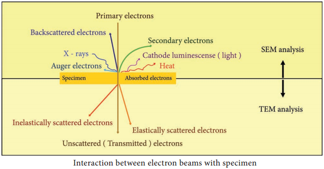
Interaction of electron beam with the sample results in different types of electrons: Elastic scattered electrons, Inelastic scattered electrons, secondary electrons and backscattered electrons. Almost all types of electron interactions can be used to retrieve information about the specimen.

Depending on the kind of radiation or emitted electrons which are used for detection, different properties of the specimen such as topography, elemental composition can be concluded. Figure 2.8 shows the interaction of the electron beam with the specimen.

In Transmission electron microscope (TEM), a beam of electrons is transmitted through an ultrathin specimen, interacting with the specimen as it passes through. An image is formed from the interaction of the transmitted unscattered electrons through the specimen.

Secondary electrons are mainly used in scanning electron microscope (SEM) for imaging the surface topography of biological specimens. The interaction of electron beam with samples results in secondary electrons and backscattered electrons that are detected by standard SEM equipment.
Electron Microscope - Definition, Principle, Parts, Uses img 1

Working Principle and Instrumentation of TEM

The optics of the TEM is similar to conventional transmission light microscope. A transmission electron microscope has the following components,

  1. Electron gun
  2. Condenser lens
  3. Specimen stage
  4. Objective lens and projector lens
  5. Screen/photographic film/Charged Coupled Device (CCD) camera

Electron Gun consists of a tungsten filament or cathode that emits electrons on receiving high voltage electric current (50,000 – 100,000 volts). A high voltage between the electron source (cathode) and an anode plate is applied leading to an electrostatic field through which the electrons are accelerated.

The emitted electrons travel through vacuum in the microscope column. Vacuum is essential to prevent strong scattering of electrons by gases. Electromagnetic condenser lenses focus the electrons into a very thin beam. Electron beam then travels through the specimen and then through the electromagnetic objective
lenses.

In a TEM microscope, the sample is located in the middle of the column. At the bottom of the microscope, unscattered electrons hit the fluorescent screen giving image of specimen with its different parts displayed in varied darkness, according to their density. The image can be studied directly, photographed or digitally
recorded. Figure 2.9 show the arrangement of components for transmission electron microscope.

Information that can be obtained using TEM include,

  • Topography: surface features, texture
  • Morphology: shape and size of the particles
  • Crystallographic arrangement of atoms
  • Composition: elements and the their relative amounts.

Electron Microscope - Definition, Principle, Parts, Uses img 2

Working Principle and Instrumentation of SEM

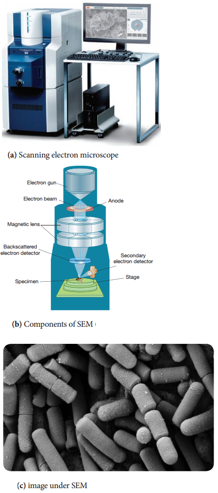
It is first built by Knoll in 1935. It is used to study the three dimensional images of the surfaces of cells, tissues or particles. The SEM allows viewing the surfaces of specimens without sectioning. The specimen is first fixed in liquid propane at-180°C and then dehydrated in alcohol at-70°C.

The dried specimen is then coated with a thin film of heavy metal, such as platinum or gold, by evaporation in a vacuum provides a reflecting surface of electrons. In SEMs, samples are positioned at the bottom of the electron column and the scattered electrons (backscattered or secondary) are captured by electron detectors.

In SEM, there are several electromagnetic lenses, including condenser lenses and one objective lens. Electromagnetic lenses are for electron probe formation, not for image formation directly, as in TEM. Two condenser lenses reduce the crossover diameter of the electron beam. The objective lens further reduces the cross-section of the electron beam and focuses the electron beam as probe on the specimen surface (Figure 2.10).

Objective lens thus functions like a condenser. This is in contrast to TEM where objective lens does the magnification. Major difference between SEM and TEM are given in Table 2.2. SEMs are equipped with an energy dispersive spectrometer (EDS) detection system which is able to detect and display most of the X-ray spectrum. The detector normally consists of semiconducting silicon or germanium. Difference between SEM and TEM.

Properties

SEM

TEM

1. Types of electrons It is based on scattered electrons that are emitted from the surface of a specimen It is based on transmitted electrons
2. Sample preparation Sample can be of any thickness and is coated with a thin layer of a heavy metal such as gold or palladium and mounted on an aluminium slab Laborious sample preparation is required. The sample has to be cut into thin sections so as to allow electrons to pass through it and are supported on TEM grids.
3. Resolution The resolution is up to 20nm TEM has much higher resolution than SEM. It can resolve objects as close as 1nm
4. Magnification The magnifying power of SEM is up to 100,000X The magnifying power of TEM is up to 5,000,000 X
5. Image formation SEM provides a 3 dimensional image. Secondary or back scattered electrons are captured, detected and displayed on computer screen TEM provides a 2 dimensional image. Transmitted electrons hit a fluroscent screen giving rise to a shadow image. The image can be studied directly by the operator or photographed with a camera.
6. Application SEM is used to study the topography and atomic composition of specimens TEM is used to study the interior of cells, the structure of protein molecule, the organization of molecules in viruses and cytoskeletal filaments and the arrangement of protein molecules in cell membranes.

Scanning transmission electron microscopy (STEM) combines the principles of transmission electron microscopy and scanning electron microscopy and can be performed on either type of instrument. Like TEM, STEM requires very thin samples and the primary electron beam is transmitted by the sample.

One of its principal advantages over TEM is in enabling the use of other of signals that cannot be spatially correlated in TEM, including secondary electrons, scattered beam electrons, characteristic X-rays, and electron energy loss.
Electron Microscope - Definition, Principle, Parts, Uses img 3