Energy of Chemical Reaction

Learninsta presents the core concepts of Microbiology with high-quality research papers and topical review articles.

Energy of Chemical Reaction

Light energy is trapped by phototrophs during photosynthesis, in which it is absorbed by bacteriochlorophyll and other pigments and converted to chemical energy for cellular work. The energy is required by the bacterium for synthesis of cell wall or membrane, synthesis of enzymes, cellular components, repair
mechanism, growth and reproduction.

Some change of energy occurs whenever bonds between atoms are formed or broken during chemical reactions. When a chemical bond is formed, energy is required. Such a chemical reaction which requires energy is called an endergonic reaction (energy is directed inward). When a bond is broken, energy is released. A chemical reaction that release energy is an exergonic reaction (energy is directed outward).

During chemical reaction energy is either released or absorbed and the quantum of energy liberated or taken up is useful energy and is referred to Free Energy Change (ΔG) of the reactions.

High Energy Phosphate

Adenosine Tri-Phosphate (ATP) is the principal energy carrying molecule of all cells and is indispensable to the life of the cell. It stores the energy released by some chemical reactions, and it provides the energy for reactions that require energy. ATP consists of an adenosine unit composed of adenine, ribose with three phosphate groups. In ATP and some other phosphorylated compounds, the outer two phosphate groups are joined by an anhydride bond.

Some of the other high energy nucleotides involved in biochemical processes are given in Table 4.1.

Table 4.1: High energy nucleotides involved in biosynthesis

Name of the Nucleotide

Biosynthesis

Uridine triphosphate (UTP)Polysaccharide
Cytidine triphosphate (CTP)Lipid
Guanidine triphosphate (GTP)Protein

Nutrients are broken from highly reduced compounds to highly oxidized compounds within the cells. Much of the energy released during oxidation reduction reactions is trapped within the cell by the formation of ATP. A phosphate group is added to ADP with the input of energy to form ATP.

ATP + H2O → ADP + pi(ΔG° = – 7.3 K cal/mol)
ATP + H2O → AMP + ppi(ΔG° = – 10.9 K cal/mol)

ATP is ideally suited for its role as an energy currency. It is formed in energy trapping and energy generating processes such as photosynthesis, fermentation, and aerobic respiration. In bacterial and archeal cells, most of the ATP is formed on the cell membrane, while in eukaryotes the reactions occur primarily in the
mitochondria (Figure 4.2).
Energy of Chemical Reaction img 1

Oxidation – Reduction Reactions

Oxidation is the removal of electrons (e) from an atom or molecule and is often an energy producing reaction. Reduction of a substrate refers to its gain or addition of one or more electrons to an atom or molecule. Oxidations and reduction are always coupled. In other words, each time one substance is oxidized, another is simultaneously reduced.
F2 + 2e → 2F
H2 + 2e → 2H+ + 2e
NAD+ + 2H+ + 2e ⇄ NADH + H+.

Microbial Metabolism

Learninsta presents the core concepts of Microbiology with high-quality research papers and topical review articles.

Microbial Metabolism

The term Metabolism refers to the sum of all bio chemical reactions that occur within a living cell. Chemical reaction either release energy or require energy. Metabolism can be viewed as an energy balancing act. It can be divided into two classes of chemical reactions namely Catabolism and Anabolism.

Catabolism:
It is called catabolic or degradative reactions because complex organic compounds are broken down into simples ones. Catabolic reactions are generally hydrolytic reactions. It is enzyme regulated chemical reaction that release energy and they are exergonic. Example: Break down of sugar into Carbon dioxide and water in cells.

Anabolism:
It is called anabolic or biosynthetic reactions because complex organic molecules are formed from simples ones. Anabolic process often involves dehydration, are bio-synthetic reactions (Figure 4.1). It is enzyme regulated energy requiring reaction and they are endergonic. Examples: Formation of proteins from amino acids.
Microbial Metabolism img 1

Catabolic reactions furnish the energy needed to drive anabolic reactions. This coupling of energy requiring and energy releasing reactions is made possible through the molecule Adenosine tri-phosphate (ATP).

Drugs Resistance Mechanisms

Learninsta presents the core concepts of Microbiology with high-quality research papers and topical review articles.

Drugs Resistance Mechanisms

Some microbes respond predictably to certain drugs making selection of treatment easy. Other microbes may vary in their responses, and laboratory tests are usually required to ensure that the selected therapy is appropriate.

Chemotherapeutic effectiveness depends upon the sensitivity of the pathogen to the agent. Antibiotic resistance, however, may develop in microbes within the population. In fact, the history of chemotherapy has been closely paralleled by the history of drug resistance.

None of the therapeutic drugs (antibiotic) inhibits all microbial pathogens and some microbial pathogens possess natural ability to resist to certain antibiotics.

Bacteria become drug resistant using several different resistance mechanisms. A particular type of resistance mechanism is not confined to a single class of drugs. Two bacteria may employ different resistance mechanisms to counter the same antibiotic.

However, bacteria acquire drugs resistance using resistance mechanisms such as reduced permeability to antibiotic, efflux (pumping) antibiotic out of the cell, drug inactivation through chemical modification, target
modification and development of a resistant biochemical pathway (Figure 3.5).
Drugs Resistance Mechanisms img 1

Antimicrobial Susceptibility Testing

Learninsta presents the core concepts of Microbiology with high-quality research papers and topical review articles.

Antimicrobial Susceptibility Testing

Antimicrobial susceptibility tests are used to determine the type and quantity of antimicrobial agents used in chemotherapy. One of the most important functions of a clinical laboratory is to determine the antimicrobial susceptibility.

Antimicrobial susceptibility of pathogens refers to the limitation of pathogens to grow in the presence of effective antibiotics. There are two methods that can be used to determine the susceptibility of a potential pathogen to antimicrobial agents. They are:

  • Disk diffusion method
  • Tube dilution method

Disc Diffusion Method (Kirby – Bauer Test)

William Kirby and Alfred Bauer, in 1966 first introduced the principle of measuring zones of inhibition around antibiotic discs to determine antimicrobial agent susceptibilities. It is a rapid, convenient method to determine the susceptibilities of microorganisms to antimicrobial agents and a most common procedure used in susceptibility testing in clinical laboratory.

Filter paper discs containing known concentrations of antimicrobial agents are placed onto the surface of an agar plate (Muller – Hinton agar medium) inoculated with the test bacterium (Figure 3.3). The plate is incubated for 16 to 18 hours, and the zones of inhibition are read around each paper disc. During the incubation periods, the antimicrobial agent diffuses through the agar, and a concentration gradient of agent is established.

At some point in this gradient, growth of the susceptible bacteria is suppressed, and no growth is observed within a circular zone around disc. The size of a zone of inhibition must be compared to a standard Table for that particular drug before accurate comparisons can be made.

Thus, enabling to classify pathogens as susceptible (S), intermediate or resistant (R) to a drug. The procedure is highly regulated and controlled by the clinical and laboratory standards institute (CLSI) and must be accompanied by a rigorous quality assurance program including performance by certified and/or licensed personnel when the results are to be reported in clinical settings.
Antimicrobial Susceptibility Testing img 1

Minimal Inhibitory Concentration (MIC) Test

The potency of an effective antimicrobial agent is expressed in terms of minimal inhibitory concentration (MIC). It is the minimum concentration of drug that will inhibit the growth of pathogen. The MIC is determined by serial dilutions of antimicrobial agents in tubes with standard amount of bacteria. Turbidity (cloudiness) after incubation indicates bacterial growth and lack of turbidity indicates that the growth of bacteria is inhibited.

E – test

This is another test to determine the minimum inhibitory concentration where a plastic strip containing a gradient of the antimicrobial agent is used (Figure 3.4). An elliptical zone of inhibitory concentration can be noted with the help of a scale printed on the strip.
Antimicrobial Susceptibility Testing img 2

The Minimal Bactericidal Concentration (MBC) Test

MBC test is similar to MIC, the minimal bactericidal concentration test is used to determine the amount of antimicrobial agent required to rather kill the pathogen. In MBC test, samples taken from MIC tubes are transferred to drug free plates. Bacterial growth in these subcultures indicates that some bacterial cells have survived antimicrobial drug. The lowest concentration of drug for which no growth occurs is the minimum bactericidal concentration.

The tube dilution method is considered accurate for determining susceptibility of a pathogen to precise quantities of antimicrobial agent. However, the method is time consuming, expensive, and not practical for use in most clinical laboratories for routine susceptibility testing.

Evaluation of Antimicrobial Chemical Agents Antibiotics

Learninsta presents the core concepts of Microbiology with high-quality research papers and topical review articles.

Evaluation of Antimicrobial Chemical Agents Antibiotics

Testing of antimicrobial agents is a complex process regulated by two different federal agencies.

The U.S. Environmental Protection Agency regulates disinfectants, where as agents used on humans and animals are under the control of the Food and Drug Administration.

Testing of antimicrobial agents often begins with an initial screening test to see if they are effective and at what concentrations.

Laboratory techniques for the evaluation of antimicrobial chemical agents are conducted by one of the following three general procedures. In each procedure, the chemical agent is tested against a specific microorganisms referred to as the test organism.
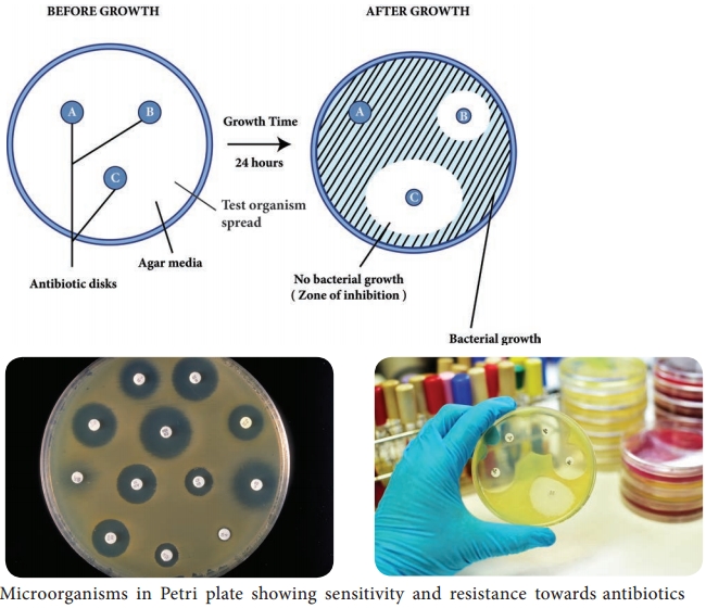
Agar Plate Method

A plate of agar medium is inoculated with the test organism and the chemical agent is placed on the surface of the medium. The chemical solution is first impregnated in absorbent papers or confined by a hollow cylinder placed on the agar surface. Following incubation, the plate is observed for a zone of inhibition around the chemical agent. This is particularly suitable for semisolid preparations.

Tube Dilution Methods

Appropriately diluted water soluble liquid substances are dispensed into sterile test tubes and are inoculated with a measured amount of the test organism. At specified intervals, a transfer is made from this tube into tubes of sterile media that are then incubated and observed for the appearance of growth.

It is necessary in this type of procedure to ascertain whether the inhibitory action is bactericidal and not bacteriostatic. This approach can also be used to determine the number of organisms killed per unit time by performing a plate count on samples taken at appropriate intervals.

Phenol Coefficient Test

Phenol coefficient is a measure of the bactericidal activity of a chemical compound in relation to phenol. Phenol coefficient is calculated by dividing the concentration of test disinfectant at which it kills the organism in 10 minutes and not in 5 minutes under the same conditions. This method is used for evaluating the efficiency of watermiscible disinfectants.

Series of 10 test tubes with 2ml of distilled water is taken (Figure 3.1). Phenol is added to first test tube and dilution is made by transferring 1ml to next tube up to 5 dilutions. Similarly commercial disinfectant is also diluted. Pure culture of test organisms, such as Staphylococcus aureus or Salmonella typhi, is added to test tubes.

Subcultures from these tubes incubated at 37°C for 48 hours are examined for the presence or absence of growth at intervals of 5, 10 and 15 minutes. The highest dilution that kills the bacteria after 10 minutes, but not after 5 minutes is used to calculate the phenol coefficient (Table 3.3),

Evaluation of Antimicrobial Chemical Agents Antibiotics img 2

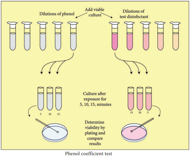
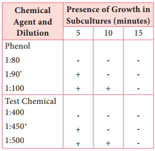
Illustration of phenol coefficient determination

Phenol dilution of 1:90 showed growth at 5 minutes but no growth at 10 minutes Test Chemical dilution of 1:450 showed growth at 5 minutes but no growth at 10 minutes phenol coefficient of test chemical as 450/90=5.

Antibiotics

The term ‘antibiotic’ was derived from ‘antibiosis’ which refers to the suppression of microorganisms due to secretion of toxic or inhibitory compounds by other microorganisms. Although antibiosis has been observed by many scientific workers fairly frequently towards the end of the nineteenth century, it was not until the discovery and development of Penicillin that a truly wide ranging search for antibiotics was initiated.

Historical Development

The first chemotherapeutic agent, discovered by Paul Ehrlich, was Salvarsan, used to treat syphilis. Alexander Fleming discovered the first antibiotic, penicillin, in 1929; its first clinical trails were done in 1940. Antibiotics are produced by species of Streptomyces, Bacillus, Penicillium and Cephalosporium.

Drugs such as the sulfonamides are sometimes called antibiotics although they are synthetic chemotherapeutic agents which are not synthesized using microbes.

Classification of Antibiotics

The antibiotics are usually classified on the basis of:-

• Target group of microorganisms
• Antimicrobial spectrum and
• Mode of action

Classification based on target group of microorganisms:-

Based on the target group, the antibiotics can be classified as antibacterial, antifungal and antiviral.

Classification based on Antimicrobial spectrum:-

Antimicrobial spectrum or antibiotic spectrum refers to the range of effectiveness of antibiotics on different kind of microorganisms, i.e. the range of different kind of microorganisms that can be inhibited, killed, or lysed by a particular type of antibiotic.

The susceptibility of microorganisms to individual antibiotic varies significantly and on account of this, the antibiotics can be classified in two groups as,

Broad – spectrum antibiotics:-

These attack different kinds of microbial pathogens and therefore find wider medical use. Antibacterial antibiotics of broad – spectrum are effective against both Gram positive and Gram negative bacteria.
They also attack pathogens belonging to Mycobacteria, Rickettsia, and Chlamydia. Similarly, broad – spectrum antifungal antibiotics attack different type of fungal pathogens.

Narrow – spectrum antibiotics:-

Narrow – spectrum antibiotics are categorized as those that are effective only against a limited variety of microbial pathogens. These antibiotics are quite valuable for the control of microbial pathogens that fail to respond to other antibiotics. For example, vancomycin is a narrow spectrum glycopeptide. It is an effective bactericidal agent for gram – positive penicillin resistant bacterial pathogens belonging to genera Staphylococcus, Bacillus, and Clostridium.

Mode of Action of Antibiotics

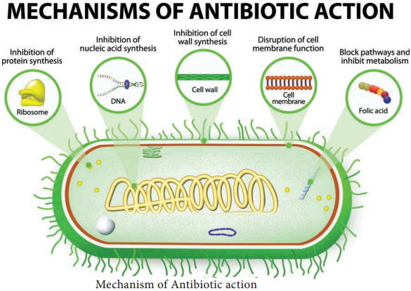
The mode of action of antibiotics varies as they damage pathogens in several ways (Flowchart 3.1). Some of the important actions of therapeutic drugs in microbial pathogens are as follows. Cell wall synthesis, Protein synthesis, Nucleic acid synthesis, Cell membrane disruption and Metabolic pathways blockage.
Evaluation of Antimicrobial Chemical Agents Antibiotics img 3

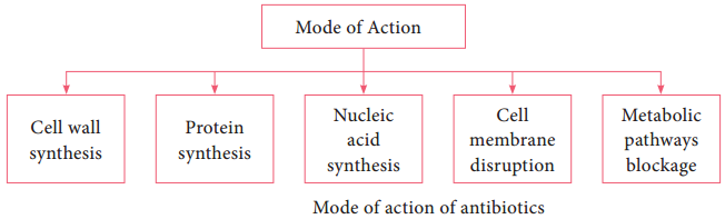
1. Inhibition of Cell Wall Synthesis

The most selective therapeutic antibiotics are those that interfere with the synthesis of bacterial cell walls. These drugs posses a high therapeutic index because bacterial cell walls have a unique structure which is not found in eukaryotic cells. The important cell wall attacking drugs are Penicillin, Cephalosporin, Ampicillin,
Methicillin and Vancomycin.

2. Inhibition of Protein Synthesis

Many therapeutic antibiotics discriminate between prokaryotic and eukaryotic ribosomes and inhibit protein synthesis. The therapeutic index of these drugs is fairly high, but not as favourable as that of cell wall synthesis inhibitors. Several of these drugs are medically useful and effective research tools because they block individual steps in protein synthesis. Some therapeutic drugs bind to 30S while others attach to 50S ribosomal subunits. Example Streptomycin, Chloramphenicol, Tetracyclin and Erythromycin.

3. Inhibition of Nucleic Acid Synthesis

Some antimicrobial drugs or antibiotics inhibit nucleic acid synthesis. These are not selectively toxic as other drugs. This is due to the fact that prokaryotic and eukaryotic nucleic acid synthesis mechanisms do not vary greatly. Example Quinolones, Novobiocin, Actinomycin and Rifampin

4. Disruption of Cell Membrane

There are some antimicrobial drugs or antibiotics that act as cell membrane disorganizing agents. Polymyxins are such drugs of clinical importance. E.g. Polymyxin B and Polymyxin E (colistin)

5. Blocking Metabolic Pathways

Some therapeutic drugs act as antimetabolites and block the functioning of metabolic pathways. They competitively inhibit the key enzymes in the metabolic pathway. Example Sulfonamides, Trimethoprim, Dapsone and Isoniazid (Figure 3.2).
Evaluation of Antimicrobial Chemical Agents Antibiotics img 4
Trimethoprim, Daspone and Isoniazid (Figure 3.2).