Rna – The Adapter Molecule

Learninsta presents the core concepts of Biology with high-quality research papers and topical review articles.

Rna – The Adapter Molecule

The transfer RNA, (tRNA) molecule of a cell acts as a vehicle that picks up the amino acids scattered through the cytoplasm and also reads specific codes of mRNA molecules. Hence it is called an adapter molecule. This term was postulated by Francis Crick.

The two dimensional clover leaf model of tRNA was proposed by Robert Holley. The secondary structure of tRNA depicted in Fig. 5.11 looks like a clover leaf. In actual structure, the tRNA is a compact molecule which looks like an inverted L. The clover leaf model of tRNA shows the presence of three arms namely DHU arm, middle arm and TΨC arm. These arms have loops such as amino acyl binding loop, anticodon loop and ribosomal binding loop at their ends.
Rna - The Adapter Molecule img 1

In addition it also shows a small lump called variable loop or extra arm. The amino acid is attached to one end (amino acid acceptor end) and the other end consists of three anticodon nucleotides. The anticodon pairs with a codon in mRNA ensuring that the correct amino acid is incorporated into the growing polypeptide chain.

Four different regions of double-stranded RNA are formed during the folding process. Modified bases are especially common in tRNA. Wobbling between anticodon and codon allows some tRNA molecules to read more than one codon.

The process of addition of amino acid to tRNA is known as aminoacylation or charging and the resultant product is called aminoacyl – tRNA (charged tRNA). Without aminoacylation tRNA is known as uncharged tRNA (Fig. 5.12).

If two such tRNAs are brought together peptide bond formation is favoured energetically. Numbers of amino acids are joined by peptide bonds to form a polypeptide chain. This aminoacylation is catalyzed by an enzyme aminoacyl – tRNA synthetase. This is an endothermic reaction and is associated with ATP hydrolysis. 20 different aminoacyl – tRNA synthetases are known. The power to recognize codon on the mRNA lies in the tRNA and not in the attached amino acid molecule.
Rna - The Adapter Molecule img 2

Translation and Its Mechansim

Learninsta presents the core concepts of Biology with high-quality research papers and topical review articles.

Translation and Its Mechansim

Translation refers to the process of polymerization of amino acids to form poly peptide chain. The decoding process is carried out by ribosomes that bind mRNA and charged tRNA molecules. The mRNA is translated, starting at the 5′ end.

After binding to mRNA, the ribosomes move along it, adding new amino acids to the growing polypeptide chain each time it reads a codon. Each codon is read by an anticodon on the corresponding tRNA. Hence the order and sequence of amino acids are defined by the sequence of bases in the mRNA.

Mechanism of Translation

The cellular factory responsible for synthesizing protein is the ribosome. The ribosome consists of structural RNAs and about 80 different proteins. In inactive state, it exists as two subunits; large subunit and small subunit.

When the subunit encounters an mRNA, the process of translation of the mRNA to protein begins. The prokaryotic ribosome (70 S) consists of two subunits, the larger subunit (50 S) and smaller subunit (30 S). The ribosomes of eukaryotes (80 S) are larger, consisting of 60 S and 40 S sub units. ‘S’ denotes the sedimentation coefficient which is expressed as Svedberg unit (S).

One of the alternative ways of dividing up a sequence of bases in DNA or RNA into codons is called reading frame. Any sequence of DNA or RNA, beginning with a start codon and which can be translated into a protein is known as an Open Reading Frame (ORF). A translational unit in mRNA is the sequence of RNA that is flanked by the start codon (AUG) and the stop codon and codes for polypeptides.

mRNA also have some additional sequences that are not translated and are referred to as Untranslated Regions (UTR). UTRs are present at both 5′ end (before start codon) and at 3′ end (after stop codon). The start codon (AUG) begins the coding sequence and is read by a special tRNA that carries methionine (met).

The initiator tRNA charged with methionine binds to the AUG start codon. In prokaryotes, N – formyl methionine (f met) is attached to the initiator tRNA whereas in eukaryotes unmodified methionine is used. The 5′ end of the mRNA of prokaryotes has a special sequence which precedes the initial AUG start codon of mRNA.

This ribosome binding site is called the Shine – Dalgarno sequence or S-D sequence. This sequences base-pairs with a region of the 16Sr RNA of the small ribosomal subunit facilitating initiation. The subunits of the ribosomes (30 S and 50 S) are usually dissociated from each other when not involved in translation (Fig. 5.13 a).
Translation and Its Mechansim img 1

Initiation of translation in E. coli begins with the formation of an initiation complex, consisting of the 30S subunits of the ribosome, a messenger RNA and the charged N-formyl methionine tRNA (fmet – t RNA fmet), three proteinaceous initiation factors (IF1, IF2, IF3), GTP (Guanine Tri Phosphate) and Mg2+.

The components that form the initiation complex interact in a series of steps. IF3 binds to the 30S and allows the 30S subunit to bind to mRNA. Another initiation protein (IF2) then enhances the binding of charged formyl methionine tRNA to the small subunit in response to the AUG triplet. This step ‘sets’ the reading frame so that all subsequent groups of three ribonucleotides are translated accurately.

The assembly of ribosomal subunits, mRNA and tRNA represent the initiation complex. Once initiation complex has been assembled, IF3 is released and allows the initiation complex to combine with the 50S ribosomal subunit to form the complete ribosome (70S). In this process a molecule of GTP is hydrolyzed providing the required energy and the initiation factors (IF1 and IF2 and GDP) are released (Fig. 5.13 b).
Translation and Its Mechansim img 2

Elongation is the second phase of translation. Once both subunits of the ribosomes are assembled with the mRNA, binding sites for two charged tRNA molecules are formed. The sites in the ribosome are referred to as the aminoacyl site (A site), the peptidyl site (P site) and the exit site (E site). The charged initiator tRNA binds to the P site.

The next step in prokaryotic translation is to position the second tRNA at the ‘A’ site of the ribosome to form hydrogen bonds between its anticodon and the second codon on the mRNA (step1). This step requires the correct transfer RNA, another GTP and two proteins called elongation factors (EF-Ts and EF-Tu).

Once the charged tRNA molecule is positioned at the A site, the enzyme peptidyl transferase catalyses the formation of peptide bonds that link the two amino acids together (step 2). At the same time, the covalent bond between the amino acid and tRNA occupying the P site is hydrolyzed (broken).

The product of this reaction is a dipeptide which is attached to the 3′ end of tRNA still residing in the A site. For elongation to be repeated, the tRNA attached to the P site, which is now uncharged is released from the large subunit. The uncharged tRNA moves through the ‘E’ site on the ribosome.

The entire mRNA-tRNA-aa1-aa2 complex shift in the direction of the ‘P’ site by a distance of three nucleotides (step 3). This step requires several elongation factors (EFs) and the energy derived from hydrolysis of GTP. This results in the third triplet of mRNA to accept another charged tRNA into the A site (step 4).

The sequence of elongation is repeated over and over (step 5 and step 6). An additional amino acid is added to the growing polypeptide, each time mRNA advances through the ribosome. Once a polypeptide chain is assembled, it emerges out from the base of the large subunit (Fig. 5.13 c).
Translation and Its Mechansim img 3

Termination is the third phase of translation. Termination of protein synthesis occurs when one of the three stop codons appears in the ‘A’ site of the ribosome. The terminal codon signals the action of GTP – dependent release factor, which cleaves the polypeptide chain from the terminal tRNA releasing it from the translational complex (step 1). The tRNA is then released from the ribosome, which then dissociates into its subunits (step 2) (Fig. 5.13 d).
Translation and Its Mechansim img 4

Genetic Code and Its Mutation

Learninsta presents the core concepts of Biology with high-quality research papers and topical review articles.

Genetic Code and Its Mutation

DNA is the genetic material that carries genetic information in a cell and from generation to generation. At this stage, an attempt will be made to determine in what manner the genetic information exists in DNA molecule? Are they written in coded language on a DNA molecule? If they occur in the language of codes what is the nature of genetic code?

The translation of proteins follows the triplet rule; a sequence of three mRNA base (a codon) designates one of the 20 different kinds of amino acids used in protein synthesis. Genetic code is the sequence relationship between nucleotide in genes (or mRNA) and the amino acids in the proteins they encode.

There are 64 possible triplets, and 61 of them are used to represent amino acids. The remaining three triplet codons are termination signals for polypeptide chains. Since there are only 20 amino acids involved in protein synthesis, most of them are encoded by more than one triplet. Two things make this multiple (degenerate) coding possible. First, there is more than one tRNA for most amino acids.

Each tRNA has a different anticodon. Second, this pairing is highly specific for the first two portions on the codon, permitting Watson and Crick base pairs (A – U and G – C) to be formed. But at the third position there is a great deal of flexibility as to which base pairs are acceptable. Most part of the genetic code is universal, being the same in prokaryotes and eukaryotes.

The order of base pairs along DNA molecule controls the kind and order of amino acids found in the proteins of an organism. This specific order of base pairs is called genetic code, the blue print establishing the kinds of proteins to be synthesized which makes and organism unique.

Marshall Nirenberg, Severo Ochoa (enzyme polynucleotide phosphorylase called Ochoa’s enzyme), Hargobind Khorana, Francis Crick and many others have contributed signifiantly to decipher the genetic code. The order in which bases are arranged in mRNA decides the order in which amino acids are arranged in proteins. Finally a checker board for genetic code was prepared (table 5.1). The salient features of genetic code are as follows:
Genetic Code img 1

1. The genetic codon is a triplet code and 61 codons code for amino acids and 3 codons do not code for any amino acid and function as stop codon (Termination).

2. The genetic code is universal. It means that all known living systems use nucleic acids and the same three base codons (triplet codon) direct the synthesis of protein from amino acids. For example, the mRNA (UUU) codon codes for phenylalanine in all cells of all organisms. Some exceptions are reported in prokaryotic, mitochondrial and chloroplast genomes. However similarities are more common than differences.

3. A non-overlapping codon means that the same letter is not used for two different codons. For instance, the nucleotide sequence GUU GUC represents only two codons.

4. It is comma less, which means that the message would be read directly from one end to the other i.e., no punctuation are needed between two codes.

5. A degenerate code means that more than one triplet codon could code for a specific amino acid. For example, codons GUU, GUC, GUA and GUG code for valine.

6. Non-ambiguous code means that one codon will code for one amino acid.

7. The code is always read in a fixed direction i.e. from 5′ → 3′ direction called polarity.

8. AUG has dual functions. It acts as a initiator codon and also codes for the amino acid methionine.

9. UAA, UAG and UGA) codons are designated as termination (stop) codons and also are known as “non sense” codons.

Mutation and genetic code

Comparative studies of mutations (sudden change in a gene) and corresponding alteration in amino acid sequence of specific protein have confirmed the validity of the genetic code. The relationship between genes and DNA are best understood by mutation studies.

The simplest type of mutation at the molecular level is a change in nucleotide that substitutes one base for another. Such changes are known as base substitutions which may occur spontaneously or due to the action of mutagens. A well studied example is sickle cell anaemia in humans which results from a point mutation of an allele of β-haemoglobin gene (βHb).

A haemoglobin molecule consists of four polypeptide chains of two types, two α chains and two β-chains. Each chain has a heme group on its surface. The heme groups are involved in the binding of oxygen. The human blood disease, sickle cell anaemia is due to abnormal haemoglobin. This abnormality in haemoglobin is due to a single base substitution at the sixth codon of the beta globin gene from GAG to GTG in β – chain of haemoglobin. It results in a change of amino acid glutamic acid to valine at the 6th position of the β – chain.

This is the classical example of point mutation that results in the change of amino acid residue glutamic acid to valine (Fig. 5.10). The mutant haemoglobin undergoes polymerisation under oxygen tension causing the change in the shape of the RBC from biconcave to a sickle shaped structure. The effect of point mutation can be understood by the following example.
Genetic Code img 2

ABC DEF GHI JKL

If we insert a letter O between DEF and GHI the arrangement would be

ABC DEF OGH IJK L

If we insert OQ at the same place the arrangement would be

ABC DEF OQG HIJ KL

The above information shows that insertion or deletion of one or two bases, changes the reading frame from the point of insertions or deletions. Such mutations are referred to as frame shift insertion or deletion mutations. This forms the genetic basis of proof that codon is a triplet and is read in a continuous manner.

Process Of Transcription and Its Unit Gene

Learninsta presents the core concepts of Biology with high-quality research papers and topical review articles.

Process Of Transcription and Its Unit Gene

Francis Crick proposed the Central dogma of protein synthesis in molecular biology states that genetic information flows as follows:
Transcription img 1

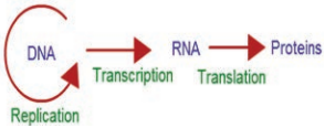
The process of copying genetic information from one strand of DNA into RNA is termed transcription. This process takes place in presence of DNA dependent RNA polymerase. In some retroviruses that contain RNA as the genetic material (e.g, HIV), the flow of information is reversed. RNA synthesizes DNA by reverse transcription, then transcribed into mRNA by transcription and then into proteins by translation.

For a cell to operate, its genes must be expressed. This means that the gene products, whether proteins or RNA molecules must be made. The RNA that carries genetic information encoding a protein from genes into the cell is known as messenger RNA (mRNA). For a gene to be transcribed, the DNA which is a double helix must be pulled apart temporarily, and RNA is synthesized by RNA polymerase.

This enzyme binds to DNA at the start of a gene and opens the double helix. Finally, RNA molecule is synthesized. The nucleotide sequence in the RNA is complementary to the DNA template strand from which it is synthesized.

Both the strands of DNA are not copied during transcription for two reasons. 1. If both the strands act as a template, they would code for RNA with diffrent sequences. This in turn would code for proteins with different amino acid sequences.

This would result in one segment of DNA coding for two different proteins, hence complicate the genetic information transfer machinery. 2. If two RNA molecules were produced simultaneously, double stranded RNA complementary to each other would be formed. This would prevent RNA from being translated
into proteins.

Transcription unit and gene

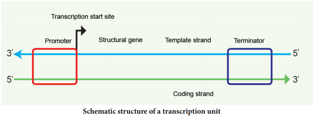
A transcriptional unit in DNA is defined by three regions, a promoter, the structural gene and a terminator. The promoter is located towards the 5′ end of the coding strand. It is a DNA sequence that provides binding site for RNA polymerase. The presence of promoter in a transcription unit, defies the template and coding strands.

The terminator region located towards the 3′ end of the coding strand contains a DNA sequence that causes the RNA polymerase to stop transcribing. In eukaryotes the promoter has AT rich regions called
TATA box (Goldberg-Hogness box) and in prokaryotes this region is called Pribnow box. Besides promoter, eukaryotes also require an enhancer.

The two strands of the DNA in the structural gene of a transcription unit have opposite polarity. DNA dependent RNA polymerase catalyses the polymerization in only one direction, the strand that has the polarity 3′ → 5′ acts as a template, and is called the template strand.

The other strand which has the polarity 5′ → 3′ has a sequence same as RNA (except thymine instead of uracil) and is displaced during transcription. This strand is called coding strand (Fig. 5.7).
Transcription img 2

The structural gene may be monocistronic (eukaryotes) or polycistronic (prokaryotes). In eukaryotes, each mRNA carries only a single gene and encodes information for only a single protein and is called monocistronic mRNA. In prokaryotes, clusters of related genes, known as operon, oftn found next to each other on the chromosome are transcribed together to give a single mRNA and hence are polycistronic.

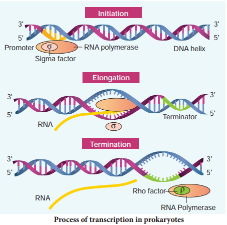
Before starting transcription, RNA polymerase binds to the promoter, a recognition sequence in front of the gene. Bacterial (prokaryotic) RNA polymerase consists of two major components, the core enzyme and the sigma subunit. The core enzyme (2α, β, β1 and ω) is responsible for RNA synthesis whereas a sigma subunit is responsible for recognition of the promoter. Promoter sequences vary in different organisms.

RNA polymerase opens up the DNA to form the transcription bubble. The core enzyme moves ahead, manufacturing RNA leaving the sigma subunit behind at the promoter region. The end of a gene is marked by a terminator sequence that forms a hair pin structure in the RNA. The sub-class of terminators require a recognition protein, known as rho (ρ), to function.

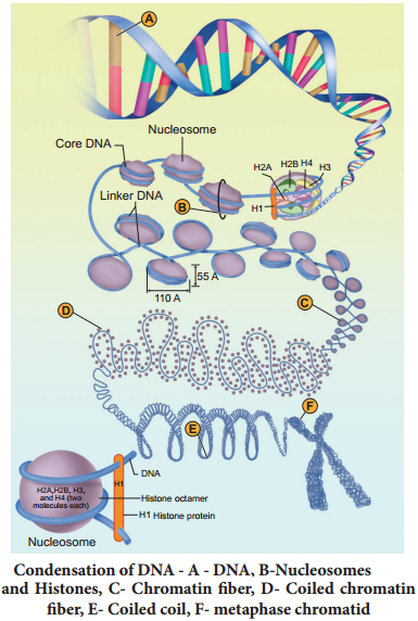
Process of transcription

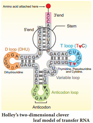
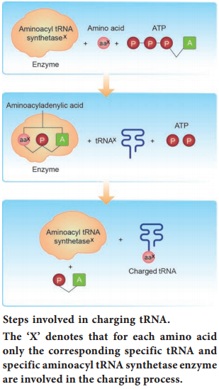
In prokaryotes, there are three major types of RNAs: mRNA, tRNA, and rRNA. All three RNAs are needed to synthesize a protein in a cell. The mRNA provides the template, tRNA brings amino acids and reads the genetic code, and rRNAs play structural and catalytic role during translation. There is a single DNA-dependent RNA polymerase that catalyses transcription of all types of RNA.

It binds to the promoter and initiates transcription (Initiation). The polymerases binding sites are called promoters. It uses nucleoside triphosphate as substrate and polymerases in a template depended fashion following the rule of complementarity. After the initiation of transcription, the polymerase continues to elongate the RNA, adding one nucleotide after another to the growing RNA chain.

Only a short stretch of RNA remains bound to the enzyme, when the polymerase reaches a terminator at the end of a gene, the nascent RNA falls off so also the RNA polymerase. The question is, how the RNA polymerases are able to catalyse the three steps initiation, elongation and termination? The RNA polymerase is only capable of catalyzing the process of elongation.

The RNA polymerase associates transiently with initiation factor sigma (σ) and termination factor rho (σ) to initiate and terminate the transcription, respectively. Association of RNA with these factors instructs the RNA polymerase either to initiate or terminate the process of transcription (Fig. 5.8).
Transcription img 3

In bacteria, since the mRNA does not require any processing to become active and also since transcription and translation take place simultaneously in the same compartment (since there is no separation of cytosol and nucleus in bacteria), many times the translation can begin much before the mRNA is fully transcribed. This is because the genetic material is not separated from other cell organelles by a nuclear membrane
consequently; transcription and translation can be coupled in bacteria.

In Eukaryotes, there are at least three RNA polymerases in the nucleus (in addition to RNA polymerase found in the organelles). There is a clear division of labour. The RNA polymerase I transcribes rRNAs (28S, 18S and 5.8S), whereas the RNA polymerase III is responsible for transcription of tRNA, 5S rRNA and snRNA.

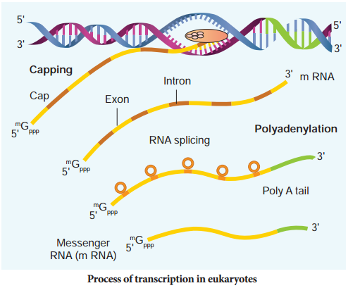
The RNA polymerase II transcribes precursor of mRNA, the hnRNA (heterogenous nuclear RNA). In eukaryotes, the monocistronic structural genes have interrupted coding sequences known as exons (expressed sequences) and noncoding sequences called introns (intervening sequences). The introns are removed by a process called splicing.

hnRNA undergoes additional processing called capping and tailing. In capping an unusual nucleotide, methyl guanosine triphosphate is added at the 5′ end, whereas adenylate residues (200-300) (Poly A) are added at the 3′ end in tailing (Fig. 5.9). Thereafter, this processed hnRNA, now called mRNA is transported out of the nucleus for translation.
Transcription img 4

The split gene feature of eukaryotic genes is almost entirely absent in prokaryotes. Originally each exon may have coded for a single polypeptide chain with a specific function. Since exon arrangement and intron removal are flexible, the exon coding for these polypeptide subunits act as domains combining in various ways to form new genes.

Single genes can produce different functional proteins by arranging their exons in several different ways through alternate splicing patterns, a mechanism known to play an important role in generating both protein and functional diversity in animals. Introns would have arosen before or after the evolution of eukaryotic gene.

If introns arose late how did they enter eukaryotic gene? Introns are mobile DNA sequences that can splice themselves out of, as well as into, specific ‘target sites’ acting like mobile transposon-like elements (that mediate transfer of genes between organisms – Horizontal Gene Transfer HGT). HGT occurs between lineages of prokaryotic cells, or from prokaryotic to eukaryotic cells and between eukaryotic cells. HGT is now hypothesized to have played a major role in the evolution of life on earth.

Packaging Of DNA Helix

Learninsta presents the core concepts of Biology with high-quality research papers and topical review articles.

Packaging Of DNA Helix

The distance between two consecutive base pairs is 0.34nm (0.34 × 10⁻⁹ m) of the DNA double helix in a typical mammalian cell. When the total number of base pairs is multiplied with the distance between two consecutive base pairs (6.6 × 109 × 0.34 × 10-9 m/bp), the length of DNA double helix is approximately 2.2 m. (The total length of the double helical DNA = total number of base pairs × distance between two consecutive base pairs).

If the length of E. coli DNA is 1.36 mm, the number of base pairs in E. coli is 4 × 10⁶bp (1.36 × 10³ m/0.34 × 10-9). The length of the DNA double helix is far greater than the dimension of a typical mammalian nucleus (approximately 10-6 m). How is such a long DNA polymer packaged in a cell?

Chromosomes are carriers of genes which are responsible for various characters from generation to generation. Du Praw (1965) proposed a single stranded model (unineme), as a long coiled molecule which is associated with histone proteins in eukaryotes. Plants and animals have more DNA than bacteria and must fold this DNA to fit into the cell nucleus.

In prokaryotes such as E. coli though they do not have defined nucleus, the DNA is not scattered throughout the cell. DNA (being negatively charged) is held with some proteins (that have positive charges) in a region called the nucleoid. The DNA as a nucleoid is organized into large loops held by protein. DNA of prokaryotes is almost circular and lacks chromatin organization, hence termed genophore.

In eukaryotes, this organization is much more complex. Chromatin is formed by a series of repeating units called nucleosomes. Kornberg proposed a model for the nucleosome, in which 2 molecules of the four histone proteins H2A, H2B, H3 and H4 are organized to form a unit of eight molecules called histone octamere. The negatively charged DNA is wrapped around the positively charged histone octamere to form a structure called nucleosome.

A typical nucleosome contains 200 bp of DNA helix. The histone octameres are in close contact and DNA is coiled on the outside of nucleosome. Neighbouring nucleosomes are connected by linker DNA (H1) that is exposed to enzymes. The DNA makes two complete turns around the histone octameres and the two turns are sealed of by an H1 molecule. Chromatin lacking H1 has a beads-on-a-string appearance in which DNA
enters and leaves the nucleosomes at random places. H1 of one nucleosome can interact with H1 of the neighbouring nucleosomes resulting in the further folding of the fire.

The chromatin fier in interphase nuclei and mitotic chromosomes have a diameter that vary between 200-300 nm and represents inactive chromatin. 30 nm fire arises from the folding of nucleosome, chains into a solenoid structure having six nucleosomes per turn. This structure is stabilized by interaction between different H1 molecules. DNA is a solenoid and packed about 40 folds.

The hierarchical nature of chromosome structure is illustrated in (Fig. 5.3). Additional set of proteins are required for packing of chromatin at higher level and are referred to as non-histone chromosomal proteins (NHC). In a typical nucleus, some regions of chromatin are loosely packed (lightly stained) and are referred to as euchromatin. The chromatin that is tightly packed (stained darkly) is called heterochromatin. Euchromatin is transcriptionally active and heterochromatin is transcriptionally inactive.
Packaging Of DNA Helix img 1