Learninsta presents the core concepts of Biology with high-quality research papers and topical review articles.
Basic Concepts Of Immunology
Immunology is the study of immune system. This system protects an individual from various infective agents. It refers to all the mechanisms used by the body for protection from environmental agents that are foreign to the body.
When the immune system does not function efficiently in an individual, it leads to infection causing disease. The overall ability of body to fight against the disease causing pathogen is called immunity. It is also called disease resistance and the lack of immunity is known as susceptibility. Immunity is highly specific.
Normally many of the responses of the immune system initiate the destruction and elimination of invading organisms and any toxic molecules produced by them. These immune reactions are destructive in nature and are made in response only to molecules that are foreign to the host and not to those of host itself. This ability to distinguish foreign molecules from self is another fundamental feature of the immune system.
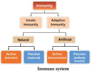
However, occasionally, it fails to make its distinction and reacts destructively against the host’s own molecules; such autoimmune diseases can be fatal to the organism. Almost all the macromolecules e.g. proteins, polysaccharides, nucleic acids, etc., as long as they are foreign to recipient organism can induce immune response. Any substance capable of eliciting immune response is called an ANTIGEN (ANTIbody GENerator). There are two broad classes of immunity responses namely, innate immunity and acquired immunity (Fig. 7.9).
Innate immunity
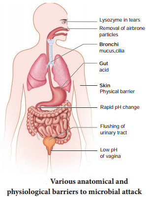
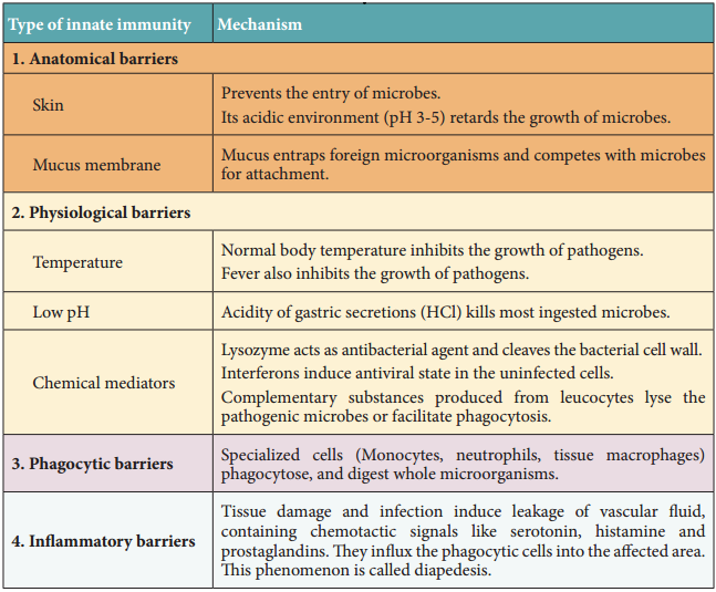
Innate immunity is the natural phenomenon of resistance to infection which an individual possesses right from the birth. The innate defense mechanisms are non-specific in the sense that they are effective against a wide range of potentially infectious agents. It is otherwise known as non-specifi immunity or natural immunity.
A number of innate defense mechanisms are operative non-specifially against a large number of microorganisms as shown in the Table 7.4 and Fig. 7.10.
Acquired immunity
The immunity that an individual acquires after birth is known as acquired immunity. It is the body’s resistance to a specific pathogen. The unique features of acquired immunity are antigenic specificity, diversity, recognition of self and non-self and immunological memory.
Components of acquired immunity
Acquired immunity has two components – cell mediated immunity (CMI) and antibody mediated immunity or humoral immunity.
1. Cell mediated immunity
When pathogens are destroyed by cells without producing antibodies, then it is known as cell mediated immune response or cell mediated immunity. This is brought about by T cells, macrophages and natural killer cells.
2. Antibody mediated immunity or humoral immunity
When pathogens are destroyed by the production of antibodies, then it is known as antibody mediated or humoral immunity. This is brought about by B cells with the help of antigen presenting cells and T helper cells. Antibody production is the characteristic feature of vertebrates only.
Types of acquired immunity
Acquired immunity may be active immunity or passive immunity (Table 7.5).
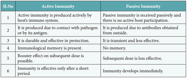
The immunological resistance developed by the organisms through the production of antibodies in their body is called active immunity. Active immunity is acquired through the use of a person’s immune responses, which lead to the development of memory cells. Active immunity results from an infection or an immunization.
Passive immunity does not require the body to produce antibodies to antigens. The antibodies are introduced from outside into the organism. This, passive immunity is acquired without the activation of a person’s immune response, and therefore there is no memory.
Immune responses
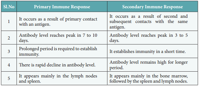
The immune responses may be primary or secondary (Table 7.6).
Primary immune response
The primary immune response occurs when a pathogen comes in contact with the immune system for the first time. During this, the immune system has to learn to recognize the antigen, produce antibody against it and eventually produce memory lymphocytes. The primary immune response is slow and short-lived.
Secondary immune response
The secondary immune response occurs when a person is exposed to the same antigen again. During this time, immunological memory has been established and the immune system can start producing antibodies immediately. Within hours after recognition of the antigen, a new army of plasma cells are generated. Within 2 to 3 days, the antibody concentration in the blood rises steeply to reach much higher level than primary response. This is also called as “booster response”.
Lymphoid organs
Immune system of an organism consists of several structurally and functionally different organs and tissues that are widely dispersed in the body. The organs involved in the origin, maturation and proliferation of lymphocytes are called lymphoid organs (Fig. 7.11).
Based on their functions, they are classified into primary or central lymphoid organs and secondary or peripheral lymphoid organs. The primary lymphoid organs provide appropriate environment for lymphocytic maturation. The secondary lymphoid organs trap antigens and make it available for mature lymphocytes, which can effectively fiht against these antigens.
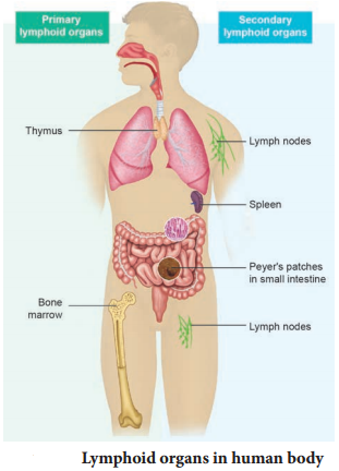
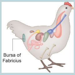
Primary lymphoid organs
Bursa of Fabricius of birds, bone marrow and thymus gland of mammals constitute the primary lymphoid organs involved in the production and early selection of lymphocytes. These lymphocytes become dedicated to a particular antigenic specificity. Only when the lymphocytes mature in the primary lymphoidal organs, they become immunocompetent cells. In mammals, B cell maturation occurs in the bone marrow and T cells maturation occurs in the thymus.
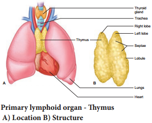
Thymus
The thymus is a flat and bilobed organ located behind the sternun, above the heart. Each lobe of the thymus contains numerous lobules, separated from each other by connective tissue called septa. Each lobule is differentiated into two compartments, the outer compartment or outer cortex, is densely packed with immature T cells called thymocytes, whereas the inner compartment or medulla is sparsely populated with
mature thymocytes.
One of its main secretions is the hormone thymosin. It stimulates the T cell to become mature and immunocompetent. By the early teens, the thymus begins to atrophy and is replaced by adipose tissue (Fig. 7.12). Thus thymus is most active during the neonatal and pre-adolescent periods.
Bone marrow
Bone marrow is a lymphoid tissue found within the spongy portion of the bone. Bone marrow contains stem cells known as haematopoietic cells. These cells have the potential to multiply through cell division and either remain as stem cells or differentiate and mature into different kinds of blood cells.
Secondary or peripheral lymphoid organs
In secondary or peripheral lymphoid organs, antigen is localized so that it can be effectively exposed to mature lymphocytes. The best examples are lymph nodes, appendix, Peyer’s patches of gastrointestinal tract, tonsils, adenoids, spleen, MALT (Mucosal-Associated Lymphoid Tissue), GALT (Gut-Associated Lymphoid Tissue), BALT (Bronchial/Tracheal-Associated Lymphoid Tissue).
Lymph node
Lymph node is a small bean-shaped structure and is part of the body’s immune system. It is the first one to encounter the antigen that enters the tissue spaces. Lymph nodes filter and trap substances that travel through the lymphatic fluid. They are packed tightly with white blood cells, namely lymphocytes and macrophages. There are hundreds of lymph nodes found throughout the body.
They are connected to one another by lymph vessels. Lymph is a clear, transparent, colourless, mobile and extracellular fluid connective tissue. As the lymph percolates through the lymph node, the particulate antigen brought in by the lymph will be trapped by the phagocytic cells, follicular and interdigitating dendritic cells.
Lymph node has three zones (Fig. 7.13). They are the cortex, paracortex and medulla. The outer most layer of the lymph node is called cortex, which consists of B-lymphocytes, macrophages, and follicular dendritic cells. The paracortex zone is beneath the cortex, which is richly populated by T lymphocytes and interdigitating dendritic cells. The inner most zone is called the medulla which is sparsely populated by lymphocytes, but many of them are plasma cells, which actively secrete antibody molecules.
As the lymph enters, it slowly percolates through the cortex, paracortex and medulla, giving sufficient chance for the phagocytic cells and dendritic cells to trap the antigen brought by the lymph. The lymph leaving a node carries enriched antibodies secreted by the medullary plasma cells against the antigens that enter the lymph node. Sometimes visible swelling of lymph nodes occurs due to active immune response and increased concentration of lymphocytes.
This swollen lymph nodes may signal an infection. There are several groups of lymph nodes. The most frequently enlarged lymph nodes are found in the neck, under the chin, in the armpits and in the groin.
Cells of the immune system
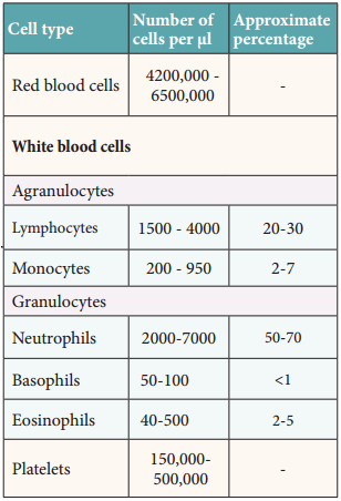
The immune system is composed of many interdependent cells that protect the body from microbial infections and the growth of tumour cells. The cellular composition of adult human blood is given in Table 7.7.
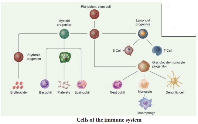
All these cells are derived from pluripotent haematopoetic stem cells. Each stem cell has the capacity to produce RBC, WBC and platelets. The only cells capable of specifially recognising and producing an immune response are the lymphocytes. The other types of white blood cells play an important role in non specific immune response, antigen presentation and cytokine production.
Lymphocytes
About 20-30% of the white blood cells are lymphocytes. They have a large nucleus filling most of the cell, surrounded by a little cytoplasm. The two main types of lymphocytes are B and T lymphocytes. Both these are produced in the bone marrow. B lymphocytes (B cells) stay in the bone marrow until they are mature. Then they circulate around the body. Some remain in the blood, while others accumulate in the lymph
nodes and spleen. T lymphocytes leave the bone marrow and mature in the thymus gland.
Once mature, T cells also accumulate in the same areas of the body as B cells. Lymphocytes have receptor proteins on their surface. When receptors on a B cell bind with an antigen, the B cell becomes activated and divides rapidly to produce plasma cells. The plasma cells produce antibodies. Some B cells do not produce antibodies but become memory cells. These cells are responsible for secondary immune response. T lymphocytes do not produce antibodies. Thy recognize antigenpresenting cells and destroy them.
The two important types of T cells are Helper T cells and Killer T cells. Helper T cells release a chemical called cytokine which activates B cells. Killer cells move around the body and destroy cells which are damaged or infected (Fig. 7.14).
Apart from these cells neutrophils and monocytes destroy foreign cells by phagocytosis. Monocytes when they mature into large cells, they are called macrophages which perform phagocytosis on any foreign organism.
Antigens
The term antigen (Ag) is used in two senses, the first to describe a molecule which generates an immune response and the second, a molecule which reacts with antibodies. In general antigens are large, complex molecular substances that can induce a detectable immune response. This an antigen is a substance that is specific to an antibody or a T-cell receptor and is often used as a synonym for immunogen.
An immunogen is a substance capable of initiating an immune response. Haptens are substance that are non-immunogenic but can react with the products of a specific immune response. Substances that can enhance the immune response to an antigen are called adjuvants. Epitope is an antigenic determinant and is the active part of an antigen. A paratope is the antigen – binding site and is a part of an antibody which
recognizes and binds to an antigen.
Types of antigens
On the basis of origin, antigens are classified into exogenous antigens and endogenous antigens. The antigens which enter the host from the outside in the form of microorganisms, pollens, drugs, or pollutants are called exogenous antigens. The antigens which are formed within the individual are endogenous antigens. The best examples are blood group antigens.
Antibodies
Antibodies are immunoglobulin (Ig) protein molecules synthesized on exposure to antigen that can combine specifically with the antigen. Whenever pathogens enter our body, the B-lymphocytes produce an army of proteins called antibodies to fight with them. Thus, they are secreted in response to an antigen (Ag) by the effect of B cells called plasma cells. The antibodies are classified into five major categories, based on their physiological and biochemical properties. They are IgG (gamma), IgM (mu), IgA (alpha), IgD (delta) and IgE (epsilon).
In the 1950s, experiments by Porter and Edelman revealed the basic structure of the immunoglobulin. An antibody molecule is Y shaped
structure that comprises of four polypeptide chains, two identical light chains (L) of molecular weight 25,000 Da (approximately 214 amino acids) and
two identical heavy chains (H) of molecular weight 50,000 Da (approximately 450 amino acids).
The polypeptide chains are linked together by di-sulphide (S-S) bonds. One light chain is attached to each heavy chain and two heavy chains are attached to each other to form a Y shaped (Fig. 7.15) structure. Hence, an antibody is represented by H2 L2. The heavy chains have a flexible hinge region at their approximate middles.
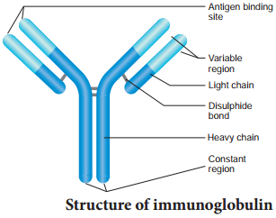
Each chain (L and H) has two terminals. They are C – terminal (Carboxyl) and amino or N-terminal. Each chain (L and H) has two regions. They have variable (V) region at one end and a much larger constant (C) region at the other end. Antibodies responding to different antigens have very different (V) regions but their (C) regions are the same in all antibodies.
In each arm of the monomer antibody, the (V) regions of the heavy and light chains combines to form an antigen – binding site shaped to ‘fit’ a specific antigenic determinant. Consequently each antibody monomer has two such antigen – binding regions. The (C) regions that forms the stem of the antibody monomer determine the antibody class and serve common functions in all antibodies. The functions of immunoglobulin are agglutination, precipitation, opsonisation, neutralization etc.,
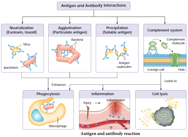
Antigen – antibody interaction
The reaction between an antigen and antibody is the basis for humoral immunity or antibody mediated immunity. The reaction between antigen and antibody occurs in three stages. During the first stage, the reaction involves the formation of antigen – antibody complex. The next stage leads to visible events like precipitation, agglutination, etc., The final stage includes destruction of antigen or its neutralization (Fig. 7.16).
Binding force of antigen – antibody reaction
The binding force between antigen and antibody is due to three factors. They are closeness between antigen and antibody, noncovalent bonds or intermolecular forces and affinity of antibody.
When antigen and antibody are closely fitted, the strength of binding is great. When they are apart binding strength is low. The bonds that hold the antigen to the antibody combining site are all non-covalent in nature. These include hydrogen bonds, electrostatic bonds, Van der Waals forces and hydrophobic bonds. Antibody affinity is the strength of the reaction between a single antigenic determinant and a single combining site on the antibody.
The chiefapplication of antigen – antibody reactions are to determine blood groups for transfusion, to study serological ascertainment of exposure to infectious agents, to develop immunoassays for the quantification of various substances, to detect the presence or absence of protein in serum and to determine the characteristics of certain immunodeficiency diseases.
Different types of antigen and antibody reactions
The reaction between soluble antigen and antibody leads to visible precipitate formation, which is called precipitin reaction. Antibodies that bring about precipitate formation on reacting with antigens are called as precipitins.
Whenever a particulate antigen interacts with its antibody, it would result in clumping or agglutination of the particulate antigen, which is called agglutination reaction. The antibody involved in bringing about agglutination reaction is called agglutinin.
Opsonisation or enhanced attachment is the process by which a pathogen is marked of ingestion and destruction by a phagocyte. Opsonisation involves the binding of an opsonin i.e., antibody, to a receptor on the pathogen’s cell membrane.
After opsonin binds to the membrane, phagocytes are attracted to the pathogen. So, opsonisation is a process in which pathogens are coated with a substance called an opsonin, marking the pathogen out for destruction by the immune system. This results in a much more efficient phagocytosis.
The neutralization reactions are the reactions of antigen-antibody that involve the elimination of harmful effects of bacterial exotoxins or a virus by specific antibodies. These neutralizing substances i.e., antibodies are known as antitoxins. This specific antibody is produced by a host cell in response to a bacterial exotoxin or corresponding toxoid (inactivated toxin).
Vaccines
A vaccine is a biological preparation that provides active acquired immunity to a particular disease and resembles a disease-causing microorganism and is often made from weakened or attenuated or killed forms of the microbes, their toxins, or one of its surface proteins.
Vaccines “teach” our body how to defend itself when viruses or bacteria, invade it. Vaccines deliver only very little amounts of inactivated or weakened viruses or bacteria, or parts of them. This allows the immune system to recognize the organism without actually experiencing the disease. Some vaccines need to be given more than once (i.e., a ‘booster’ vaccination) to make sure the immune system can overcome a real infection in the future.
Vaccine initiates the immunization process. The vaccines are classified as first, second and third generation vaccines. First generation vaccine is further subdivided into live attenuated vaccine, killed vaccine and toxoids (Fig. 7.17). Live attenuated vaccines use the weakened (attenuated), aged, less virulent form of the virus. E.g. Measles, mumps and rubella (MMR) vaccine and the Varicella (chickenpox) vaccine, Killed (inactivated) vaccines are killed or inactivated by heat and other methods. E.g. Salk’s polio vaccine.
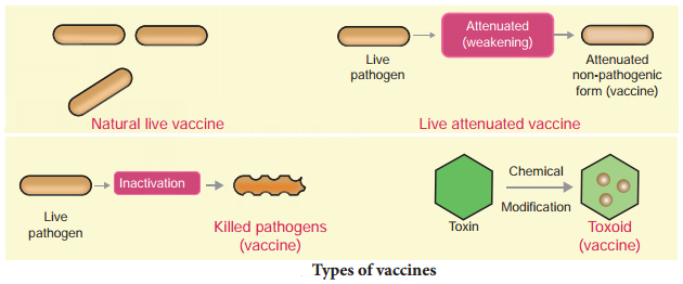
Toxoid vaccines contain a toxin or chemical secreted by the bacteria or virus. They make us immune to the harmful effects of the infection, instead of to the infection itself. E.g. DPT vaccine (Diphtheria, Pertussis and Tetanus).
Second generation vaccine contains the pure surface antigen of the pathogen. E.g.Hepatitis-B vaccine. Third generation vaccine contains the purest and the highest potency vaccines which are synthetic in generation. The latest revolution in vaccine is DNA vaccine or recombinant vaccine (Refer Chapter- 9 for details).
Vaccination and immunization
“Vaccination is the process of administrating a vaccine into the body or the act of introducing a vaccine into the body to produce immunity to a specific disease.” Immunization is the process of the body building up immunity to a particular disease.
Immunization describes the actual changes in the body after receiving a vaccine. Vaccines work by fighting the pathogen and then recording it in their memory system to ensure that the next time this pathogen enters the body, it is eliminated far quickly. Once, the body is able to fight against the disease, it is believed to have built the immunity for it, also known as the body being immunized against the disease.
Hypersensitivity
Some of the individuals are very sensitive to some particles present in the environment. The exaggerated response of the immune system to certain antigens present in the environment is called allergy (allo-altered, erg-reaction). The substances to which such an immune response is produced are called allergens. An allergen is an antigen that causes an allergic reaction. Allergic reactions begin within few seconds after the contact with the allergen and last about half an hour.
The common examples of allergens are mites in dust, pollens and some proteins in insect venom. Hay fever and asthma are some common examples of allergy. Symptoms of allergic reactions include sneezing, watery eyes, running nose and difficulty in breathing.
Allergy is a form of over active immune response mediated by IgE and mast cells. It can also be due to the release of chemicals like histamine and serotonin from the mast cells. Anaphylaxis is the classical immediate hypersensitivity reaction. It is a sudden, systematic, severe and immediate hypersensitivity reaction occurring as a result of rapid generalized mast-cell degranulation.