Exchange of Gases in Respiratory Pigments, Methaemoglobin

Learninsta presents the core concepts of Biology with high-quality research papers and topical review articles.

Exchange of Gases in Respiratory Pigments, Methaemoglobin

The primary site for the exchange of gases is the alveoli. The uptake of O2 and the release of CO2 occur between the blood and tissues by simple diffusion driven by partial pressure gradient of O2 and CO2. Partial pressure is the pressure contributed by an individual gas in a mixture of gases.

It is represented as pO2 for oxygen and pCO2 for carbon-dioxide. Due to pressure gradients, O2 from the alveoli enters into the blood and reaches the tissues. CO2 enters into the blood from the tissues and reaches alveoli for elimination. As the solubility of CO2 is 20-25 times higher than that of O2, the partial pressure of CO2 is much higher than that of O2 (Table 6.1 and Figure 6.6).

Respiratory Pigments

Haemoglobin

Haemoglobin belongs to the class of conjugated protein. The iron containing pigment portion haem constitutes only 4% and the rest colourless protein globin belongs to histone classs. Haemoglobin has a molecular weight of 68,000 daltons and contains four atoms of iron, each of which can combine with a molecule of oxygen.

Methaemoglobin

If the iron component of the haem moieties is in the ferric state, than the normal ferrous state, it is called methaemoglobin. Methaemoglobin does not bind O2. Normally RBC contains less than 1% methaemoglobin.
Exchange of Gases img 1
Exchange of Gases img 2
Table 6.1 Partial pressure of Oxygen and Carbon dioxide (in mmHg) in comparison to those gases in the atmosphere.

Mechanism of Breathing in Human Beings

Learninsta presents the core concepts of Biology with high-quality research papers and topical review articles.

Mechanism of Breathing in Human Beings

The movement of air between the atmosphere and the lungs is known as ventilation or breathing. Inspiration and expiration are the two phases of breathing. Inspiration is the movement of atmospheric air into the lungs and expiration is the movement of alveolar air that diffuse out of the lungs. (Figure 6.4)
Muscle Tissue img 1

Lungs do not contain muscle fires but expands and contracts by the movement of the ribs and diaphragm. The diaphragm is a sheet of tissue which separates the thorax from the abdomen. In a relaxed state, the diaphragm is domed shaped. Ribs are moved by the intercostal muscles.

External and internal intercostal muscles found between the ribs and the diaphragm helps in creating pressure gradients. Inspiration occurs if the pressure inside the lungs (intrapulmonary pressure) is less than the atmospheric pressure.
Muscle Tissue img 2

Inspiraton is initiated by the contraction of the diaphragm muscles and external intercostal muscles, which pulls the ribs and sternum upwards and outwards and increases the volume of the thoracic chamber in the dorso-ventral axis, forcing the lungs to expand the pulmonary volume.

The increase in pulmonary volume and decrease in the intrapulmonary pressure forces the fresh air from outside to enter the air passages into the lungs to equalize the pressure. This process is called inspiration.

Relaxation of the diaphragm allows the diaphragm and sternum to return to its dome shape and the internal intercostal muscles contract, pulling the ribs downward reducing the thoracic volume and pulmonary volume. This results in an increase in the intrapulmonary pressure slightly above the atmospheric pressure causing the expulsion of air from the lungs. This process is called expiration.

On an average, a healthy human breathes 12-16 times/minute. An instrument called Spirometer is used to measure the volume of air involved in breathing movements for clinical assessment of a person’s pulmonary function.

Respiratory Volumes and Capacities

The volume of air present in various phases of respiration is denoted as

Respiratory Volumes: (Figure 6.5)

Tidal Volume (TV)

Tidal volume is the amount of air inspired or expired with each normal breath. It is approximately 500 mL., i.e. a normal human adult can inspire or expire approximately 6000 to 8000mL of air per minute. During vigorous exercise, the tidal volume is about 4-10 times higher.

Inspiratory Reserve Volume (IRV)

Additional volume of air a person can inspire by forceful inspiration is called Inspiratory Reserve Volume. The normal value is 2500-3000 mL.

Expiratory Reserve Volume (ERV)

Additional volume of air a person can forcefully exhale by forceful expiration is called Expiratory Reserve Volume. The normal value is 1000-1100 mL.

Residual Volume (RV)

The volume of air remaining in the lungs after a forceful expiration. It is approximately 1100-1200 mL.

Respiratory Capacities:

Vital capacity (VC) the maximum volume of air that can be moved out during a single breath following a maximal inspiration. A person first inspires maximally then expires maximally. VC = ERV + TV + IRV

Inspiratory Capacity (IC)

The total volume of air a person can inhale after normal expiration. It includes tidal volume and inspiratory reserve volume. IC = TV + IRV

Expiratory Capacity (EC)

The total volume of air a person can exhale after normal inspiration. It includes tidal volume and expiratory reserve volume. EC = TV + ERV
Muscle Tissue img 3

Total Lung Capacity (TLC)

The total volume of air which the lungs can accommodate after forced inspiration is called Total Lung Capacity. This includes the vital capacity and the residual volume. It is approximately 6000mL. TLC = VC + RV

Minute Respiratory Volume

The amount of air that moves into the respiratory passage per minute is called minute respiratory volume.
Normal TV = 500mL; Normal respiratory rate = 12 times/minute Therefore, minute respiratory volume = 6 Litres/minute (for a normal healthy man).

Dead Space

Some of the inspired air never reaches the gas exchange areas but fills the respiratory passages where exchange of gases does not occur. This air space is called dead space. Dead space is not involved in gaseous exchange. It amounts to approximately 150mL.

Respiratory Function, Facts Organs & Anatomy

Learninsta presents the core concepts of Biology with high-quality research papers and topical review articles.

Respiratory Function, Facts Organs & Anatomy

The five primary functions of the respiratory system are: –

  • To exchange O2 and CO2 between the atmosphere and the blood.
  • To maintain homeostatic regulation of body pH.
  • To protect us from inhaled pathogens and pollutants.
  • To maintain the vocal cords for normal communication (vocalization).
  • To remove the heat produced during cellular respiration.

Your lungs are part of the respiratory system, a group of organs and tissues that work together to help you breathe. The respiratory system’s main job is to move fresh air into your body while removing waste gases.

There are Five Functions of the Respiratory System

Gas Exchange – oxygen and carbon dioxide.
Breathing – movement of air.
Sound Production.
Oldfactory Assistance – sense of smell.
Protection – from dust and microbes entering body through mucus production, cilia, and coughing.

Allows you to talk and to smell. Brings air to body temperature and moisturizes it to the humidity level your body needs. Delivers oxygen to the cells in your body. Removes waste gases, including carbon dioxide, from the body when you exhale.

Respiratory failure is a serious condition that develops when the lungs can’t get enough oxygen into the blood. Buildup of carbon dioxide can also damage the tissues and organs and further impair oxygenation of blood and, as a result, slow oxygen delivery to the tissues.

Inside the lungs, oxygen is exchanged for carbon dioxide waste through the process called external respiration. This respiratory process takes place through hundreds of millions of microscopic sacs called alveoli. Oxygen from inhaled air diffuses from the alveoli into pulmonary capillaries surrounding them.

The bronchial tubes divide into smaller air passages called bronchi, and then into bronchioles. The bronchioles end in tiny air sacs called alveoli, where oxygen is transferred from the inhaled air to the blood. After absorbing oxygen, the blood leaves the lungs and is carried to the heart.

The oxygen we inhale is used to breakdown glucose into carbon dioxide and water. Energy is released in the process. The breakdown of glucose occurs in the cells of an organism (cellular respiration). If the breakdown occurs without the use of oxygen, the respiration is called anaerobic respiration.

Respiratory Organs in Various Organisms

Learninsta presents the core concepts of Biology with high-quality research papers and topical review articles.

Respiratory Organs in Various Organisms

Different animals have different organs for exchange of gases, depending upon their habitats and levels of organization. The amount of dissolved oxygen is very low in water compared to the amount of oxygen in the air. So the rate of breathing in aquatic organisms is much faster than land animals.

In animals like sponges, coelenterates and flatworms exchange of gases takes place through the body surface by simple diffusion. Earthworms use their moist skin, whereas insects have tracheal tubes. Gills are used as respiratory organs in most of the aquatic Arthropods and Molluscs.

Among vertebrates, fishes use gills whereas amphibians, reptiles, birds and mammals have well vascularised lungs. Frogs spend most of their time in water and also use their moist skin for respiration along with lungs and bucco pharynx.

Human Respiratory System

The respiratory system includes external nostrils, nasal cavity, pharynx, larynx, trachea, bronchi and bronchioles and lungs which contain alveoli (Figure 6.1). The parts starting from the external nostrils up to the terminal bronchioles constitute the conducting zone, whereas the alveoli and the ducts are called the respiratory zone. The parts of the conducting zone, humidifies and warms the incoming air.
Respiratory Organs in Various Organisms img 1

In human beings, air enters the upper respiratory tract through the external nostrils. The air passing through the nostrils is filtered by fine hairs and mucus lining the passage. The external nostrils lead to the nasal chamber which opens into the nasopharynx which opens through the glottis of the larynx region into the trachea. The ciliated epithelial cells lining the trachea, bronchi and bronchioles secrete mucus.

Mucus membrane lining the airway contains goblet cells which secrete mucus, a slimy material rich in glycoprotein. Microorganisms and dust particles attach in the mucus films and are carried upwards to pass down the gullet during normal swallowing. During swallowing a thin elastic flap called epiglottis prevents the food from entering into the larynx and avoids choking of food.

The trachea is semiflexible tube supported by multiple cartilaginous rings which extends up to the midthoracic cavity and at the level of the 5th thoracic vertebra where it divides into right and left primary bronchi, one bronchus to each lung. Within the lungs the bronchi divides repeatedly into secondary and tertiary bronchi and further divides into terminal bronchioles and respiratory bronchioles.

Bronchi have ‘C’ shaped curved cartilage plates to ensure that the air passage does not collapse or burst as the air pressure changes during breathing. The bronchioles are without cartilaginous rings and have rigidity that prevent them from collapsing but are surrounded by smooth muscle which contracts or relaxes to adjust the diameter of these airways.

The fine respiratory bronchioles terminate into highly vascularised thin walled pouch like air sacs called alveoli meant for gaseous exchange (Figure 6.2, 6.3). The diffusion membrane of alveolus is made up of three layers – the thin squamous epithelial cells of the alveoli, the endothelium of the alveolar capillaries and the basement substance found in between them. The thin squamous epithelial cells of the alveoli are composed of Type I and Type II cells. Type I cells are very thin so that gases can diffuse rapidly through them. Type II cells are thicker, synthesize and secrete a substance called Surfactant.

The lungs are light spongy tissues enclosed in the thoracic cavity surrounded by an airtight space. The thoracic cavity is bound dorsally by the vertebral column and ventrally by the sternum, laterally by the ribs and on the lower side by the dome shaped diaphragm.

The lungs are covered by double walled pleural membrane containing a several layers of elastic connective tissues and capillaries, which encloses the pleural fluid. Pleural fluid reduces friction when the lungs expand and contract.
Respiratory Organs in Various Organisms img 2
Respiratory Organs in Various Organisms img 3

Characteristic Features of Respiratory Surface:

  • Surface area must be very large and richly supplied with blood vessels
  • Should be extremely thin and kept moist
  • Should be in direct contact with the environment
  • Should be permeable to respiratory gases

The Steps Involved in Respiration are:-

  • The exchange of air between the atmosphere and the lungs.
  • The exchange of O2 and CO2 between the lungs and the blood.
  • Transport of O2 and CO2 by the blood.
  • Exchange of gases between the blood and the cells.
  • Uptake of O2 by the cells for various activities and the release of CO2.

Nutritional and Digestive Disorders

Learninsta presents the core concepts of Biology with high-quality research papers and topical review articles.

Nutritional and Digestive Disorders

Intestinal tract is more prone to bacterial, viral and parasitic worm infections. This infection may cause inflammation of the inner lining of colon called colitis. The most common symptoms of colitis are rectal bleeding, abdominal cramps, and diarrhoea.

Protein Energy Malnutrition: (PEM)

Growing children require more amount of protein for their growth and development. Protein deficient diet during early stage of children may lead to protein energy malnutrition such as Marasmus and Kwashiorkor. Symptoms are dry skin, pot-belly, oedema in the legs and face, stunted growth, changes in hair colour, weakness and irritability.

Marasmus is an acute form of protein malnutrition. This condition is due to a diet with inadequate carbohydrate and protein. Such children are suffer from diarrhoea, body becomes lean and weak (emaciated) with reduced fat and muscle tissue with thin and folded skin.

Indigestion:

It is a digestive disorder in which the food is not properly digested leading to a feeling of fullness of stomach. It may be due to inadequate enzyme secretion, anxiety, food poisoning, over eating, and spicy food.

Constipation:

In this condition, the faeces are retained within the rectum because of irregular bowel movement due to poor intake of fire in the diet and lack of physical activities.

Vomiting:

It is reverse peristalsis. Harmful substances and contaminated food from stomach are ejected through the mouth. This action is controlled by the vomit centre located in the medulla oblongata. A feeling of nausea precedes vomiting.

Jaundice:

It is the condition in which liver is affected and the defective liver fails to break down haemoglobin and to remove bile pigments from the blood. Deposition of these pigments changes the colour of eye and skin yellow. Sometimes, jaundice is caused due to hepatitis viral infections.

Liver Cirrhosis:

Chronic disease of liver results in degeneration and destruction of liver cells resulting in abnormal blood vessel and bile duct leading to the formation of fibrosis. It is also called deserted liver or scarred liver. It is caused due to infection, consumption of poison, malnutrition and alcoholism.

Gall Stones:

Any alteration in the composition of the bile can cause the formation of stones in the gall bladder. The stones are mostly formed of crystallized cholesterol in the bile. The gall stone causes obstruction in the cystic duct, hepatic duct and also hepato-pancreatic duct causing pain, jaundice and pancreatitis.

Appendicitis:

It is the inflammation of the vermiform appendix, leading to severe abdominal pain. The treatment involves the removal of appendix by surgery. If treatment is delayed the appendix may rupture and results in infection of the abdomen, called peritonitis.

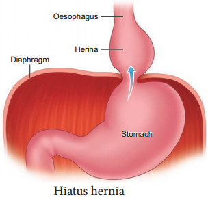
Hiatus Hernia (Diaphragmatic hernia):

It is a structural abnormality in which superior part of the stomach protrudes slightly above the diaphragm. The exact cause of hiatus hernias is not known. In some people, injury or other damage may weaken muscle tissue, by applying too much pressure (repeatedly) on the muscles around the stomach while coughing, vomiting, and straining during bowel movement and lifting heavy object.

Heart burn is also common in those with a hiatus hernia. In this condition, stomach contents travel back into the oesophagus or even into oral cavity and causes pain in the centre of the chest due to the eroding nature of acidity (Figure 5.10).
Nutritional and Digestive Disorders img 1

Diarrhoea:

It is the most common gastrointestinal disorder worldwide. It is sometimes caused by bacteria or viral infections through food or water. When the colon is infected, the lining of the intestine is damaged by the pathogens, thereby the colon is unable to absorb fluid.

The abnormal frequency of bowel movement and increased liquidity of the faecal discharge is known as diarrhoea. Unless the condition is treated, dehydration can occur. Treatment is known as oral hydration therapy. This involves drinking plenty of fluids – sipping small amounts of water at a time to rehydrate the body.

Peptic Ulcer:

It refers to an eroded area of the tissue lining (mucosa) in the stomach or duodenum. Duodenal ulcer occurs in people in the age group of 25 – 45 years. Gastric ulcer is more common in persons above the age of 50 years.

Ulcer is mostly due to infections caused by the bacterium Helicobacter pylori. It may also be caused due to uncontrolled usage of aspirin or certain antiinflammatory drugs. Ulcer may also be caused due to smoking, alcohol, caffine and psychological stress.

Obesity:

It is caused due to the storage of excess of body fat in adipose tissue. It may induce hypertension, atherosclerotic heart disease and diabetes. Obesity may be genetic or due to excess intake of food, endocrine and metabolic disorders.

Degree of obesity is assessed by body mass index (BMI). A normal BMI range for adult is 19-25 above 25 is considered as obese. BMI is calculated as body weight in Kg, divided by the square of body height in meters. For example, a 50 Kg person with a height of 160 cms would have a BMI of 19.5. That is BMI = 50/(1.6)2 = 19.5