Body Fluids – Types, Composition & Functions Of Body Fluids, Circulation

Learninsta presents the core concepts of Biology with high-quality research papers and topical review articles.

Body Fluids – Types, Composition & Functions Of Body Fluids, Circulation

The body fluid consists of water and substances dissolved in them. There are two types of body fluids, the intracellular fluid present inside the cells and the extracellular fluid present outside the cells. The three types of extracellular fluids are the interstitial fluid or tissue fluid (surrounds the cell), the plasma (fluid component of the blood) and lymph.

The blood flowing into the capillary from an arteriole has a high hydrostatic pressure. This pressure is brought about by the pumping action of the blood and it tends to force water and small molecules out through the permeable walls of the capillary into the tissue fluid.

The volume of fluid which leaves the capillary to form tissue fluid is the result of two pressure (hydrostatic pressure and Oncotic pressure). At the anterior end of the capillary bed, the water potential is lesser than hydrostatic pressure inside the capilary bed which is enough to push fluid into the tissues.

The tissue fluid has low concentration of protien than that of plasma. At the venous end of the capillary bed, the water potential is greater than the hydrostatic pressure and the fluid from the tissues flows into the capillary and water is drawn back into the blood, taking with it waste products produced by the cells.

Composition Of Blood

Blood is the most common body fluid that transports substances from one part of the body to the other. Blood is a connective tissue consisting of plasma (fluid matrix) and formed elements. The plasma constitutes 55% of the total blood volume. The remaining 45% is the formed elements that consist of blood cells. The average blood volume is about 5000ml (5L) in an adult weighing 70 Kg.

Plasma

Plasma mainly consists of water (80-92%) in which the plasma proteins, inorganic constituents (0.9%), organic constituents (0.1%) and respiratory gases are dissolved. The four main types of plasma proteins synthesized in the liver are albumin, globulin, prothrombin and firinogen. Albumin maintains the osmotic pressure of the blood. Globulin facilitates the transport of ions, hormones, lipids and assists in immune function. Both Prothrombin and Fibrinogen are involved in blood clotting.

Organic constituents include urea, amino acids, glucose, fats and vitamins and the inorganic constituents include chlorides, carbonates and phosphates of potassium, sodium, calcium and magnesium. The composition of plasma is not always constant. Immediately after a meal, the blood in the hepatic portal vein has a very high concentration of glucose as it is transporting glucose from the intestine to the liver where it is stored.

The concentration of the glucose in the blood gradually falls after sometime as most of the glucose is absorbed. If too much of protein is consumed, the body cannot store the excess amino acids formed from the digestion of proteins. The liver breaks down the excess amino acids and produces urea. Blood in the hepatic vein has a high concentration of urea than the blood in other vessels namely, hepatic portal vein and hepatic artery.

Formed Elements

Red blood cells/corpuscles (erythrocytes), white blood cells/corpuscles (Leucocytes) and platelets are collectively called formed elements.

Red Blood Cells

Red blood cells are abundant than the other blood cells. There are about 5 million to 5.5 millions of RBC mm-3 of blood in a healthy man and 4.5-5.0 millions of RBC mm-3 in healthy women. The RBCs are very small with the diameter of about 7µm (micrometer). The structure of RBC is shown in Figure 7.1.
Body Fluids img 1

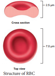
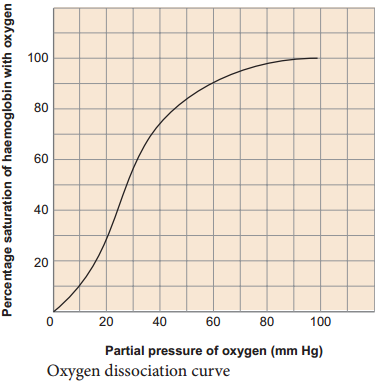
The red colour of the RBC is due to the presence of a respiratory pigment, haemoglobin dissolved in the cytoplasm. Haemoglobin plays an important role in the transport of respiratory gases and facilitates the exchange of gases with the fluid outside the cell (tissue fluid). The biconcave shaped RBCs increases the surface area to volume ratio, hence oxygen diffuses quickly in and out of the cell. The RBCs are devoid of nucleus, mitochondria, ribosomes and endoplasmic reticulum.

The absence of these organelles accommodates more haemoglobin thereby maximising the oxygen carrying capacity of the cell. The average life span of RBCs in a healthy individual is about 120 days after which they are destroyed in the spleen (graveyard/cemetery of RBCs) and the iron component returns to the bone marrow for reuse.

Erythropoietin is a hormone secreted by the kidneys in response to low oxygen and helps in diffrentiation of stem cells of the bone marrow to erythrocytes (erythropoiesis) in adults. The ratio of red blood cells to blood plasma is expressed as Haematocrit (packed cell volume).

White blood cells (leucocytes) are colourless, amoeboid, nucleated cells devoid of haemoglobin and other pigments. Approximately 6000 to 8000 per cubic mm of WBCs are seen in the blood of an average healthy individual.

The different types of WBCs are shown in Figure 7.2. Depending on the presence or absence of granules, WBCs are divided into two types, granulocytes and agranulocytes. Granulocytes are characterised by the presence of granules in the cytoplasm and are differentiated in the bone marrow. The granulocytes include neutrophils, eosinophils and basophils.
Body Fluids img 2

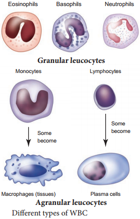
Neutrophils are also called heterophils or polymorphonuclear (cells with 3-4 lobes of nucleus connected with delicate threads) cells which constitute about 60% – 65% of the total WBCs. They are phagocytic in nature and appear in large numbers in and around the infected tissues.

Eosinophils have distinctly bilobed nucleus and the lobes are joined by thin strands. They are non-phagocytic and constitute about 2-3% of the total WBCs. Eosinophils increase during certain types of parasitic infections and allergic reactions.

Basophils are less numerous than any other type of WBCs constituting 0.5%-1.0% of the total number of leucocytes. The cytoplasmic granules are large sized, but fewer than eosinophils. Nucleus is large sized and constricted into several lobes but not joined by delicate threads. Basophils secrete substances such as heparin, serotonin and histamines. They are also involved in inflammatory reactions.

Agranulocytes are characterised by the absence of granules in the cytoplasm and are diffrentiated in the lymph glands and spleen. These are of two types, lymphocytes and monocytes. Lymphocytes constitute 28% of WBCs.

These have large round nucleus and small amount of cytoplasm. The two types of lymphocytes are B and T cells. Both B and T cells are responsible for the immune responses of the body. B cells produce antibodies to neutralize the harmful effects of foreign substances and T cells are involved in cell mediated immunity.

Monocytes (Macrophages) are phagocytic cells that are similar to mast cells and have kidney shaped nucleus. They constitute 1-3% of the total WBCs. The macrophages of the central nervous system are the ‘microglia’, in the sinusoids of the liver they are called ‘Kupffer cells’ and in the pulmonary region they are the ‘alveolar macrophages’.

Platelets are also called thrombocytes that are produced from megakaryocytes (special cells in bone marrow) and lack nuclei. Blood normally contains 1, 50,000 – 3, 50,000 platelets mm-3 of blood. They secrete substances involved in coagulation or clotting of blood. The reduction in platelet number can lead to clotting disorders that result in excessive loss of blood from the body.

Blood Groups

Commonly two types of blood groupings are done. They are ABO and Rh which are widely used all over the world.

ABO Blood Grouping

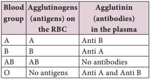
Depending on the presence or absence of surface antigens on the RBCs, blood group in individual belongs to four different types namely, A, B, AB and O. The plasma of A, B and O individuals have natural antibodies (agglutinins) in them. Surface antigens are called agglutinogens. The antibodies (agglutinin) acting on agglutinogen A is called anti A and the agglutinin acting on agglutinogen B is called anti B. Agglutinogens
are absent in O blood group.

Agglutinogens A and B are present in AB blood group and do not contain anti A and anti B in them. Distribution of antigens and antibodies in blood groups are shown in Table 7.1. A, B and O are major allelic genes in ABO systems.

All agglutinogens contain sucrose, D-galactose, N-acetyl glucosamine and 11 terminal amino acids. The attachments of the terminal amino acids are dependent on the gene products of A and B. The reaction is
catalysed by glycosyl transferase.

Table 7.1 Distribution of antigens and antibodies in different blood groups
Body Fluids img 3

Rh factor is a protein (D antigen) present on the surface of the red blood cells in majority (80%) of humans. This protein is similar to the protein present in Rhesus monkey, hence the term Rh. Individuals who carry the antigen D on the surface of the red blood cells are Rh+ (Rh positive) and the individuals who do not carry antigen D, are Rh (Rh negative). Rh factor compatibility is also checked before blood transfusion.

When a pregnant women is Rh and the foetus is Rh+ incompatibility (mismatch) is observed. During
the first pregnancy, the Rh antigens of the foetus does not get exposed to the mother’s blood as both their blood are separated by placenta.

However, small amount of the foetal antigen becomes exposed to the mother’s blood during the birth of the first child. The mother’s blood starts to synthesize D antibodies. But during subsequent pregnancies the Rh antibodies from the mother (Rh) enters the foetal circulation and destroys the foetal RBCs.

This becomes fatal to the foetus because the child suffers from anaemia and jaundice. This condition is called erythroblastosis foetalis. This condition can be avoided by administration of anti D antibodies (Rhocum) to the mother immediately after the first child birth.

Coagulation Of Blood

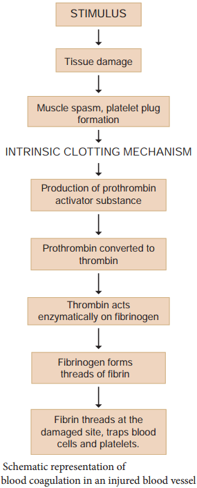
If you cut your finger or when you get yourself hurt, your wound bleeds for some time after which it stops to bleed. This is because the blood clots or coagulates in response to trauma. The mechanism by which excessive blood loss is prevented by the formation of clot is called blood coagulation or clotting of blood. Schematic representation of blood coagulation is shown Figure 7.3.

The clotting process begins when the endothelium of the blood vessel is damaged and the connective tissue in its wall is exposed to the blood. Platelets adhere to collagen fibres in the connective tissue and release substances that form the platelet plug which provides emergency protection against blood loss. Clotting factors released from the clumped platelets or damaged cells mix with clotting factors in the plasma. The protein called prothrombin is converted to its active form called thrombin in the presence of calcium and vitamin K.

Thrombin helps in the conversion of fibrinogen to fibrin threads. The threads of fibrins become interlinked into a patch that traps blood cell and seals the injured vessel until the wound is healed. After sometime fibrin fibrils contract, squeezing out a strawcoloured fluid through a meshwork called serum (Plasma without fibrinogen is called serum). Heparin is an anticoagulant produced in small quantities by mast cells of connective tissue which prevents coagulation in small blood vessels.
Body Fluids img 4

Composition Of Lymph and Its Functions

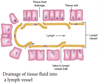
About 90% of fluid that leaks from capillaries eventually seeps back into the capillaries and the remaining 10% is collected and returned to blood system by means of a series of tubules known as lymph vessels or lymphatics.

The fluid inside the lymphatics is called lymph. The lymphatic system consists of a complex network of thin walled ducts (lymphatic vessels), filtering bodies (lymph nodes) and a large number of lymphocytic cell concentrations in various lymphoid organs.

The lymphatic vessels have smooth walls that run parallel to the blood vessels, in the skin, along the respiratory and digestive tracts. These vessels serve as return ducts for the fluids that are continually diffusing out of the blood capillaries into the body tissues. The end of a vessel is shown in Figure 7.4.
Body Fluids img 5

Lymph fluid must pass through the lymph nodes before it is returned to the blood. The lymph nodes that filter the fluid from the lymphatic vessels of the skin are highly concentrated in the neck, inguinal, axillaries, respiratory and digestive tracts.

The lymph fluid flowing out of the lymph nodes flow into large collecting duct which finally drains into larger veins that runs beneath the collar bone, the subclavian vein and is emptied into the blood stream. The narrow passages in the lymph nodes are the sinusoids that are lined with macrophages.

The lymph nodes successfully prevent the invading microorganisms from reaching the blood stream. Cells found in the lymphatics are the lymphocytes. Lymphocytes collected in the lymphatic fluid are carried via the arterial blood and are recycled back to the lymph. Fats are absorbed through lymph in the lacteals present in the villi of the intestinal wall.

Health Effects of Smoking

Learninsta presents the core concepts of Biology with high-quality research papers and topical review articles.

Health Effects of Smoking

Today due to curiosity, excitement or adventure youngsters start to smoke and later get addicted to smoking. Research says about 80% of the lung cancer is due to cigarette smoking. Smoking is inhaling the smoke from burning tobacco. There are thousands of known chemicals which includes nicotine, tar, carbon monoxide, ammonia, sulphur – dioxide and even small quantities of arsenic.

Carbon monoxide and nicotine damage the cardiovascular system and tar damages the gaseous exchange system. Nicotine is the chemical that causes addiction and is a stimulant which makes the heart beat faster and the narrowing of blood vessels results in raised blood pressure and coronary heart diseases.

Presence of carbon monoxide reduces oxygen supply. Lung cancer, cancer of the mouth and larynx is more common in smokers than non-smokers. Smoking also causes cancer of the stomach, pancreas and bladder and lowers sperm count in men. Smoking can cause lung diseases by damaging the airways and alveoli and results in emphysema and chronic bronchitis.

These two diseases along with asthma are often referred as Chronic Obstructive Pulmonary Disease (COPD). When a person smokes, nearly 85% of the smoke released is inhaled by the smoker himself and others in the vicinity, called passive smokers, are also affected. Guidance or counselling should be done in such users to withdraw this habit.

Smoking causes cancer, heart disease, stroke, lung diseases, diabetes, and chronic obstructive pulmonary disease (COPD), which includes emphysema and chronic bronchitis. Smoking also increases risk for tuberculosis, certain eye diseases, and problems of the immune system, including rheumatoid arthritis.

Smoking can cause lung disease by damaging your airways and the small air sacs (alveoli) found in your lungs. Lung diseases caused by smoking include COPD, which includes emphysema and chronic bronchitis. Cigarette smoking causes most cases of lung cancer.

Lung Cancer. More people die from lung cancer than any other type of cancer. COPD (chronic obstructive pulmonary disease) COPD is an obstructive lung disease that makes it hard to breathe.

  • Heart Disease
  • Stroke
  • Asthma
  • Reproductive Effects in Women
  • Premature, Low Birth-Weight Babies
  • Diabetes

Immediate Effects of Smoking

  • Initial stimulation, then reduction in activity of brain and nervous system
  • Increased alertness and concentration
  • Feelings of mild euphoria
  • Feelings of relaxation
  • Increased blood pressure and heart rate
  • Decreased blood flow to fingers and toes
  • Decreased skin temperature
  • Bad breath

Nicotine that is inhaled in cigarette smoke is absorbed by the lungs into the bloodstream and quickly goes to the heart and brain. Many smokers report that they enjoy the ritual of smoking. They also say that smoking gives them a pleasurable feeling. Smoking relieves their nicotine withdrawal symptoms.

The bottom line. So, smoking probably doesn’t make you poop, at least not directly. There’s a whole host of other factors that might be responsible for this sensation of urgency to visit the toilet after smoking. But smoking does have a major impact on your gut health.

One of the ingredients in tobacco is a mood-altering drug called nicotine. Nicotine reaches your brain in mere seconds and makes you feel more energized for a while. But as that effect wears off, you feel tired and crave more. Nicotine is extremely habit-forming, which is why people find smoking so difficult to quit.

Disorders of the Respiratory System

Learninsta presents the core concepts of Biology with high-quality research papers and topical review articles.

Disorders of the Respiratory System

Respiratory system is highly affected by environmental, occupational, personal and social factors. These factors may be responsible for a number of respiratory disorders. Some of the disorders are discussed here.

Asthma

It is characterized by narrowing and inflammation of bronchi and bronchioles and difficulty in breathing. Common allergens for asthma are dust, drugs, pollen grains, certain food items like fish, prawn and certain fruits etc.

Emphysema

Emphysema is chronic breathlessness caused by gradual breakdown of the thin walls of the alveoli decreasing the total surface area of a gaseous exchange. i.e., widening of the alveoli is called emphysema. The major cause for this disease is cigarette smoking, which reduces the respiratory surface of the alveolar walls.

Bronchitis

The bronchi when it gets inflated due to pollution smoke and cigarette smoking, causes bronchitis. The symptoms are cough, shortness of breath and sputum in the lungs.

Pneumonia

Inflammation of the lungs due to infection caused by bacteria or virus is called pneumonia. The common symptoms are sputum production, nasal congestion, shortness of breath, sore throat etc.

Tuberculosis

Tuberculosis is caused by Mycobacterium tuberculae. This infection mainly occurs in the lungs and bones. Collection of fluid between the lungs and the chest wall is the main complication of this disease.

Occupational Respiratory Disorders

The disorders due to one’s occupation of working in industries like grinding or stone breaking, construction sites, cotton industries, etc. Dust produced affects the respiratory tracts. Long exposure can give rise to inflammation leading to fibrosis.

Silicosis and asbestosis are occupational respiratory diseases resulting from inhalation of particle of silica from sand grinding and asbestos into the respiratory tract. Workers, working in such industries must wear protective masks.

Problems in Oxygen Transport

Learninsta presents the core concepts of Biology with high-quality research papers and topical review articles.

Problems in Oxygen Transport

When a person travels quickly from sea level to elevations above 8000ft, where the atmospheric pressure and partial pressure of oxygen are lowered, the individual responds with symptoms of acute mountain sickness (AMS) – headache, shortness of breath, nausea and dizziness due to poor binding of O2 with haemoglobin.

When the person moves on a long-term basis to mountains from sea level is the body begins to make respiratory and haematopoietic adjustments. To overcome this situation kidneys accelerate production of the hormone erythropoietin, which stimulates the bone marrow to produce more RBCs.

When a person descends deep into the sea, the pressure in the surrounding water increases which causes the lungs to decrease in volume. This decrease in volume increases the partial pressure of the gases within the lungs. This effect can be beneficial, because it tends to drive additional oxygen into the circulation, but this benefit also has a risk, the increased pressure can also drive nitrogen gas into the circulation.

This increase in blood nitrogen content can lead to a condition called nitrogen narcosis. When the diver ascends to the surface too quickly a condition called ‘bends’ or decompression sickness occurs and nitrogen comes out of solution while still in the blood forming bubbles.

Small bubbles in the blood are not harmful, but large bubbles can lodge in small capillaries, blocking blood flow or can press on nerve endings. Decompression sickness is associated with pain in joints and muscles and neurological problems including stroke. The risk of nitrogen narcosis and bends is common in scuba divers.

During carbon-dioxide poisoning, the demand for oxygen increases. As the O2 level in the blood decreases it leads to suffocation and the skin turns bluish black.

In terms of O2 transport, decreased arterial blood oxygenation (hypoxemia) is the primary limitation, and thus, the problem resides with the respiratory system. First, additional capillaries open to reduce diffusion distances and increase the surface area for oxygen exchange; oxygen extraction subsequently increases.

Carbon dioxide levels, blood pH, body temperature, environmental factors, and diseases can all affect oxygen’s carrying capacity and delivery. A decrease in the oxygen-carrying ability of hemoglobin is observed with an increase in carbon dioxide and temperature, as well as a decrease in pH within the body.

Oxygen is transported in the blood in two ways: A small amount of O2 (1.5 percent) is carried in the plasma as a dissolved gas. Most oxygen (98.5 percent) carried in the blood is bound to the protein hemoglobin in red blood cells. A fully saturated oxyhemoglobin (HbO2) has four O2 molecules attached.

The blood hemoglobin concentration is determinant of oxygen delivery. In anemic patients, oxygen delivery decreases and oxygen extraction is increased. This leads to decreased venous hemoglobin saturation and a lower tissue oxygen saturation.

Since carbon dioxide reacts with water to form carbonic acid, an increase in CO2 results in a decrease in blood pH, resulting in hemoglobin proteins releasing their load of oxygen. Conversely, a decrease in carbon dioxide provokes an increase in pH, which results in hemoglobin picking up more oxygen.

Oxygen delivery (DO2) represents the amount of oxygen transported to tissues and is defined as the product of cardiac output (CO) and oxygen content. Normal value is 520 to 570 mL/min/m2. Oxygen delivery can be improved by increasing cardiac output, oxygen saturation, or hemoglobin.

Carbon dioxide is transported in the blood from the tissue to the lungs in three ways:

  • Dissolved in solution
  • Buffered with water as carbonic acid
  • Bound to proteins, particularly haemoglobin.

Approximately 75% of carbon dioxide is transport in the red blood cell and 25% in the plasma. Tissue oxygenation may be impaired either by a low arterial oxygen saturation, as in cyanotic congenital heart disease, or by a reduced blood flow, as occurs with myocardial failure or with obstruction to the left heart (aortic atresia). Anaerobic metabolism results in the accumulation of lactic acid in the tissues.

Transport of Various Types of Respiratory Gases

Learninsta presents the core concepts of Biology with high-quality research papers and topical review articles.

Transport of Various Types of Respiratory Gases

Transport of Oxygen

Molecular oxygen is carried in blood in two ways bound to haemoglobin within the red blood cells and dissolved in plasma. Oxygen is poorly soluble in water, so only 3% of the oxygen is transported in the dissolved form. 97% of oxygen binds with haemoglobin in a reversible manner to form oxyhaemoglobin (HbO2).

The rate at which haemoglobin binds with O2 is regulated by the partial pressure of O2. Each haemoglobin
carries maximum of four molecules of oxygen. In the alveoli high pO2, low pCO2, low temperature and less H+ concentration, favours the formation of oxyhaemoglobin, whereas in the tissues low pO2, high pCO2, high H+ and high temperature favours the dissociation of oxygen from oxyhaemoglobin.
Transport of Various Types of Respiratory Gases img 1

A sigmoid curve (S-shaped) is obtained when percentage saturation of haemoglobin with oxygen is plotted against pO2. This curve is called oxygenhaemoglobin dissociation curve (Figure 6.7). This S-shaped curve has a steep slope for pO2 values between 10 and 50mmHg and then flattens between 70 and 100 mm Hg. Under normal physiological conditions, every 100mL of oxygenated blood can deliver about 5mL of O2 to the tissues.

Transport of Carbon – Dioxide

Blood transports CO2 from the tissue cells to the lungs in three ways

(i) Dissolved in Plasma

About 7 – 10% of CO2 is transported in a dissolved form in the plasma.

(ii) Bound to Haemoglobin

About 20 – 25% of dissolved CO2 is bound and carried in the RBCs as carbaminohaemoglobin (Hb CO2)
CO2 ⇄ Hb Hb CO2

(iii) As Bicarbonate Ions in Plasma

About 70% of CO2 is transported as bicarbonate ions. This is influenced by pCO2 and the degree of haemoglobin oxygenation. RBCs contain a high concentration of the enzyme, carbonic anhydrase, whereas small amounts of carbonic anhydrase is present in the plasma.

At the tissues the pCO2 is high due to catabolism and diffuses into the blood to form HCO3and H+ ions. When CO2 diffuses into the RBCs, it combines with water forming carbonic acid (H2CO3) catalyzed by carbonic anhydrase. Carbonic acid is unstable and dissociates into hydrogen and bicarbonate ions. Carbonic anhydrase facilitates the reaction in both directions.
Transport of Various Types of Respiratory Gases img 2

The HCO3 moves quickly from the RBCs into the plasma, where it is carried to the lungs. At the alveolar site where pCO2 is low, the reaction is reversed leading to the formation of CO2 and water. Thus CO2 trapped as HCO3 at the tissue level it is transported to the alveoli and released out as CO2. Every 100mL of deoxygenated blood delivers 4mL of CO2 to the alveoli for elimination.