Learninsta presents the core concepts of Microbiology with high-quality research papers and topical review articles.
Tissue Flagellates – Leishmania Donovani
The genus is named after the scientist Leishman, who first described the parasite in London in May 1903.
Geographical Distribution:
Leishmania species is found in the Mediterranean, the Middle East, Africa and Asia including India.
Habitat:
Leishmania donovani is an obligate intracellular parasite of human and other mammalian hosts. They are always found as intracellular amastigotes in the reticuloendothelial cells of the spleen, bone marrow, liver, intestinal mucosa and mesenteric lymph nodes of hosts.
Morphology:
The parasite exists in two forms:
Amastigote:
It is the form found in human and other mammalian hosts. They are found inside monocytes, polymorphonuclear leucocytes or endothelial cells. They are small, round to oval bodies measuring 2-3µm in length (Figure 8.8). They are also known as LD (Leishman donovan) bodies.
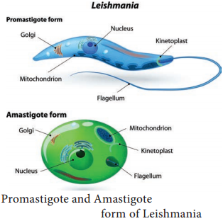
Promastigote:
These forms are found in the mid-gut of sand fly and in the culture media. The fully developed promastigotes are long, slender and spindle – shaped. They measure 15µm to 25µm in length and 1.5µm to 3.5µm in breadth. A single nucleus is situated at the centre. The kinetoplast lies near the anterior end. The flagellum is single, delicate and measures 15µm-28µm (Figure 8.8).
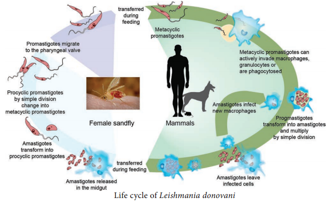
Life – Cycle of Leishmania donovani
Leishmania donovani completes its life cycle in two different hosts. The complete life cycle is given in Figure 8.9.
Host | Forms |
| Human and other mammals (Example: Dogs) | Amastigote |
| Sandfly of Genus Phlebotomus | Promastigote |
Development in Human
The parasite is transmitted to human and other vertebrate hosts by the bite of blood sucking female sandfly. During the blood meal, the sandfly deposists promastigotes on surface of the skin. These promastigotes are immediately phagocytosed by fixed macrophages of the host, in which they are transformed into amastigotes. The amastigotes multiply by binary fission within the macrophages.
As many as 50 to 200 amastigotes may be present inside the enlarged cell. These are called LD bodies. The rupture of cell releases amastigotes in large numbers which inturn are free to infect other cells. Free amastigotes are subsequently carried by circulation. These forms invade monocytes of the blood and macrophages of the spleen, liver, bone marrow, lymph nodes and other tissues of the reticuloendothelial cells.
Development in sandfly
Female sandfly during a blood meal ingest free, as well as intracellular amastigotes in the blood. In the mid gut of the sandfly, the amastigotes are transformed within 72 hours to flagellated promastigotes. These promastigotes multiply by binary fission. After a period of 6 to 9 days, these forms migrate from the midgut to the pharynx and buccal cavity of sandfly. Bite of the infected sandfly transmits infection to susceptible persons and the life – cycle is repeated.
Pathogenesis
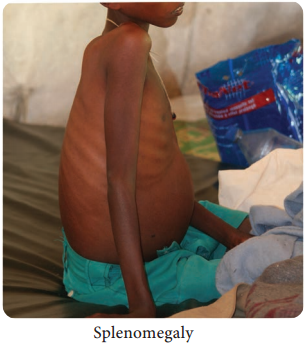
Leishmania donovani causes visceral Leishmaniasis. The disease is also known as Dum – Dum fever, Asian fever, Assam fever, or infantile splenomegaly. Leishmaniasis is a disease of the reticuloendothelial system. Proliferation and destruction of reticuloendothelial cells of the internal organs are responsible for the pathological changes in visceral leishmaniasis.
Spleen, liver and lymphnodes are enlarged in this condition. Bone marrow is dark red in colour and shows extensive proliferation of reticuloendothelial cells. Kidney shows cloudy swelling and is invaded by macrophages parasitized by amastigotes.
Clinical Features
Incubation period:
It is usually 3-6 months but can be months or years.
Visceral Leishmaniasis is a serious and fatal systemic disease. In India, the disease is called Kala – azar meaning “black disease”. The disease is characterized by the presence of fever, hepatosplenomegaly (Figure 8.10) (the simultaneous enlargement of both liver and the spleen), hypergammaglobulinemia (a condition in which increased levels of a certain immunoglobulin in blood serum), Leucopenia, Thrombocytopenia (deficiency of platelets in the blood), Cachexia (a condition that causes extreme weight loss) with marked anemia, emaciation and loss of weight.
Epistaxis (bleeding from nose) and bleeding from gums are common. In Indian patients, the skin on the hands, feet, abdomen, around the mouth and fore – head becomes grayish and dark coloured. This hypo – pigmentation of the skin is unique in Indian patients giving the disease name Kala – azar.
Post kala – azar dermal leishmaniasis
(PKDL):
It is a non – ulcerative lesion of the skin, which is seen after completion of treatment of the kala – azar. This condition is characterized by multiple, hypopigmented, erythematous macules involving the face and trunk (Figure 8.11).
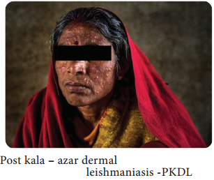
In Indian forms, PKDL appears after a latent period of 2 years and may even persist as long as 20years, creating a persistent human reservoir of infection.
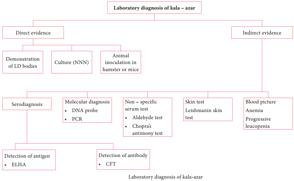
Laboratory diagnosis
Specimens:
Aspiration from spleen, bone marrow, lymph node, liver biopsy and peripheral blood.
Methods of examination:
This includes, microscopy and culture
1. Direct microscopy
The amastigotes of Leishmania donovani (known as LD bodies) can be demonstrated in the smears of spleen, bone marrow, liver, lymph node and peripheral blood stained in Leishman, Giemsa or wright stains. Splenic aspiration is the most sensitive method to detect LD bodies. Examination of peripheral blood smear and buffy coat smear is more commonly used to find LD bodies in the circulating monocytes.
2. Culture
Promastigotes are found in the culture media. Tissue samples and aspirates are inoculated in the NNN (Novy-MacNeal-Nicolle) medium for demonstration of promastigotes. Laboratory diagnosis of kala – azar is briefly discussed in Flowchart 8.5.
Treatment:
Pentavalent antimonials are the drugs of choice. Pentamidine, Amphotericin B and Miltefosine (oral drug) are recommended.
Prevention and Control
Integrated insecticidal spraying (DDT and Malathion) to reduce sandfly population. Reduction of reservoir by killing all the infected dogs. Personal prophylaxis by using anti – sandfly measures like using thick clothes, bed nets, window mesh or insect repellants and keeping the environment clean. No vaccine is available against kala – azar.