Learninsta presents the core concepts of Microbiology with high-quality research papers and topical review articles.
Corynebacterium diphtheriae
Several species of the genus Corynebacterium are normal flora of skin, upper respiratory tract (URT), urogenital and intestinal tract. The most important member of the genus is C. diphtheriae the causative agent of diphtheria, a localized inflammation of the throat with greyish white pseudomembrane and a generalized toxemia due to the secretion and dissemination of a highly potent toxin.
The name Corynebacterium diphtheria is derived from Greek word ‘Coryne’ – “Club shaped swellings” or “Knotted rod” ‘Diphthera’ – Leather.
Morphology
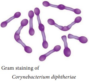
They are Gram positive slender rods, pleomorphic club shape or coryneform bacterium Non – motile, non – sporing and non – capsulated (Figure 7.9 a & b).
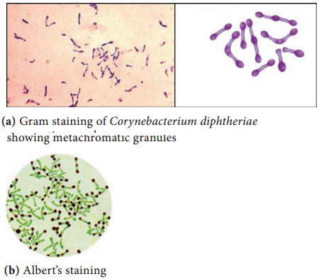
The bacilli are arranged in a characteristic fashion in angular fashion resembling the letters V or L. This has been called Chinese letter or cuneiform arrangement (Figure 7.10).
They are club shaped due to the presence of metachromatic granules at one or both ends. These granules are composed of polymetaphosphates and represent energy storage depots.
Cultural Characteristics
- They are aerobic and facultative anaerobe. Optimum temperature is 37°C and pH 7.2.
- They grow on the following media and show the characteristic colony morphology (Table 7.5).
Table 7.5: Colony Morphology of Corynebacterium diphtheriae on cultural media
Media | Colony Morphology |
| Loeffler’s Serum slope | They glow on this medium very rapidly. Colonies appear after 6-8 hours of incubation. The colonies are small, circular white or creamy and glistening. |
| Tellurite Blood Agar | Grey or black colonies. Based on colony morphology on tellurite medium, three main biotypes – Gravis, Intermedius and Mitis. |
Toxin
• The pathogenicity is due to production of a very powerful exotoxin by virulent strains of diphtheria bacilli.
• The toxigenicity of diphtheria bacillus depends on the presence of a tox<sup>+</sup> gene which can be transferred from one bacterium to another by lysogenic bacteriophages, of which beta phage is the most important.
Properties
The diphtheria toxin is a heat – labile protein and has a molecular weight of about 62,000 Dalton. It consists of two fragments
- Fragment A (24,000 Dalton) – It has all enzymatic activity.
- Fragment B (38,000 Dalton) – It is responsible for binding the toxin to the target cells.
Mode of Action
The toxin acts by inhibiting protein synthesis, specifically fragment A inhibits polypeptide chain elongation in the presence of NAD by inactivating the elongation factor (EF – 2) the toxin has special affinity for myocardium, adrenal gland and nerve endings.
Pathogenicity
Source of infection – Airborne droplets
Route of entry – Upper respiratory tract
Incubation period – 3 – 4 days
Site of infection – Faucial (nasal, otitis, conjunctival, laryngeal, genital) diphtheria is most commonly seen in children of 2-10 years.
Faucial diphtheria is the most common type. The infection is confined to humans only. The toxin has both local (flowchart 7.3) as well as systemic effects.
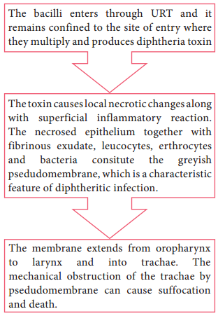
Flowchart 7.3: Localized effect of diphtheria toxin
Systemic effects
The toxin diffuses into the blood stream and causes toxemia. It has got affinity for cardiac muscle, adrenal and nerve endings. It acts on the cells of these tissues.
Clinical Manifestations
- Laryngeal obstruction, asphyxia (it is a condition of severe deficient supply of oxygen, causing suffocation).
- Diphtheritic myocarditis (inflammation of heart muscle), polyneuropathy (damage of multiple peripheral nerves), paralysis of palatine (the top part of the inside of the mouth) and ciliary muscles.
- Degenerative changes in adrenal glands, kidney and liver may occur.
Specimen:
Two swabs from the lesions are collected. One swab is used for smear preparationand other swab for inoculation on culturemedia.
Direct microscopy:
Smears are stained with both Gram stain and Albert stain.
- Gram Staining – Gram positive slender rods were observed.
- Albert staining – Club shaped with metachromatic granules were observed.
Culture:
The swabis inoculated on Loeffler’s serum slope, after overnight incubation at 37°C, the plates were observed for characteristic colonies, which are identified by gram staining.
Prophylaxis
Diphtheria can be controlled by immunization. Three methods of immunization are available (Table 7.6).
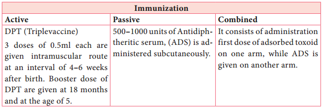
Table 7.6: Immunization for diphtheria
Treatment
The specific treatment for diphtheria consists of administration of antitoxin with dose of 20,000-100,000 units of ADS intramuscularly and antibiotic therapy using penicillin.