Lipid Catabolism

Learninsta presents the core concepts of Microbiology with high-quality research papers and topical review articles.

Lipid Catabolism

Microorganisms frequently use lipids such as triglyceride or triacylglycerol (esters of glycerol and fatty acids) as common reserve energy sources. These can be hydrolyzed to glycerol and fatty acid by microbial lipases. The glycerol is then phosphorylated and oxidized to Dihydroxyacetone phosphate and then catabolized in the Glycolysis pathway.

Fatty acids from triacylglycerols and other lipids are often oxidized in the β-oxidation pathway. In this pathway fatty acids are degraded to acetyl CoA (2C segment), then it enters into the TCA cycle.

Lipid catabolism comprises two major spatially and temporarily separated steps, namely lipolysis, which releases fatty acids and head groups and is catalyzed by lipases at membranes or lipid droplets, and degradation of fatty acids to acetyl-CoA, which occurs in peroxisomes through the β-oxidation pathway in green.

The released fatty acids are catabolized in a process called β-oxidation, which sequentially removes two-carbon acetyl groups from the ends of fatty acid chains, reducing NAD+ and FAD to produce NADH and FADH2, respectively, whose electrons can be used to make ATP by oxidative phosphorylation.

Lipid metabolism begins in the intestine where ingested triglycerides are broken down into smaller chain fatty acids and subsequently into monoglyceride molecules by pancreatic lipases, enzymes that break down fats after they are emulsified by bile salts.

Lipid metabolism is the process that most of the fat ingested by the body is emulsified into small particles by bile and then the lipase secreted by the pancreas and small intestine hydrolyzes the fatty acids in the fat into free fatty acids and monoglycerides.

Electron Transport Chain

Learninsta presents the core concepts of Microbiology with high-quality research papers and topical review articles.

Electron Transport Chain

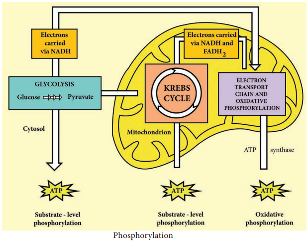
An electron transport chain consists of a sequence of carrier molecules that are capable of oxidation and reduction. In and FADH2 to acceptor such as molecular Oxygen. In the process, protons are pumped from the mitochondrial matrix to the inner membrane space, and eventually combine with O2 and H+ to form water (Figure 4.6).

As the electrons flow through the chain, much of their free energy is conserved in the form of ATP. The process by which energy from electron transport is used to make ATP is called as oxidative phosphorylation.

Respiratory chain is an electron transport chain where a pair of electrons or hydrogen atoms containing electron from the substrate oxidized is coupled to reduction of oxygen to water.

The mitochondrial system is arranged Eukaryotic cell, the ETC is contained in the inner membrane of mitochondria or chloroplast membrane, whereas in prokaryotic cells, it is found in plasma membrane or cytoplasmic membrane.

The ETC is carried out through a series of electron transporters embedded in the inner mitochondrial membrane that transfer electrons from electron donors NADH into three complexes of electron carriers.
They are:

1. Flavoproteins:
These proteins contain flavin, a coenzyme derived from riboflavin (Vit B12). One important flavoprotein is flavin mono nucleotide.

2. Ubiquinones (coenzyme Q):
These are small non protein carriers.

3. Cytochromes:
These are proteins with iron containing group, capable of existing alternately as reduced (Fe2+) and oxidized form (Fe3+). Cytochromes involved in ETC include cyt (b),cyt c1, cyt c, cyt a, cyt a3.

The first step in electron transport chain is the transfer of high energy electrons from NADH to FMN. This transfer actually involves the passage of hydrogen atom with 2e to FMN, which then picks up an additional H+ from the surrounding aqueous medium.
Electron Transport Chain img 1

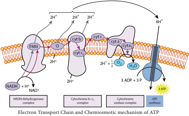
As a result of the first transfer, NADH is oxidized to NAD+, and FMN is reduced to FMNH2.

In the second step, FMNH2 passes 2 H+ to the other side of the mitochondrial membrane and passes 2 e to coenzyme Q. As a result, FMNH2 is oxidized to FMN. Coenzyme Q also picks up additional 2H+ from the surrounding aqueous and releases to other side of the membrane.

In the next step, electrons are passed successively from coenzyme Q to cyt b1, cyt c1, cyt c, cyt a, cyt a3.
Each cytochrome in the chain is reduced, as it picks up electrons and is oxidized as it gives up electrons. The last cytochrome cyt a3 passes its electrons to molecular O2 which picks up protons from the surrounding medium to form H2O.

FADH2 derived from the Krebs cycle is another source of electrons. Thus at the end of ETC, NADH pumps three protons (synthesizes 3ATPs) whereas FADH2 pumps only two protons (synthesizes 2ATPs).

Chemiosmotic Mechanism of ATP

Chemiosmotic mechanism of ATP synthesis was first proposed by the Biochemist, Peter Mitchell in 1961. In ETC, when energetic electrons from NADH pass down the carriers, some of the carriers (proton pumps) in the chain pump [actively transport] protons across the membrane to inner membrane space.

Thus in addition to a concentration gradient, an electrical charge gradient is created. The resulting electro chemical gradient has potential energy called proton motive force.

The proton diffuses across the membrane through protein channels that contain an enzyme called ATP synthase. When this flow occurs, energy is released and is used by the enzyme to synthesize ATP from ADP and phosphate.

At the end of the chain, electrons join with protons and O2 in the matrix fluid to form H2O. Thus O2 is the final electron acceptor. ETC also operates in photophosphorylation and is located in thylakoid membrane of Cyanobacteria (BGA), and of eukaryotic chloroplasts. Overview of Aerobic respiration (Figure 4.7):
Electron Transport Chain img 2

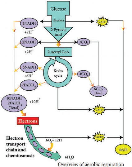
1. Electron transport chain regenerates NAD and FAD which can be used again in Glycolysis and Krebs cycle.

2. Various electrons transfer in the electron transport chain generates about 34 ATP, (10 NADH = 10 × 3 = 30 + 2 FADH2 = 2 × 2 = 4).

3. A total of 38 ATP molecules is generated from one molecule of glucose oxidized in prokaryotes, whereas in eukaryotes, 36 molecules of ATP is generated because in eukaryotes, some energy is lost when electrons are shuttled across the mitochondrial membranes that separate Glycolysis (in the cytoplasm) from the electron transport chain (Table 4.2). There is no such separation exists in prokaryotes.
C6H12O6 + 6CO2 + 38ADP + 38Pi → 6CO2 + 6H2O + 38 ATP

Glycolysis

1. Oxidation of glucose to Pyruvic acid.
2. Production of 2 NADH

Preparatory step

2 ATP (substrate level phosphorylation)
6 ATP (Oxidative phosphorylation in ETC)
Preparatory step

1. Formation of acetyl CoA produces 2NADH

6 ATP (Oxidative phosphorylation in ETC)
Krebs cycle

1. Oxidation of succinyl CoA to succinic acid
2. Production of 6 NADH
3. Production of 2 FADH

2 ATP (Substrate level phosphorylation)
18 ATP (Oxidative phosphorylation in ETC)
4 ATP (Oxidative phosphorylation in ETC)
Total 38 ATP  

1 NADH = 3 ATPs and 1 FADH2 = 2 ATP

Tricarboxylic Acid Cycle

Learninsta presents the core concepts of Microbiology with high-quality research papers and topical review articles.

Tricarboxylic Acid Cycle

TCA cycle was first elucidated by Sir Hans Adolf Krebs, a German Biochemist in 1937. It is also known as Tricarboxylic acid cycle, Citric acid cycle or Amphibolic cycle. In prokaryotic cells, the citric acid cycle occurs in the cytoplasm; in eukaryotic cells it takes place in the matrix of the mitochondria.

The process oxidizes glucose derivatives, fatty acids, and amino acids to carbon dioxide (CO2) through a series of enzyme controlled steps. The purpose of the Krebs cycle is to collect high energy electrons from these fuels by oxidizing them, which are transported by activated electron carriers such as NADH and FADH2 to electron transport chain.

The Krebs cycle is also the source for the precursor of many other molecules and is therefore an amphibolic pathway (both anabolic and catabolic reactions take place in this cycle) and therefore, it can be used for both the synthesis and degradation of bio molecules.
Tricarboxylic Acid Cycle img 1

Pyruvate cannot enter the Krebs cycle directly. In a preparatory step, it must lose one molecule of CO2 and becomes a two-carbon compound. This process is called decarboxylation. The two-carbon compound, called acetyl group, attaches to coenzyme A through a high-energy bond, the resulting is a complex known as acetyl coenzyme (acetyl CoA).

During this reaction, pyruvic acid is also oxidized and NAD+ is reduced to NADH by pyruvate dehydrogenase complex (PDHC). This multi enzyme complex is responsible for the conversion of pyruvate to acetyl-coA. Therefore PDHC contribute to linking the Glycolysis pathway to the citric acid pathway.

The Krebs cycle generates a pool of chemical energy (ATP, NADH, and FADH2) from the oxidation of Pyruvic acid and it loses one carbon atom as CO2 and reduces NAD+ to NADH. The resulting two carbon acetyl molecule is joined to Co enzyme A. Acetyl CoA transfers its acetyl group to a 4C compound (oxaloactate) to make a 6C compound (Citrate) and the Coenzyme A is released which goes back to the link reaction to form another molecule of acetyl CoA. Oxaloacetate is both the first reactant and the product of the metabolic pathway (creating a loop).

After citrate has been formed, the cycle machinery continues through seven distinct enzyme catalyzed reactions that produce in order isocitrate, α – ketoglutarate, succinyl CoA, succinate, fumarate, malate and oxaloacetate.

At the end of Krebs cycle, each pyruvic acid produces 2 CO2, 1 ATP (substrate level phosphorylation), 3 NADH and 1 FADH2. Then NADH and FADH2 can be oxidized by electron transport chain to provide more ATPs.

Carbohydrate Catabolism

Learninsta presents the core concepts of Microbiology with high-quality research papers and topical review articles.

Carbohydrate Catabolism

Most microorganisms oxidize carbohydrates as their primary source of cellular energy. Carbohydrate catabolism is the breakdown of carbohydrate molecule to produce energy and is therefore of great importance in cell metabolism. Glucose is the most common carbohydrate energy source used by cells.

To produce energy from glucose, microorganism use two general processes namely Respiration and Fermentation.

Cellular Respiration

Respiration is defined as an ATP generating process in which organic molecules are oxidized and the final electron acceptor is an inorganic compound.

In aerobic respiration, the final electron acceptor is Oxygen and in anaerobic respiration the final electron acceptor is an inorganic molecule like NO3, SO42- other than Oxygen.

The aerobic respiration of glucose typically occurs in three principal stages. They are Glycolysis Krebs cycle
Electron transport chain.

Glycolysis

Glycolysis is the process of splitting of sugar molecule, where the glucose is enzymatically degraded to produce ATP. Glycolysis is the oxidation of glucose to pyruvic acid with simultaneous production of some ATP and energy containing NADH. It takes place in the cytoplasm of both prokaryotic and eukaryotic cells.
Glycolysis occurs in the extra mitochondrial part of the cell cytoplasm.

Glycolysis was discovered by Emden, Meyerhof and Parnas. So, this cycle is shortly termed as EMP pathway, in honour of these pioneer workers. This cycle occurs in animals, plants and large number of microorganisms. Glycolysis does not require oxygen, it can occur under aerobic or anaerobic condition. Glycolysis is a sequence of ten enzyme catalyzed reactions.

Aerobic condition

Carbohydrate Catabolism img 1

Since glucose is a six carbon molecule and pyruvate is a three carbon molecule, two molecules of pyruvate are produced for each molecule of glucose that enters Glycolysis. Net energy production from each glucose molecule is two ATP molecules The Glycolysis pathway consists of two phases. They are

  1. The preparatory/Investment phase, where ATP is consumed
  2. The pay off phase where ATP is produced (Figure 4.4).

Carbohydrate Catabolism img 2

1. In the preparatory stage, two molecules of ATP are utilized and then glucose is phosphorylated, restructured, and split into two 3 carbon compounds namely Glyceraldehyde-3-phosphate and Dihydroxyacetone phosphate.

2. In pay off phase or energy conserving stage, the two 3 carbon molecules are oxidized in several steps to 2 molecules of pyruvic acid and two molecules of NAD+ are reduced to NADH, thus four molecules of ATP are formed by substrate level phosphorylation.

Two molecules of ATP are needed to initiate Glycolysis and four molecules of ATP are generated at the end of the process. Therefore, the net gain of Glycolysis is two ATP for each molecule of glucose oxidized.

Alternatives to Glycolysis

Many bacteria have another pathway in addition to Glycolysis for the oxidation of glucose. Some of the common pathways that occur in most of the bacteria are

• Pentose phosphate pathway (PPP) or Hexose Mono Phosphate shunt
• Entner – Doudoroff Pathway

Generation of ATP

Learninsta presents the core concepts of Microbiology with high-quality research papers and topical review articles.

Generation of ATP

Much of energy released during oxidation reduction reaction is trapped within the cell by the formation of ATP. A phosphate group is added ADP with the input of energy to form ATP. The addition of a phosphate to a chemical compound is called phosphorylation. Organism uses three different mechanisms of phosphorylation to generate ATP from ADP.

Substrate Level Phosphorylation

It is a metabolic reaction that results in the formation of ATP or GTP by the direct transfer of a phosphoryl group to ADP or GDP from another phosphorylated compound.

Oxidative Phosphorylation

In this reaction, electrons are transferred from organic compounds to molecules of Oxygen (O2) or other inorganic molecules through a series of different electron carriers (Example: NAD+ and FAD). Then the electrons are passed through a series of different electron carriers to oxygen. The process of oxidative phosphorylation occurs during electron transport chain (Figure 4.3).
Generation of ATP img 1

Photophosphorylation

It occurs only in photosynthetic cells which contain light trapping pigments. Example: In photosynthesis, photosynthetic pigment, Chlorophyll is involved in the synthesis of organic molecules especially sugars, with the energy of light from the energy poor building blocks like Carbon dioxide and water. In phototropic bacteria (purple, green sulphur bacteria, Cyanobacteria), photosynthetic pigments bateriochlorophylls are involved in ATP production.