Neisseria meningitidis (Meningococcus)

Learninsta presents the core concepts of Microbiology with high-quality research papers and topical review articles.

Neisseria meningitidis (Meningococcus)

The genus Neisseria is included in the family Neisseriaceae (Figure 7.6). It contains two important pathogens Neisseria meningitidis and Neisseria gonorrhoeae, both the species are strict human pathogens. N. meningitides causes meningococcal meningitis (formerly known as cerebrospinal fever).

The word Meningitis is derived from Greek word ‘meninx’ means membrane and ‘it is’ means inflammation. It is an inflammation of meanings of brain or spinal cord. Bacterial meningitis is a much more severe disease than viral meningitis.

Morphology

They are Gram negative diplococci (0.6µm-0.8µm in size) arranged typically in pairs, with adjacent sides flattened. They are non – motile, capsulated (Fresh isolates). Cocci are generally intracellular when isolated from lesions (Figure 7.7).
Neisseria meningitidis (Meningococcus) img 1

Cultural Characteristic

They are strict aerobes, but growth is facilitated by 5-10% CO<sub>2</sub> and high humidity. The optimum temperature is 35°C-36°C and optimum pH is 7.4-7.6. They are fastidious pathogens, growth occurs on media enriched with blood or serum. They grow on the following media and show the characteristic colony morphology (Table 7.4).
Neisseria meningitidis (Meningococcus) img 2

Table 7. 4: Colony morphology of Neisseria Meningitides on media

Name of the Media

Colony Morphology

Chocolate agarColonies are large, colorless to grey opaque colonies.
Mueller Hinton agarColonies are small, round, convex grey, translucent with entire edges. The colonies are butyrous in consistency and easily emulsified.

Pathogenesis

N. meningitidis is the causative agent of meningococcal meningitis, also known as pyogenic or septic meningitis. Infection is most common in children and young adults. Meningococci are strict human pathogens. Human nasopharynx is the reservoir of N.meningitidis. The pathogenesis is dicussed in the
flowchart 7.2.

Source of infection – Airborne droplets
Route of entry – Nasopharynx
Site of infection – Meninges
Incubation period – 3 days

Flowchart 7.2: Pathogenesis of Neisseria Meningitides
Neisseria meningitidis (Meningococcus) img 3

Laboratory Diagnosis

Specimens:

CSF, blood, nasopharyngeal scrapings from petechiae lesions are the specimens collected from pyogenic meningitis patients.

Direct Microscopy:

CSF is centrifuged, and smear is prepared from the deposit for gram staining. Meningococci are Gram negative diplococci, present mainly inside polymorphs and many pus cells are also seen.

Culture:

The centrifuged deposit of CSF is inoculated on chocolate agar. The plate is incubated at 36°C under 5-10% CO2 for 18-24 hours. After incubation period, meningococcusis identified by gram staining, colony morphology and biochemical reactions. N. meningitides is catalase and oxidase positive (Figure 7.8).
Neisseria meningitidis (Meningococcus) img 4

Treatment and Prophylaxis

Penicillin – G is the drug of choice. In penicillin allergic cases, chloramphenicol is recommended.

  • Monovalent and polyvalent vaccines (capsular polysaccharide) induce good immunity in older children and adults.
  • Conjugate vaccines are used for children below the age of 2 years.

Streptococcus pyogenes (Flesh eating Bacteria)

Learninsta presents the core concepts of Microbiology with high-quality research papers and topical review articles.

Streptococcus pyogenes (Flesh eating Bacteria)

The genus Streptococcus includes a large and varied group of bacteria. They inhabit various sites, notably the upper respiratory tract. However, some species of which Streptococcus pyogenes is the most important and are highly pathogenic. The name Streptococcus is derived from Greek word ‘Streptos’ which means twisted or coiled.

Morphology

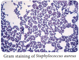
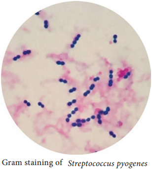
  • They are Gram positive, spherical or oval cocci and arranged in chains (0.6µm-1µm)
  • They are non – motile, non – sporing. Some strains are capsulated (Figure 7.4).

Streptococcus pyogenes (Flesh eating Bacteria) img 1

Cultural Characteristics

  • They are aerobe and facultative anaerobe. Optimum temperature is 37°C and pH is 7.4 to 7.6
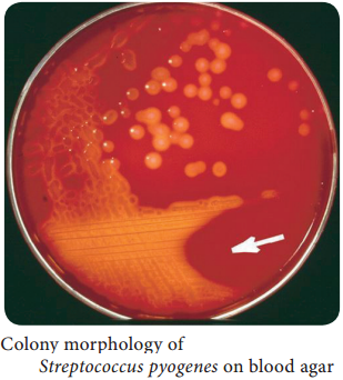
  • They grow only in media enriched with blood or serum. It is cultivated on blood agar. On blood agar, the colonies are small, circular, semitransparent, low convex, with an area of clear hemolysis around colonies (Figure 7.5).
  • Crystal violet blood agar – a selective medium for Streptococcus pyogenes.

Streptococcus pyogenes (Flesh eating Bacteria) img 2

Antigenic Structure

Capsule:

It inhibits phagocytosis

Cell wall:

The outer layer of cell wall consists of protein and lipoteichoic acid which helps in attachment to the host cell. Middle layer of cell wall consists of Group Specific C – Carbohydrate that is used for Lancefield grouping. Inner layer of cell wall is made up of peptidoglycan which has pyrogenic and thrombolytic activity.

Toxins and Enzymes:

Streptococcus pyogenes produces several exotoxins and enzymes which contribute to its virulence.

Toxins and Hemolysins:

Streptococci produces two types of hemolysins which are Streptolysin O and Streptolysin S.

Erythrogenic toxin: (Pyrogenic exotoxin)

The induction of fever is the primary effect of this toxin and it is responsible for the rash of scarlet fever.

Enzymes:

The various enzyme of Streptococcus pyogenes which exhibits virulence activity are listed in Table 7.3.

Enzymes of Streptococcus pyogens and its virulence nature

Enzymes

Virulence nature

Streptokinase (fibrinolysin)It promotes the lysis of human fibrin clot by catalyzing the conversion of plasminogen into plasmin. It facilitates the spread of infection by breaking down the fibrin barrier around the lesions.
DeoxyribonucleasesIt liquefy the highly viscous DNA that accumulate in thick pusand responsible for thin serous character of streptococcal exudates
HyaluronidaseIt breaks down hyaluronic acid of the tissues and favours spread of streptococcal lesion along intercellular spaces.
Other enzymesNADase, lipase, amylase, esterase, phosphates and other enzymes.

Pathogenesis

Streptococcus pyogenes is intrinsically a much more dangerous pathogen than Staphylococcus aureus and has a much greater tendency to spread in the tissues.

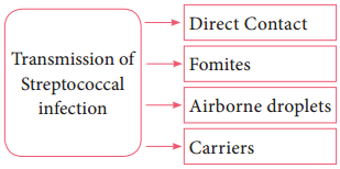
Mode of transmission:

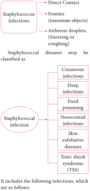
Streptococcal infections are transmitted by the following ways:
Streptococcus pyogenes (Flesh eating Bacteria) img 3

Streptococcal diseases may be broadly classified, and it is shown in flowchart 7.1
Streptococcus pyogenes (Flesh eating Bacteria) img 4

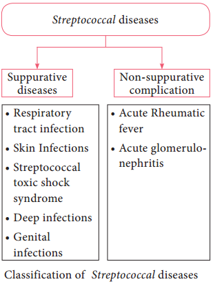
Suppurative Infections

1. Respiratory tract infection

a. Streptococcal sore throat:

Sore throat (acute tonsillitis and pharyngitis) is the most common streptococcal diseases. Tonsillitis is more common in older children and adults. The pathogen may spread from throat to the surrounding tissues leading to suppurative (pus – formation) complication such as cervical adenitis (inflammation of a lymph node in the neck) otitis media (inflammation of middle ear), quinsy (ulcers of tonsils) Ludwig’s angina (purulent inflammation around the sub maxillary glands) mastoiditis (inflammation of mastoid process).

b. Scarlet fever:

The disease consists of combination of sore throat and a generalized erythematous (redness of skin or mucous membranes) rash.

2. Skin infections

a. Erysipeals:

It is an acute spreading lesion. The skin shows massive brawny oedema with erythema it is seen in elderly persons or elders.

b. Impetigo: (Streptococcal pyoderma)

It is a skin infection that occurs most often in young children. It consists of superficial blisters that break down and eroded areas whose surface is covered with pus. It is the main cause leading to acute glomerulonephritis in children.

c. Necrotizing fasciitis:

It is an invasive, infection characterized by inflammation and necrosis of the skin, subcutaneous fat and fascia. It is a life-threatening infection.

3. Streptococcal toxic shock syndrome

Streptococcal pyrogenic exotoxin leads to streptococcal toxic shock syndrome (TSS). It is a condition in which the entire organ system collapses, leading to death.

4. Genital infections

Streptococcus pyogenes is an important cause of puerperal sepsis or child bed fever (infection occur when bacteria infect the uterus following child birth)

5. Deep infection

Streptococcus pyogenes may cause pyaemia (blood poisoning characterized by pus forming pathogens in the blood) septicemia (A condition in which bacteria circulate and actively multiply in the bloodstream) abscess in internal organs such as brain, lung, liver and kidney.

Non – Suppurative Complication

Streptococcus pyogenes infections are sometimes followed by two important non – suppurative complications which are, acute rheumatic fever and acute glomerulonephritis. These complications occur 1-4 weeks after the acute infection and it is believed to be the result of hypersensitivity to some streptococcal
components.

1. Rheumatic fever

It is often preceded by sore throat and most serious complication of haemolytic streptococcal infection. The mechanism by which Streptococci produce rheumatic fever is still not clear. A common cross – reacting antigen exist in some group A streptococci and heart, therefore, antibodies produced in response to the streptococcal infection could cross react with myocardial and heart valve tissue, causing cellular destruction.

2. Acute glomerulonephritis

It is often preceded by the skin infection. It is caused by only a few “nephritogenic types (strains)”. It develops because some components of glomerular basement membrane are antigenically similar to the cell membranes of nephritogenic streptococci.

The antibodies Formed against Streptococci cross react with glomerular basement membrane and damage. Some patients develop chronic glomerulonephritis with ultimate kidney failure.

Laboratory Diagnosis

Specimens:

Clinical specimens are collected according to the site of lesion. Throat swab, pus or blood is obtained for culture and serum for serology.

Direct Microscopy:

Gram stained smears of clinical specimens is done, where Gram positive cocci in chains were observed. It is indicative of streptococcal infection.

Culture:

The clinical specimen is inoculated on blood agar medium and incubated at 37° C for 18-24 hours. After incubation period, blood agar medium was observed for zone of beta – haemolysis around colonies.

Catalase test:

Streptococci are catalase negative which is an important test to differentiate Streptococci from Staphylococci.

Serology:

Serological tests are done for rheumatic fever and glomerulonephritis. It is established by demonstrating high levels of antibody to streptococci toxins. The standard test is antistreptolysin Otitration. ASOtitres higher than 200 units are indicative of prior Streptococcal infection.

Treatment and Prophylaxis

  • Penicillin G is the drug of choice.
  • In patients allergic to penicillin, erythromycin or cephalexin is used.
  • Antibiotics have no effect on established glomerulonephritis and rheumatic fever.
  • Prophylaxis is indicated only in the prevention of rheumatic fever, it prevents streptococcal reinjection and further damage to the heart.
  • Penicillin is given for a long period in children who have developed early signs of rheumatic fever.

Medical Bacteriology of Staphylococcus aureus

Learninsta presents the core concepts of Microbiology with high-quality research papers and topical review articles.

Medical Bacteriology of Staphylococcus aureus

The genus Staphylococcus is included in the family Micrococcaceae. Staphylococcus is a normal flora of skin and mucous membranes, but it accounts for human infections, which is known as staph infection.

The name Staphylococcus was derived from a Greek word, ‘staphyle’ means bunch of grapes and ‘kokkos’ means berry. Staphylococcus aureus is a pathogenic species that causes pyogenic infections in human.

Morphology

  • Staphylococci are gram positive spherical cocci, (0.8µm-1.0µm in diameter) arranged characteristically in grape like clusters (Figure 7.1).
  • They are non-motile and non-sporing and few strains are capsulated.

Medical Bacteriology of Staphylococcus aureus img 1

Cultural Characteristics

  • They are aerobes and facultative anaerobes, optimal temperature is 37°C and optimum pH is 7.4-7.6.
  • They grow on the following media and shows the characteristic colony morphology (Table 7.1 & Figure 7.2).

Staphylococci aureus colony morphology on various media

MediaColony Morphology
Nutrient AgarColonies are circular, smooth, convex, opaque and produces golden yellow pigment (most strains).
Blood AgarBeta haemolysis
Mannitol salt Agar (MSA)It is a selective medium for S. aureus produces yellow colored colonies due to fermentation of mannitol.

Medical Bacteriology of Staphylococcus aureus img 2

Virulence Factors

  1. Peptidoglycan → It is a polysaccharide polymer. It activates complement and induces the release of inflammatory cytokines.
  2. Teichoic acid → it facilitates adhesion of cocci to the host cell surface.
  3. Protein A → It is chemotactic, antiphagocytic, anticomplementary and induce platelet injury.

4. Toxins:

  • Hemolysins – It is an exotoxin, those lysis red blood cells. They are of four types namely α-lysin, β-lysin, γ-lysin and delta lysin.
  • Leucocidin – It damages PMNL (polymorphonuclear leucocytes) and macrophages.
  • Enterotoxin – It is responsible for manifestations of Staphylococcus food poisoning.
  • Exfoliative toxin – This toxin causes epidermal splitting resulting in blistering diseases.
  • Toxic shock syndrome toxin – TSST is responsible for toxic shock syndrome.

5. Enzymes:

S. aureus produces several enzymes, which are related to virulence of the bacteria.

  • Coagulase – It clots human plasma and converts fibrinogen into fibrin.
  • Staphylokinase – It has fibrinolytic activity.
  • Hyaluronidase – It hydrolyzes hyaluronic acid of connective tissue, thus facilitates the spread of the pathogens to adjacent cells.
  • Other enzymes – S. aureus also produces lipase, nucleases and proteases.

Pathogenicity

S. aureus is an opportunistic pathogen which causes infection most commonly at sites of lowered host resistance. (Example: damaged skin) Mode of Transmission: Staphylococcus infections are transmitted by the following ways.

Medical Bacteriology of Staphylococcus aureus img 3

It includes the following infections, which are as follows:

Cutaneous infections:

Wound (injury), burn infections (tissue injury caused by heat), pustules (A small elevated skin lesions containing pus), furuncles (boil forms around a hair follicle and containspus), styes (a painful swelling of hair follicle at eyelids), carbuncles (painful cluster of boils of the skin), Impetigo (skin infection with vesicles, pustules which ruptures), pemphigus neonatorum (an auto immune diseases that affect skin and
mucous membranes)

Deep infections:

It includes Osteomyelitis (inflammation of bones), tonsillitis (inflammation of tonsils), pharyngitis (inflammation of pharynx) sinusitis (inflammation of sinuses), periostitis (inflammation of membrane covering bones), bronchopneumonia (inflammation of lungs), empyema (collection of pus in the body cavity), septicemia (blood poisoning caused by bacteria and its toxins), meningitis (inflammation of meninge), endocarditis (inflammation of endocardium), breast and renal abscess.

Food Poisoning:

Staphylococcal food poisoning may follow 2-6 hours after the ingestion of contaminated food (preformed enterotoxin). It leads to nausea, vomiting and diarrhea.

Nosocomial infection:

S. aureus is a leading cause of hospital acquired infections. It is the primary cause of lower respiratory tract (LRT) infections and surgical site infections and the second leading cause of nosocomial bacteremia, pneumonia, and Cardiovascular infections.

Exfoliative diseases:

These diseases are produced due to the production of epidermolytic toxin. The toxin separates the outer layer of epidermis from the underlying tissues leading to blistering disease. The most dramatic manifestation of this toxin is scalded skin syndrome. The patient develops painful rash which slough off and skin surface resembles scalding.

Laboratory Diagnosis

Specimens:

The clinical specimens are collected according to the nature of Staphylococcal infections, which is given in the (Table 7.2).

Table 7.2: Clinical specimen collected for Staphylococcal infections

Infections

Clinical Specimens

Supportive lesionsPus
Respiratory infectionsSputum
SepticemiaBlood
MeningitisCSF
Food poisoiningFaeces, food or vomitus

Specimens should be transported immediately to the laboratory and processed.

Direct Microscopy:

Gram stained smears of clinical specimens is done, where gram positive cocci in clusters were observed.

Culture:

The collected specimen is inoculated on selective media-MSA and the media incubated at 37°C for 18-24 hours. Next day culture plates are examined for bacterial colonies, which are identified by gram staining, colony morphology and biochemical tests such as
Medical Bacteriology of Staphylococcus aureus img 4

a. Catalase test:
The genusStaphylococci are catalase positive. This test distinguishes Staphylococcus from Streptococcus (catalase negative).

b. Coagulase test:
This test helps in differentiating a pathogenic strain from non-pathogenic strain. S. aureus is coagulasepositive (Figure 7.3).

Treatment

Benzyl penicillin is the most effective antibiotic. Cloxacillin is used against beta lactamase. Producing strains (β-lactamase is produced by few strains of S. aureus which cleaves β-lactam ring of penicillin). Vancomycin is used against MRSA (Methicillin Resistant Staphylococcus aureus) strains.

Topical applications:

For mild superficial lesions, topical applications of bacitracin or chlorhexidine is recommended.

Control measures:

Proper sterilization of medical instruments must be done. Intake of antibiotics must be taken under proper medical advice. The detection of source & carriers among hospital staff, their isolation and treatment should be practiced.

Routes of Entry

Learninsta presents the core concepts of Microbiology with high-quality research papers and topical review articles.

Routes of Entry

To establish an infection, pathogen must first enter the host. Normal defense mechanisms and barriers (For example Skin, mucus, ciliated epithelium, lysozyme) make it difficult for the pathogen to enter the body.

Sometimes these barriers are break through for example cut in the skin, wound, tumor, ulcer which provides portal of entry for the bacteria. Some bacterial pathogens have the means to overcome the barriers through various virulence factors and invade the body.

Certain bacteria are infective when introduced through optimal route. The various route of entry of pathogens, which are cut or abrasion or wound (skin), Ingestion, Inhalation, arthropod bite, sexual transmission and congenital transmission.

These are already explained in the XI Standard text book. The various bacterial pathogens, its pathogenesis clinical symptoms, laboratory diagnosis, control, prophylaxis and treatment with appropriate antibiotics are discussed below.

The way a substance is able to enter the body such as ingestion (mouth), inhalation (lungs) or absorption (cintact).

There are four major routes by which a chemical may enter the body:

  • Inhalation (breathing).
  • Skin (or eye) contact.
  • Swallowing (ingestion or eating).
  • Injection.

Pathogenic Attributes of Medical Bacteriology

Learninsta presents the core concepts of Microbiology with high-quality research papers and topical review articles.

Pathogenic Attributes of Medical Bacteriology

The host-parasite relationship is determined by the interaction between host factors and the infecting pathogens.Pathogenicity refers to the ability of a pathogen to produce disease. Virulence is the ability of the pathogen to cause disease.

Adhesion, invasiveness (Streptococcal infections), Bacterial toxins (endotoxins and exotoxins), capsule enzymes (proteases, collagenase, coagulase and other enzymes). These are already explained in the XI Standard text book.

Pathogenicity refers to the ability of an organism to cause disease (ie, harm the host). This ability represents a genetic component of the pathogen and the overt damage done to the host is a property of the host-pathogen interactions. Commensals and opportunistic pathogens lack this inherent ability to cause disease.

Pathogen types. There are different types of pathogens, but we’re going to focus on the four most common types: viruses, bacteria, fungi, and parasites.

The definition of a pathogenic organism is an organism capable of causing disease in its host. A human pathogen is capable of causing illness in humans. Common examples of pathogenic organisms include specific strains of bacteria like Salmonella, Listeria and E. coli, and viruses such as Cryptosporidium.

Decrease your risk of infecting yourself or others:

  1. Wash your hands often.
  2. Get vaccinated.
  3. Use antibiotics sensibly.
  4. Stay at home if you have signs and symptoms of an infection.
  5. Be smart about food preparation.
  6. Disinfect the ‘hot zones’ in your residence.
  7. Practice safer sex.
  8. Don’t share personal items.