Life Cycle of Entamoeba Histolytica

Learninsta presents the core concepts of Microbiology with high-quality research papers and topical review articles.

Life Cycle of Entamoeba Histolytica

Geographical Distribution

It is Worldwide in distribution they are more common in the tropics than elsewhere. It is found wherever sanitation is poor.

Habitat

Trophozoites of E.histolytica live in the mucous and submucous layers of the large intestine of human.

Morphology

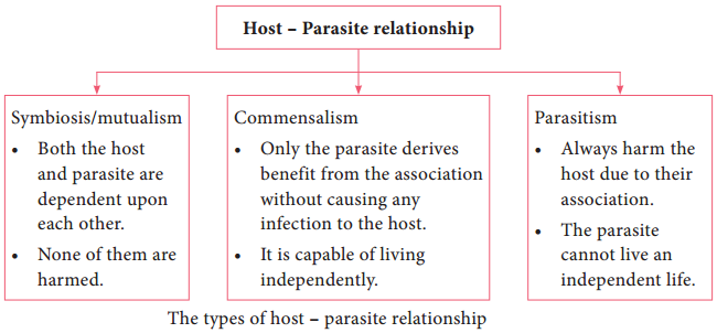
E. histolytica occurs in 3 forms as Trophozoite, Precyst and Cyst.

Trophozoite:

It is the growing or feeding stage of the parasite. It is the only form present in tissues. It has no fixed shape. They vary in size from 18 to 40µ, average being 20 to 30µ. The cytoplasm is usually described as outer ectoplasm and inner endoplasm (Figure 8.1). The endoplasm contains nucleus, food vacuoles, erythrocytes, occasionally leucocytes and tissue debris.

The nucleus is characterised by evenly arranged chromatin on the nuclear membrane and the presence of a small, compact, centrally located karoyosome (It is a DNA containing body, situated peripherally or centrally within the nucleus). Trophozoites exhibits active crawling or gliding motility by forming finger-like projections called Pseudopodia.

The trophozoite reproduce by binary fission in every 8 hours. Trophozoites survives upto 5 hours at 37°C and are killed by heat, drying and chemical sterilization. Even if live trophozoites from freshly passed stools are ingested, they are rapidly destroyed in stomach and cannot initiate infection. Therefore, the infections is not
usually transmitted by trophozoites.

Precyst

Trophozoites undergo encystment in the intestinal lumen. Encystment does not occur in the tissue or in feces outside the body. Precyst is smaller in size about 10 – 20 µm in size. It is round or oval in shape. The endoplasm is free of red blood cells and other ingested food particles (Figure 8.1). The nuclear structure retains the characteristics of the trophozoite.

Cyst

Precyst secretes a highly refractive cyst wall around it and becomes a cyst. A mature cyst is a quadrinucleate spherical body. The cyst begins as a uninucleate body but soon divides by binary fission and develops into binucleate and quadrinucleate bodies (Figure 8.1).
Life Cycle of Entamoeba Histolytica img 1

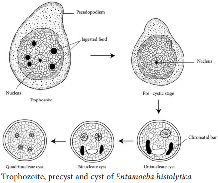
The cytoplasm of the cyst is clear and hyaline (translucent) and the nuclear structure retain the characteristic of the trophozoites. The mature quadrinucleate cyst, passed in the stool, does not undergo any further development and remain alive for several months in the soil or in environment where they were deposited. The mature quadrinucleate cysts are the infective forms of the parasite.

Life – Cycle of Entamoeba histolytica

E. histolytica passes its life cycle only in one host, the human.

Infective form:
Mature quadrinucleate cyst.

Mode of transmission:
Ingestion of food and water contaminated with cyst.

The cysts that are swallowed along with food and water enter into the alimentary canal. The cyst wall is resistant to action of gastric juice. The cysts pass through the stomach undamaged and enters the small intestine (Figure 8.2).
Life Cycle of Entamoeba Histolytica img 2

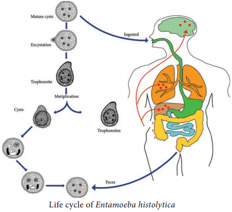
When the cyst reaches caecum or lower part of the ileum, due to alkaline medium, the cyst wall is damaged by trypsin leading to excystation.

The cytoplasm gets detached from the cyst wall and an amoeboid movement appear causing a tear in the cyst wall, through which quadrinucleate amoeba is liberated. This stage is called the metacyst.

The nuclei in the metacyst immediately undergo division to form 8 nuclei, each of which gets surrounded by its own cytoplasm to become 8 small amoebulae or metacystic trophozoites.

These metacystic trophozoites are carried to the caecum and colon. They invade the tissues and lodge in the submucous tissue of the large intestine which is their normal habitat.

Trophozoite grow and multiply by binary fission. The trophozoite phase of the parasite is responsible for producing the characteristic lesion of amoebiasis.

Some of the trophozoites in colon develop into precystic forms and cysts, which are passed in feces to repeat the cycle.
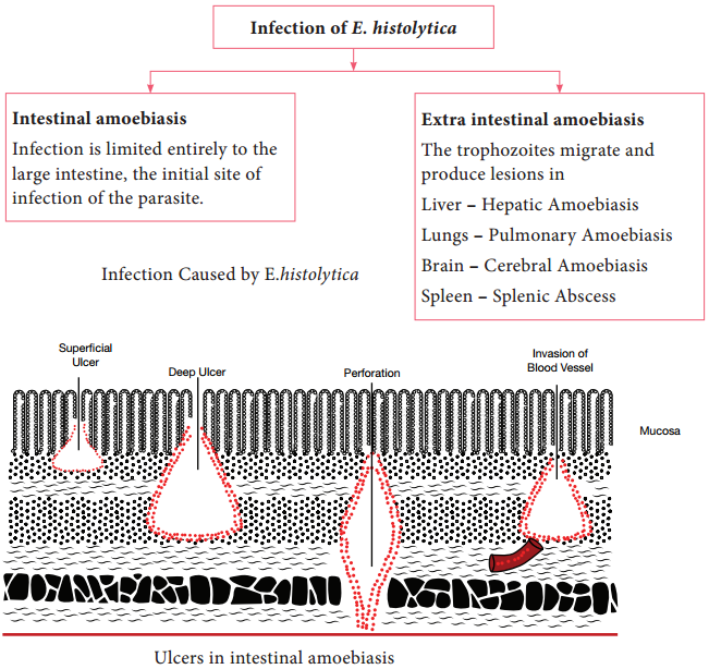
Pathogenesis

E. histolytica causes intestinal and extra intestinal amoebiasis (Flowchart 8.3).

E. histolytica can live in the intestine without causing symptoms. But, they can also cause severe disease. These amoebas may invade the wall of the intestine leading to amoebic dysentery, an illness that causes intestinal ulcers, bleeding, increased mucus production and diarrhoea. The ulcers are strictly confined to the large intestine being most numerous in the caecum and next in the sigmoid-rectal regions.

The lesions may be generalized or localised. A typical amoebic ulcer varies from pin’s head to one inch or more in diameter in size. The shape of ulcer may be round or oval.

On vertical section, the ulcer appears like flask, with mouth and neck being narrow and base being large and rounded (Figure 8.3 shows the flask – shaped ulcer). The base of ulcer is generally formed by the muscular coat and filled up by the necrotic material. The ulcers generally do not extend deeper than submucosal layer.
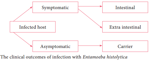
Life Cycle of Entamoeba Histolytica img 3

Clinical Features

Incubation period is highly variable, but is generally 4 to 5 days. A wide spectrum, from asymptomatic infection (luminal amoebiasis), to invasive intestinal amoebiasis (dysentery, colitis, appendicitis, toxic mega colon, amoebomas), to invasive extraintestinal amoebiasis occurs. Flowchart 8.4 classifies the clinical
outcomes of infection with Entamoeba histolytica. Only about 10% to 20% of people who are infected with E. histolytica become sick from the infection.
Life Cycle of Entamoeba Histolytica img 4

The typical manifestation of intestinal amoebiasis is amoebic dysentery. The symptoms are often quite mild and can include loose feaces, stomach pain and stomach cramping. In acute amoebic dysentery, the symptoms include abdominal pain, bloody stool, fever, tenderness, rectal tenesmus and hepatomegaly (enlargement of liver).

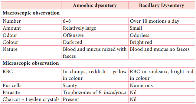
People affected may develop anemia due to loss of blood. On clinical and laboratory ground, amoebic dysentery should be differentiated from bacillary dysentery. A Table 8.1 shows the difference between the stools of amoebic and bacillary dysentery. Table 8.1: Difference between the stools of amoebic and bacillary dysentery.
Life Cycle of Entamoeba Histolytica img 5

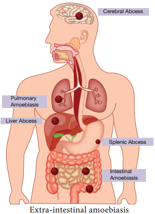
Extra intestinal amoebiasis

1. Hepatic amoebiasis:

This is the most common form of extra intestinal invasive amoebiasis. Liver abscess may be multiple or more often solitary, usually located in the upper right lobe of the liver (Figure 8.4). Amoebic liver abscess (ALA) contains an odour less and thick chocolate brown pus called anchovy sauce pus.

ALA is associated with an abrupt onset of high fever, right upper abdominal pain and tenderness. Anorexia (loss of appetite for food), nausea (the sensation to vomit), vomiting, fatigue (extreme tiredness) and weight loss are also frequent.

2. Pulmonary Amoebiasis:

It is very rare, but this may occur by direct hematogenous spread from the colon. The patient presents with severe chest pain and have dyspnoea (shortness of breath). The sputum of patient is chocolate brown. Amoebic trophozoites may be demonstrated in the sputum.

3. Cerebral amoebiasis:

The condition is unusual. In cerebral amoebiasis, the abscess is single, small and is located in the cerebral hemisphere. The patient may die of rupture or involvement of cerebellam within 12-72 hours. Biopsy of the brain shows the amoebic trophozoites.

4. Cutaneous amoebiasis:

It can be caused by perforation of an amoebic abscess or surgical wound infected with amoebae. It is less frequent condition.

5. Genitourinary Amoebiasis:

This condition includes amoebiasis of the kidney and genital organs. Amoebiasis of the genital organs is a rare condition. Lesions of amoebiasis is shown in Figure 8.5.
Life Cycle of Entamoeba Histolytica img 6

Laboratory Diagnosis

Specimens:
Stool is the specimen of choice. Other specimens collected includes blood, rectal exudates and rectal ulcer tissue collected from the base by endoscopies.

Methods in examination of stool

A. Direct wet mount examination of stool:

Demonstration of mature quadrinucleate cysts or trophozites in stool is diagnostic of intestinal amoebiasis. The wet mount of stool is prepared in the saline, iodine or lacto phenol cotton blue.

B. Examination of stool after concentration:

Demonstration of amoebic cysts by Formalin – ether is the method of choice.

C. Examination of stained stool smears:

Staining by iron haematoxylin, Periodic Acid – Schiff (PAS) stains demonstrate the presence of both trophozoites and cyst.

Amoebic liver abscess (ALA):
Demonstration of amoebic trophozoites in the aspirated liver pus establishes the diagnosis of ALA.

Serology:
Detection of amoebic antigens in the serum by Enzyme Linked Immunosorbent Assay (ELISA).

Molecular diagnosis:
PCR (Polymerase chain reaction) is employed to detect amoebic genome in the aspirated liver pus for the diagnosis of ALA.

Imaging methods:
X – Ray magnetic resonance imaging (MRI) scan and computerized Axial Tomography (CAT) Scan are the imaging methods used.

Treatment:
Eradication of amoebae by the use of amoebicidal drugs and replacement of fluid and electrolyte is the treatment for amoebiasis. Listed below the drugs used in the treatment for amoebiasis.

  • Paramomycin and iodoquinol acts in the intestinal lumen but not in tissues.
  • Emetine, chloroquine are effective in systemic infection. They act only on trophozoites.

Metronidazole is the drug of choice which acts as both luminal and tissue amoebicides. It is low in toxicity and is effective against intestinal as well as extra -intestinal amoebic infections.

Prevention and Control

  • Proper sanitation is the key to avoid amoebiasis. Washing hands with soap and water after using the bathrooms and before handling food.
  • Drinking safe and boiled water.
  • Avoid eating unwashed fruits and vegetables.
  • Prevention of water supplies from faecal contamination.
  • Early rapid detection of diseased people and subsequent treatment with amoebicidal drugs. No vaccine is available yet against amoebiasis in humans.

Medical Parasitology of Parasite and Host

Learninsta presents the core concepts of Microbiology with high-quality research papers and topical review articles.

Medical Parasitology of Parasite and Host

Parasites are living organisms, which depend on living host for their nourishment and survival. They multiply or undergo development in the host. Host is defined as an organism, which harbors the parasite, provides nourishment and gives shelter to parasite. Host is relatively larger than the parasite.

Association between Host and Parasite

The relationship between host and the parasite can be of the following types:

  • Symbiosis
  • Commensalism, and
  • Parasitism.

Flowchart 8.1 describes the types of host – parasite relationshipMedical Parasitology of Parasite and Host img 1

Types and Classification of Parasite

According to the nature of the host – parasite interaction and the environmental factors, the parasite may be one of the following,

Ectoparasite:

These parasites live on the outer surface or in the superficial tissues of the host (Example: Lice). The infection by these parasites is called infestation.

Endoparasite:

The parasite which lives within the host is called Endoparasite. Invasion by the parasite is called Infection. Most of the protozoan and helminthic parasites causing human diseases are endoparasites.

Endoparasites can be further classified as:

Obligate parasite:
This parasite is completely dependent on its host and cannot survive without it.

Example:
Hookworms.

Facultative parasite:
This parasite may either live as free living form or as a parasite when the opportunity arises.

Example:
Naegleria fowleri.

Opportunistic parasite:
This parasite is capable of producing disease in an immune deficient host (like AIDS and cancer patients).

Example: Toxoplasma gondii.

Zoonotic Parasite:
This parasite primarily infects animals and is transmittable to humans.

Example:
Fasciola species.

Accidental parasite:
This parasite infect an unusual host are known as accidental parasites.

Example:
Echinococcus granulosus infects man accidentally.

Wandering or Aberrant parasites:

Parasites which infect a host migrate to the site where it cannot live or develop further are called aberrant parasites.

Example:
Dog roundworm infecting humans.

Types of Host

Definite host:

The host which harbour the adult parasites or in which parasites undergo sexual method of reproduction is referred to as a definite host. The definite host may be a human or any other living organism. Example: Mosquito acts as a definite host for Plasmodium spp. in Malaria.

Intermediate host:

The host in which the larval stages of the parasite live or in which asexual reproduction of parasite takes place is called the intermediate host.

Example:
Man acts as an intermediate host for Plasmodium spp. in Malaria.

Reservoir host:

The host which harbour the parasite and acts has an important source of infection to other susceptible hosts is known as reservoir host. It is also called temporary host.

Example:
Dog is the reservoir host for disease kala azar.

Natural host:
The host which is naturally infected with a certain species of parasite, is called natural host.

Example:
Pig is the natural host of Balantidium coli.

Paratenic host or transport host:

some parasites enter a host in which they do not undergo any development but remains alive till they gain entry into the definitive host or intermediate host. Such a host is termed as paratenic host or transport host or carrier host.

Classification of Medical Parasitology

The most acceptable taxonomic classification of human parasites includes Endoparasites and Ectoparasite. Endoparasites are subclassified into protozoan parasite (unicellular organisms) and helminthic parasite (multicellular organism). Parasites of medical Importance come under the Kingdom called Protista and Animalia. Protista includes the microscopic single – celled eukaryotes known as protozoa.

In contrast, helminths are microscopic, multicellular worms possessing well differentiated tissues and complex organs belonging to the kingdom Animalia. Classification of medically important parasites is given in Flowchart 8.2.

Life Cycle of Parasites

Direct life cycle

The life cycle of parasite that requires only single host to complete its development, is called direct life cycle.

Example:
Entamoeba histolytica requires only human host to complete its life cycle.

Indirect life cycle

The life cycle of parasite that requires two or more species of hosts to complete its development, the life cycle is called as indirect life cycle.

Example:
Malarial parasite (Plasmodium spp.) requires both human host and mosquito to complete its life cycle.

Transmission of Parasites

It depends upon Source or reservoir of infection, and mode of transmission.

1. Sources of infection

A. Human:

Human is the source or reservoir for a majority of parasitic infection. The condition in which the infection is transmitted from one infected human to another human is called anthroponoses.

B. Animals:

Animals act as the source of infection in many parasitic diseases. The condition where infection is transmitted from animals to humans is called zoonoses.

2. Mode of transmission

A. Oral transmission:

This is through ingestion of contaminated food, water, vegetables, soiled fingers or fomites contaminated by faeces that contain the infective stage of parasite. This mode of transmission is referred to as faecal-oral route. Example: Cysts of Entamoeba histolytica.

B. Skin transmission:

This is another important route. The infective larvae of hookworm enter the skin of persons walking bare footed on contaminated soil.

C. Vector transmission:

It could be a biological or a mechanical means. Many parasitic diseases are transmitted by insect bite.

Example:
sandfly is vector for Leishmania.

D. Direct transmission by person to person contact. Frequently, Entamoeba, Giardia andTrichomonas are transmitted by sexual contact among homosexuals.

E. Vertical transmission:

It is the transmission from mother to fetus.

Example:
Toxoplasmosis.

So far, we have learnt about the general introduction and classification of parasites. Now, let us learn a few important human parasites in detail.

Introduction to Protozoa

General characteristics of protozoa:

  1. They are microscopic unicellular eukaryotes.
  2. The single cell has a relatively complex internal structure and it performs various complex metabolic activities such as digestion, reproduction, respiration and excretion.
  3. Each cell consists of nucleus and cytoplasm.
  4. A protozoa parasite during its life cycle may exist in two stages such as trophozoite and cyst.

Amoebae

Amoebae are structurally simple protozoans which have no fixed shape. The cytoplasm of amoeba is bounded by a membrane and can be differentiated into an outer ectoplasm and inner endoplasm. Pseudopodia (false foot) are formed by the amoebae by throwing out ectoplasm followed by endoplasm. These are employed for locomotion and engulfment of food by phagocytosis.

Reproduction occurs by fission and budding. Amoebae are classified as either free living or intestinal amoebae.

An Overview of Leptospira Interrogans

Learninsta presents the core concepts of Microbiology with high-quality research papers and topical review articles.

An Overview Leptospira Interrogans

Spirochaetes of the genus Leptospira are actively motile, delicate and possess numerous closely wound spirals with characteristic hooked ends. Several Leptospires are saprophytes, while many are potential pathogens of rodents, domestic animals and humans. The genus Leptospira consists of two important
species, which are Leptospira interrogans and Leptospira biflexa.

Leptospira interrrogans is the causative agent of leptospirosis, a zoonotic disease. The word Leptospira is derived from Latin word ‘Leptos’ = fine or thin and ‘spira’ = Coil and interrogans = Question mark (The shape of this spirochete accounts for its name)

Morphology

  • They are spiral bacteria (5-20µm × 0.1µm) with numerous closely set coils. Their ends are hooked and resemble umbrella handles.
  • They are actively motile by rotatory movements. They cannot be seen under light microscope due to its thinness, best observed by dark fieldmicroscopy (Figure 7.24), phase contrast and electron microscope.
  • They stain poorly with aniline dyes, it may be stained with giemsa stain or silver impregnation techniques.

Leptospira Interrogans img 1

Antigenic Structure

Leptospires show considerable antigenic cross reaction.

  1. Genus – Specific somatic antigen – It is present in all members of the genus.
  2. Surface antigens – This antigen is used to classify Leptospira into serogroups and serotypes.

Pathogenicity

Source of infection:

Contaminated water

Route of entry:

Through cuts or abrasions on skin or mucosa

Incubation period:

6-8 days

  • Leptospira interrogans causes a zoonotic disease named Leptospirosis. It is transmitted to humans by direct or indirect contact with water, contaminated by urine of carrier animals (rat and dog).
  • Leptospira enter the body through cuts or abrasions on skin or through mucous membranes of the mouth, nose or conjunctiva.
  • After an incubation period of 6-8 days. There is onset of febrile (related to fever) illness with Leptospira in blood (Septicemic phase) which lasts for 3-7 days.
  • The organisms disappear from the blood and invades liver, kidney, spleen, meninges producing meningeal irritation such as headache, vomiting.
  • The pathogen persists in the internal organs and most abundantly in the kidney. Severe Leptospirosis (Weil’s disease) is associated with Fever, conjunctivitis (inflammation of conjunctiva), albuminuria (presence of albumin in the urine), jaundice and hemorrhage. It is a fatal illness with hepatorenal (Kidney failure with severe liver damage).

Clinical manifestations

  • In severe cases, vomiting, headache, irregular fever and intense infection of the eyes.
  • Jaundice, Albuminuria (The presence of protein Albumin in the urine) and purpuric hemorrhages sometimes occur on skin and mucosa.

Laboratory Diagnosis

The diagnosis of Leptospirosis is made by the following ways

  • Direct microscopy of blood or urine
  • Isolation of pathogen by culture
  • Serological tests.

Direct Microscopy

Blood:

Leptospira can be observed in the blood by dark – filed microscope. Blood examination is useful in first week as Leptospira disappear from blood after 8 days.

Urine:

Leptospira may be present in urine in the 22nd week of the disease and intermittently thereafterup to 6 weeks. Centrifuged deposit of urine may be observed by Dark filed microscopy.

Culture:

Blood (1st week) and urine (22nd – 6  week) can be cultured in Korthof ’s medium. Media are incubated at 37°C for 2 days and then left at room temperature for 2 weeks. Culturesare examined every third day for the presence of Leptospira under DFM.

Serological tests

It is very useful method of diagnosis two types of serological tests are used, which are,

a. Screening tests:

These tests are genus – specific and done using reactive genus specific antigen (non – pathogenic L. biflexapatoc I strain).

Screening test includes – CFT, ELISA, SEL, HAT indirect IF these tests are capable to detect IgM and IgG leptospiral antibodies.

b. Serotype specific tests:

These tests identify the infecting serovar by demonstrating specific antibodies.

  • Macroscopic agglutination test
  • Microscopic agglutination test

Treatment and Preventions

  • Leptospira are sensitive to penicillin and tetracycline.
  • Preventive measures include rodent control, disinfection of water.

Treponema Pallidum of Medical Bacteriology

Learninsta presents the core concepts of Microbiology with high-quality research papers and topical review articles.

Treponema Pallidum of Medical Bacteriology

Treponema pallidum is included in the family Spirochaetaceae. They are slender spirochaetes with fine spirals having pointed ends. Some of them are pathogenic for humans, while others are normal flora in mouth and genitalia.

These pathogens are strict parasites and the diseases caused by Treponema are called Treponematoses. Treponema pallidum is the causative agent of syphilis. The name Treponema pallidum is derived from Greek words, which means, Trepos – Turn Nema – Thread and palladium – Pale staining.

Morphology

It is thin, delicate spirochete with tapering ends, about 10µm long and 0.1-0.2 µm wide. It has about ten regular spirals, which are sharp and angular, at regular intervals of about 1µm. They are actively motile (endoflagella), exhibiting rotation around the long axis, backward and forward movements and flexion of the
whole body.

It cannot be seen under light microscope and does not take ordinary bacterial stains. It can be seen under the dark ground (Figure 7.22) or phase contrast microscope.It can be stained by silver impregnation method.
Treponema Pallidum of Medical Bacteriology img 1

Culture

  • Pathogenic Treponema cannot be grown in artificial culture media.

Pathogenesis

Source of infection – Human beings (patients)
Mode of transmission – Sexual contact
Site of entry – Through minute abrasions/cuts on skin or mucosa

Incubation period – 10 – 90 days

  • Treponema pallidum causes venereal syphilis, which is acquired by sexual contact. The pathogen enters the human body through cut on the skin or mucosa of genital areas.
  • The clinical disease sets in after an incubation period of about a month. There are 3 clinical stage of venereal syphilis, namely – primary, secondary and tertiary syphilis.

Primary syphilis

  • A papule appears on the genital area that ulcerates, forming a chancre of primary syphilis called hard chancre.
  • The chancre is covered by thick exudates, very rich in spirochetes.
  • The regional lymph nodes are swollen, discrete, rubbery and non – tender.
  • Even before the chancre appears, the spirochetes spread from the site of entry into the lymph and bloodstream, so the patient may be infectious during the late incubation period.
  • The chancre invariably heals within 10-40 days, even without treatment, leaving a thin scar.

Secondary syphilis

  • Secondary syphilis sets in 1-3 months after the primary lesion heals. During this interval, the patient is asymptomatic.
  • The secondary lesions are due to widespread multiplication of the spirochetes and dissemination through the blood.
  • Secondary syphilis is characterized by appearance of papular skin rashes, mucous patches in the oropharynx and condylomata (a raised growth on the skin resembling a wart).
  • The lesions are abundant in spirochetes and the patient is most infectious during the secondary stage.
  • There may be retinitis (inflammation of the retina of the eye), meningitis, periostitis, and arthritis.
  • Secondary lesions usually undergo spontaneous healing, in some cases taking as long as 4 or 5 years.
  • After the secondary lesions disappear, there is a period of dormant known as latent syphilis the patient does not show any clinical symptoms but with positive serology.

Tertiary syphilis

  • After several years, manifestations of tertiary syphilis appear. These consist of cardiovascular lesions including aneurysms (enlargement of an artery), gummata (a small rubbery granuloma that has a necrotic centre) and meningovascular manifestations. Tertiary lesions contain few spirochetes.
  • In few cases, neurosyphilis such as tabesdorsalis or general paralysis of the insane develops. These are known as late tertiary or quaternary syphilis.

Congenital syphilis

In congenital syphilis, the infection is transmitted from mother to fetus by crossing the placental barrier.

Non – Venereal syphilis

It may occur in doctors or nurses due to contact with patients lesion during examination. The primary chancre occurs usually on the fingers.

Laboratory Diagnosis

The diagnosis of syphilis includes

  1. Demonstration of Treponemes
  2. Serological tests

Specimen:
Exudates are collected from the chancre. Blood (serum) is collected for serology.

Demonstration of Treponemes

a. Dark ground microscopy:

The wet film is prepared with exudates and examined under dark ground microscope. Under dark field examination Treponema pallidum appears motile spiral organism.

b. Treponemes in tissues:

It can be demonstrated by silver impregnation method of staining.

Serological tests

Non – Treponemal tests – In the standard tests for syphilis includes;

  • VDRL – Venereal Diseases Research Laboratory test.
  • RPR – Rapid Plasma Reagain (Figure 7.23).

VDRL or RPR tests are used for serological screening for syphilis and more useful for the assessment of cure following treatment.
Treponema Pallidum of Medical Bacteriology img 2

Treponemal Tests:

The treponemal tests includes

  • TPHA – Treponema pallidum hemagglutination assay
  • FTA – ABS – Fluorescent treponemal antibody absorption test.

These two tests are used to confirm the diagnosis.

Treatment and Preventive Measure

In early syphilis

  1. Benzathine benzyl penicillin, 24 lakhs units intramuscularly in a single dose.
  2. Alternatively, doxycycline 100 mg twice a day orally for 15 days.

In late syphilis

Benzathine benzyl penicillin 24 lakhs units, intramuscularly once weekly for 3 weeks.

  1. Avoiding sexual contact with an infected individual.
  2. Use of sex barriers (condoms).

Mycobacterium Tuberculosis (Tubercle Bacillus)

Learninsta presents the core concepts of Microbiology with high-quality research papers and topical review articles.

Mycobacterium Tuberculosis (Tubercle Bacillus)

The genus Mycobacterium is distinguished by its thick, complex, lipidrich waxy cell walls. This high lipid content (Mycolic acids) imparts the characteristic of acid fastness or resistance to decolorization by a strong acid after staining with carbol fuchsin. Many of the Mycobacterial species are saprophytes but several species are highly significant human pathogens.

Mycobacterium tuberculosis is the causative agent of tuberculosis (TB) It is a killer disease and ranks as one of the most serious infection diseases of the developing countries. TB is primarily a disease of the lungs but may spread to other sites of the body.

The name Mycobacterium tuberculosis is derived form,

  • Mycobacterium (Greek) – Fungus like bacterium
  • Tuberculosis (Latin) – Swelling or Knob

Morphology

They are acid fast bacilli, slightly curved rods, it may occur singly or in small clumps. They are non-motile, non-sporing, and non-capsulated.

Cultural Characteristics

They are obligate aerobe, optimum temperature is 37°C and optimum pH is 6.4-7.0. The pathogen grows on an enriched culture media – Lowenstein Jensen medium.

The colonies appear in about 2-3 weeks. The colonies are dry, rough, raised, irregular colonies with a wrinkled surface. Initially creamy white and becoming yellowish later (Figure 7.19).
Mycobacterium Tuberculosis (Tubercle Bacillus) img 1

Pathogenesis

Human tuberculosis is divisible into two form, they are Primary TB and Secondary TB. The pathogenesis of Primary Tuberculosis is described in flowchart 7.8.

Source of infection – Airborne droplets
Route of entry – Respiratory tract
Incubation period – 3 – 6 weeks.
Mycobacterium Tuberculosis (Tubercle Bacillus) img 2

Secondary TB – (Post primary TB)

It is caused by reactivation of the primary lesion or by exogenous reinfection. Granulomas of secondary TB most often occur in the apex of the lungs. The necrotic element of the reaction causes tissue destruction and the formation of large area of caseation termed tuberculomas.

The presence of caseous necrosis and cavities are two important clinical manifestations of secondary TB. The cavities may rupture into blood vessels, spreading the bacilli throughout the body and break into airways, releasing the pathogen in aerosols and sputum – called as open tuberculosis (Figure 7.20).
Mycobacterium Tuberculosis (Tubercle Bacillus) img 3

Clinical Symptoms

It includes, cough that lasts for more than 2-3 weeks, weight loss, fever, night sweat and loss of appetite.

Laboratory Diagnosis

Specimen:

In case of pulmonary tuberculosis the most usual specimen is sputum.

Direct Microscopy:

Smear is made from the sputum specimen and stained by Ziehl – Neelson technique. It is examined under oil immersion objective lens. The acid fast bacilli appear as bright red bacilli against a blue background.

Culture:

The specimen is inoculated onto LJ – medium and incubated at 37°C for 2 weeks the tubercle bacilli usually grow in 2-8 weeks. The bacterial growth is confirmed by Ziehl – Neelsonstaining.

1. Tuberculin Skin test

Mantoux test:

This method has been used routinely. In this test 0.1 ml of PPD (Purified protein derivative) containing 5 TU (Tuberculin unit) is injected intradermally on the flexor aspect of forearm (Figure 7.21) The site is examined after 48-72 hours and induration are measured (diameter in mm).

Positive test:

Indurations of diameter d10 mm or more is considered positive.

Negative test:

Indurations of 5 mm or less is negative.

2. Gene Xpert MTB

It is an automated diagnosis test it detects DNA sequences specific for M. tuberculosis and rifampicin resistance by PCR. Results can be obtained within 2 hours.

Treatment

The antitubercular drugs include two types of agents which are;

Bactericidal agents – Rifampicin (R), Isoniazid (H), Pyrazinamide (z), Streptomycin.
Bacteriostatic agents – Ethambutol (E).

The regimen for treating TB consists of an intensive phase of 2 months of isoniazid, rifampin, pyrazinamide and ethambutol, followed by a continuation phase of 4 months of isonizid and Rifampin.

Prophylaxis and Control Measures

The BCG (Bacille – Calmette – Guerin) administered by intradermal injection of the live attenuated vaccine. The immunity may last for about 10 years. The prevention of TB can be done by the following general measures such as

  1. Adequate nutrition.
  2. Practicing good hygiene (washing hands)
  3. Health education.
  4. Cover the mouth with a tissue when you cough or sneeze.