Overview of an Transplantation

Learninsta presents the core concepts of Microbiology with high-quality research papers and topical review articles.

Overview of an Transplantation

Transfer of living cells, tissues or organs from one part of the body to another or from one individual to another is known as transplantation. A tissue or organ that is removed from one site and placed to another site usually in a same or different individual is called graft. The individual who provides the graft is called donor and the individual who receives the graft is called host or recipient.

If the graft is placed into its normal anatomic location, the procedure is called orthotopic transplantation. If the graft is placed in a different site it is called heterotopic transplantation. Transplantation is the only form of treatment for most end-stage organ failure.

In clinical practice, transplantation is used to overcome a functional and anatomic deficit in the recipient. Transplantation of kidneys, hearts, livers, lungs, pancreas and bone marrow are widely done today.

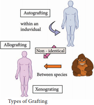
Methods of Transplantation

Auto grafting:
The transfer of self tissue from one body site to another in the same individual

Allografting:
The transfer of organs or tissues from human to human

Xenografting:
The transfer of tissue from one species to another (Figure 11.11).
Overview of an Transplantation img 1

Graft Acceptance

When transplantation is made between genetically identical individuals the graft survives and lives as healthy as it is in the original places. When the graft tissue remains alive, it is said to be accepted and the process is called graft acceptance.

Graft Rejection

When transplantation is made between genetically distinct individual the graft tissue dies and decays. When the graft tissue dies, the graft is said to be rejected and the process is called graft rejection. It is of two types. They are:-

  1. Host Verses Graft Reaction
  2. Graft Verses Host Rejection.

Host Verses Graft Reaction (HVG)

The graft tissue antigens induce an immune response in the host. This type of immune response is called host versus graft reaction.

Allograft Rejection

Types of allograft rejection

  • Acute rejection-Quick graft rejection. It is due to stimulation of thymocytes and B lymphocytes
  • Hyperacute rejection-It is a very quick rejection. It is due to pre-existing humoral antibodies in the serum of the host as a result of presensitization with previous grafts.
  • Insidious rejection-It is a secret rejection due to deposition of immune complex on the tissues like glomerulus membrane that can be demonstrated in kidney by immune fluorescence.

Mechanism of Allograft Rejection

Immunological contact

When tissue is implanted as graft, its antigen can pass into local lymph nodes of the host. The graft antigens then make contact with the lymphocytes of the host. Production of sensitized T cells and cytotoxic antibodies are produced in the host. This brings about graft rejection.

First set rejection

When the graft is made between genetically different individuals, the graft gets blood supply from the host and it appears to be normal for the first 3 days. But on the 5th day, sensitized T cells, macrophages and a few plasma cells invade the graft. Inflammation starts in the graft. This leads to necrosis. It is similar to the primary immune response to an antigen.

Second set rejection

When a graft is implanted in an individual who has already rejected a graft is second set rejection. This is similar to the secondary immune response of our body.

Cell mediated cytotoxic reaction

The 1st set of rejection of allograft is brought about mainly by CMI response. In this process the cells involved in the cytotoxic mediated immunity involves. On stimulation of these cells interferon causes the lysis of the graft.

Antibody mediated cytotoxic reaction

The 22nd set rejection of graft is brought about mainly by HMI response. This is one of the hyperacute rejection brought about by the antibodies. Complement, macrophages, mast cells, platelets, B cells bring about this reaction.

Graft versus Host Rejection (GVH)

Sometimes the graft tissue elicits an immune response against the host antigens. This immune response is called graft versus host reaction. It occurs when:

  • Graft remains inside the host and the host should not reject the graft.
  • The graft should have immune competent T cells.
  • The transplantation antigens of the host should be different from that of the graft.

Mechanism of the graft rejection

The graft lymphocytes aggregate in the host lymphoid organs and are stimulated by the lymphocytes of the host. The stimulated lymphocytes produce lymphokines. Lymphocytes in turn activate the host T cell. Activated T cell further activates the B cells. The stimulated B cell reacts with the self antigen and causes the damage.

How to prevent graft rejection?

Before transplantation the following things should be done to avoid graft rejection.

  • Perform blood grouping and Rh grouping
  • HLA typing should be done
  • Immuno suppressive drugs should be administered
  • Suitable donor should be chosen

Hypersensitivity Types and its Classification

Learninsta presents the core concepts of Microbiology with high-quality research papers and topical review articles.

Hypersensitivity Types and its Classification

Hypersensitivity is defined as the exaggerated immunological response leading to severe symptoms and even death in a sensitized individual when exposed for the second time. It is commonly termed as allergy. The substances causing allergic/hypersensitivity is known as allergens. Example: Drugs, food stuffs, infectious microorganisms, blood transfusion and contact chemicals.

Classification of Hypersensitivity (Coombs and Gell Classification)

Type I:
Immediate (Atopic or anaphylactic) Hypersensitivity

Type II:
Antibody-dependent Hypersensitivity

Type III:
Immune complex mediated Hypersensitivity

Type IV:
Cell mediated or delayed Hypersensitivity

Type I:
Immediate (Atopic or anaphylactic) Hypersensitivity

This type of hypersensitivity is an allergic reaction provoked by the re-exposure to a specific antigen. The antigen can make its entry through ingestion, inhalation, injection or direct contact. The reaction may involve skin, eyes, nasopharynx and gastrointestinal tract. The reaction is mediated by IgE antibodies (Figure 11.7).
Hypersensitivity Types and its Classification img 1

IgE has very high affinity for its receptor on mast cells and basophils. Cross linking of IgE receptor is important in mast cell trigerring. Mast cell degranulation is preceded by increased Ca++ influx.

Basophils and mast cells release pharmacologically active substances such as histamines and tryptase. This causes inflammatory response. The response is immediate (within seconds to minutes). Hence, it is termed as immediate hypersensitivity. The reaction is either local or systemic.

Hay Fever

Allergic rhinitis is commonly known as hay fever. Allergic rhinitis develops when the body’s immune system becomes sensitized and overreacts to something in the environment like pollen grains, strong odour of perfumes, dust etc that typically causes no problem in most people. When a sensitive person inhales an allergen the body’s immune system may react with the symptoms such as sneezing, cough and
puffy swollen eyelids.

Type II Hypersensitivity: Antibody dependent hypersensitivity

In this type of hypersensitivity reactions the antibodies produced by the immune response binds to antigens on the patient’s own cell surfaces. It is also known as cytotoxic hypersensitivity and may affect variety of organs or tissues. Ig G and Ig M antibodies bind to these antigens and form complexes. This inturn activates the classical complement pathway and eliminates the cells presenting the foreign antigen. The reaction takes hours to day (Figure 11.8).
Hypersensitivity Types and its Classification img 2

Drug induced haemolytic anaemia Certain drugs such as penicillin, cephalosporin and streptomycin can absorb non-specifically to protein on surface of RBC forming complex similar to hapten-carrier complex. In some patients these complex induce formation of antibodies, which binds to drugs on RBC and induce complement mediated lysis of RBC and thus produce progressive anaemia. This drug induced haemolytic anaemia is an example of Type II hypersensitivity reaction.

Type III Hypersensitivity: Immune complex mediated hypersensitivity

When a huge amount of antigen enters into the body, the body produces higher concentrations of antibodies. These antigens and antibodies combine together to form insoluble complex called immune complex. These complexes are not completely removed by macrophages.

These get attached to minute capillaries of tissues and organs such as kidneys, lung and skin (Figure 11.9). These antigen-antibody complexes activate the classical complement pathway leading to vasodilation. The complement proteins and antigen-antibody complexes attract leucocytes to the area. The leukocytes discharge their killing agents and promote massive inflammation. This can lead to tissue death and haemorrhage.
Hypersensitivity Types and its Classification img 3

Arthus reaction

It was first observed by Arthus. It is a local immune complex reaction occurring in the skin. Horse serum and egg albumin are the antigens that induce the arthus reaction. It is characterized by erythema, induration, oedema, haemorrhage and necrosis. This reaction occurs when antibody is found in excess. It appears in 2-8 hours after injection and persists for about 12-24 hours (Table 11.1).

Table 11.1: Difference between Immediate Hypersensitivity and Delayed Hypersensitivity

Immediate Hypersensitivity

Delayed Hypersenstivity

1. It appears and disappers rapidly1. It appears slowly and last longer.
2. It is induced by antigens or haptens by any route2. Induced by infection, injection of antigen intra dermally or with adjuvants of by skin contact.
3. The reaction is antibody mediated B-cell response3. The reaction is T-cell mediated response.
4. Passive transfer is possible with serum4. Cannot be transferred with serum but can be transferred by lymphocytes
5. Desensitization is easy, but does not last long5. Desensitization is difficult but long lasting.

It is often called as delayed hypersensitivity reaction as the reaction takes two to three days to develop. Type IV hypersensitivity is involved in the pathogenesis of many autoimmune and infectious diseases such as tuberculosis and leprosy. T lymphocytes, monocytes and macrophages are involved in the reaction. Cytotoxic T Cells cause direct damage whereas the T helper cells secrete cytokines and activate monocytes and macrophages and cause the bulk damage (Figure 11.10).

Type IV hypersensitivity: Cell Mediated Delayed Hypersensitivity

Tuberculin reaction (Mantoux Reaction)

When a small dose of tuberculin is injected intra dermally in an individual already having tubercle bacilli, the reaction occurs. It is due to the interaction of sensitized T cell and tubercle bacterium. The reaction is manifested on the skin very late only after 48-72 hours.

Immunology of Western Blot Techniques Principle and its Applications

Learninsta presents the core concepts of Microbiology with high-quality research papers and topical review articles.

Immunology of Western Blot Techniques

Macromolecules immobilized or fixed on nitrocellulose membrane i.e., blotted can be subjected to a variety of analytical techniques more easily. Southern blotting was the first blotting technique developed which made the analysis and recording of DNA easy.

Later the technique was extended for analysis of RNA and proteins and they have acquired the jargon terms Northern and Western Blotting respectively.

Western blotting is also known as immunoblotting because it uses antibodies to detect the protein. Western blotting is a quantitative test to determine the amount of protein in sample.

Principle

Western blotting technique is used for the identification of a particular protein from the mixture of a proteins. In this method, the proteins are first extracted from the sample. Extracted proteins are subjected to Poly Acryl – amide Gel Electrophoresis (PAGE).

Transfer of proteins from poly acryl amide to the nitrocellulose paper is achieved by applying electric field. When radio labelled specific antibody is added on such membrane it binds to the specific complementary protein. Finally the proteins on the membrane can be detected by staining or through ELISA technique.

Steps

Step I:
Extraction of Protein

The most common protein sample used for Western blotting is cell lysate. The protein from the cell is generally extracted by mechanical means or by adding chemicals which can lyse the cell. The extraction step is termed as tissue preparation.

Protease inhibitor is used to prevent the denaturing of proteins. Using spectroscopy the concentration of the protein sample is analysed and diluted in loading buffer containing glycerol. This will help the sample to sink in the well. Bromothymol blue is used as tracking dye and is used to monitor the movement of the sample.

Step II:
Gel electrophoresis

The protein sample is loaded in well of SDS-PAGE (Sodium dodecyl sulfatepoly-acryl amide gel electrophoresis). The proteins are separated on the basis of electric charge, isoelectric point, molecular weight, or combination of all these. Proteins are negatively charged, so they move toward positive (anode) pole as electric current is applied. Smaller proteins move faster than the larger proteins.

Step III:
Blotting

Blotting refers to the transfer of the protein from the gel to the nitrocellulose paper by capillary action. Electro blotting is done nowadays to speed up the process. In electro-blotting nitrocellulose membrane is sandwich between gel and cassette of filter paper and then electric current is passed through the gel causing transfer of protein to the membrane.

Step IV:
Blocking

The nitrocellulose membrane is nonspecifically saturated or masked by using casein or Bovine serum albumin (BSA) before adding the primary antibody. This blocking step is very important in western blotting as antibodies are also proteins and they are likely to bind to the nitrocellulose paper.

Step V:
Treatment with primary and secondary antibody

The primary antibody is specific to desired protein so it forms Ag-Ab complex. The secondary antibody is enzyme labelled and is against primary antibody (antiantibody) so it can bind with Ag-Ab complex. Alkaline phosphatase or Horseradish peroxidase (HRP) is labelled
with secondary antibody.

Step VI:
Treatment with suitable substrate

Finally, the reaction mixture is incubated with specific substrate. The enzyme convert the substrate to give visible coloured product, so band of colour can be visualized in the membrane (Figure 11.6).
Western Blot Techniques img 1

Application

  1. The size and concentration of protein in given sample is determined by western blotting.
  2. It is used in the detection of antibody against virus or bacteria in serum and helps in the disease diagnosis.
  3. Western blotting technique is the confirmatory test for HIV. It detects anti HIV antibody in patient’s serum.
  4. Useful to detect defective proteins.

Antigen Antibody Reactions and its types | Working principle, Applications

Learninsta presents the core concepts of Microbiology with high-quality research papers and topical review articles.

Antigen Antibody Reactions and its types | Working principle, Applications

The interaction between antigen and antibody is called antigen-antibody reactions. It is abbreviated as Ag-Ab reaction. This reaction is the basis of humoral immunity. The antigen and the antibody react to form immune complex.

Ag + Ab …………….. Ag – Ab complex The reaction between antigen and antibody is highly specific. It is compared to the lock and key system. The part of the antigen that combines with the antibody is called epitope or antigenic determinant. The part of antibody which combines with the antigen is called paratope or antigen determining site. Most of the antibodies have two binding sites and IgM has 5-10 binding sites.

Immunofluorescence

When antibodies are mixed with the fluorescent dyes such as fluorescein or rhodamine, they emit radiation. This phenomenon of emitting radiation by antibodies labelled with fluorescent dye is called immuno fluorescence. This reaction is well observed under fluorescent microscope. It is used to locate and identify antigens in tissues.

Types of Immunofluorescence

  • Direct method
  • Indirect method

Direct Method

In this method, the antibody labelled with fluorescent dye is directly applied on the tissue section. The labelled antibody binds with specific antigen. This can be observed under the fluorescent microscope.

Indirect Method

In this method, unlabelled antibodies are directly applied on the tissue sectionswhich bind with the specific antigens. Then the antibody labelled with the fluorescent dye is added to the tissue. Anti-antibody specifically binds with already added or linked unlabelled antibody (Figure 11.1).
Antigen Antibody Reactions img 1

ELISA (Enzyme Linked Immuno Sorbent Assay)

ELISA (Enzyme-Linked Immuno Sorbent Assay) is a plate-based assay technique designed for detecting and quantifying substances such as peptides, proteins, antibodies and hormones. It is also known as Enzyme Immuno Assay (EIA).

In 1971, after the descriptions of Peter Perlmann and Eva Engvall at Stockholm University in Sweden, ELISA has become the system of choice when assaying soluble antigens and antibodies. All assays for antibody production depend upon the measurement of interaction of elicited antibody with antigen.

Principle

The principle of ELISA is very simple. The test is generally conducted in micro titre plates. (Figure 11.2 Micro titre plate).
Antigen Antibody Reactions img 2

If the antigen is to be detected the antibody is fixed in the micro titre plate and vice versa. Test sample is added in the microtitre plate, if there is presence of Ag or Ab in the test sample, there will be Ag-Ab reactions (with immobilized Ab or Ag). Later enzyme labelled antibody is added in the reaction mixture, which will combine with either test antigen or Fc portion of test antibody.

The enzyme system consists of:

1. An enzyme:

Horse Radish Peroxidase(HRP), alkaline phosphatase which is labelled or linked, to a specific antibody.

2. A specific substrate:

  • O-Phenyl-diaminedihydrochloride for peroxidase
  • P nitrophenyl Phosphate – for Alkaline Phosphatase

Substrate is added after the antigenantibody reaction. The enzyme hydrolyses the substrate to give a yellow colour compound in case of alkaline phosphatase (Figure 11.3). The intensity of the colour is proportional to the amount of antibody or antigen present in the test sample, which can be quantified using ELISA reader
(Figure 11.4 ELISA reader)
Antigen Antibody Reactions img 3
Antigen Antibody Reactions img 4

Types

There are four kinds of ELISA assay tests. They are: Direct ELISA, Indirect ELISA, Sandwich ELISA and Competitive ELISA (Figure 11.5).
Antigen Antibody Reactions img 5

i. Direct ELISA

An antigen is immobilized in the well of an ELISA plate. The antigen is then detected by an antibody directly conjugated to an enzyme such as HRP. Direct ELISA detection is much faster than other ELISA techniques as fewer steps are required.

The assay is also less prone to error since fewer reagents and steps are needed, i.e. no potentially cross-reacting secondary antibody needed. Finally, the direct ELISA technique is typically used when the immune response to an antigen needs to be analyzed.

ii. Indirect ELISA

Indirect ELISA is used to detect antibody. A known antigen is coated on the micro titre plate. If the patient’s serum contains antibody specific to the antigen, the antibody will bind to the antigen.

After incubation the wells are washed and the enzyme labelled anti Human Gamma Globulin (HGG) is added to the well. AntiHGG can react with antigen antibody complex. The substrate for the enzyme is added finally which is hydrolysed by the enzyme which develops a colour.

iii. Sandwich ELISA

Sandwich ELISA is used to detect antigen. A known antibody is coated on the micro titre plate. A test antigen is added to each well and allowed to react with the bound antibody. If the patient’s serum contains antigen specific to the antibody, the antigen will bind to the antibody.

Specifically bound antigen and antibody will remain in the wells even after washing. The second antibody is added and allowed to react with bound antigen. Substrate is added to measure colour reaction.

iv. Competitive ELISA

It is used for the detection of antigens. Antibody is first incubated with a sample-containing antigen. The antigen and antibody complex is added to the antigen coated microtitre well. If more antigen present in the sample, the less free antibody will be available to bind to the antigen coated well.

Addition of an enzyme conjugated secondary antibody specific to the primary antibody can be used to determine the amount of primary antibody bound to the well. It is a quantitative test for the antigen detection.

Application

An ELISA test may be used to diagnose:

HIV, Lyme disease, pernicious anaemia, Rocky Mountain spotted fever, rotavirus, squamous cell carcinoma, syphilis, toxoplasmosis, varicella-zoster virus, which causes chickenpox and Zika virus.

Overview of Arbo Virus and its Various Types

Learninsta presents the core concepts of Microbiology with high-quality research papers and topical review articles.

Overview of Arbo Virus and its Various Types

Arbo Viruses (arthropod – borne viruses) are viruses of vertebrates biologically transmitted by hematophagous insect vectors. They multiply in blood sucking insects and are transmitted by bite to vertebrate hosts. Arbo viruses are worldwide in distribution.

Arbo viruses have been named according to the disease caused (yellow fever), the place of isolation of the virus (kyasanur forest disease) or the local name for the disease (chikungunya). They are classified into Toga, Flavi, Bunya, Reo and Rhabdovirus families.

Arbo viruses have a very wide host range including many species of animals and birds. The most important arbo virus vectors are mosquitoes, followed by ticks. The virus enters the body through the bite of the insect vector.

After multiplication in the reticuloendothelial system, viremia of varying duration occurs, or the virus is transported to the target organs such as central nervous system in encephalitis, the liver in yellow fever and the capillary endothelium in hermorrhagic fever.

Clinical syndromes are fever with or without rash, encephalitis, hemorrhagic fever, systemic disease and yellow fever. Diagnosis may be established by virus isolation or serology.

Samples (Blood, CSF) are inoculated intra cerebrally into sucking mice. The animal develop fatal encephalitis. Viruses may be isolated in tissue cultures or in eggs. Isolates are identified by hemagglutination inhibition, complement fixation, gel precipitation, immunofluorescence and ELISA. Virus isolated from insect vectors and from reservoir animal.

Toga Viruses

Toga viruses are spherical enveloped viruses with a diameter of 50-70nm. Single stranded RNA genome. The virus replicates in the cyloplasm of the host cell and released by budding through host cell membranes. The name Toga Virus is derived from ‘toga’ meaning the Roman Mantle refers to the viral envelope.

The genus Alpha Virus was formerly classified as Group A arbo viruses which explains the name Alpha Virus. The genus Alpha Virus contains 32 species of which 13 infect humans. All are mosquito borne.

Chikungunya Virus

The virus was first isolated from human patients of Aedes aegypti mosquitoes (Figure 10.9) from Tanzania in 1952. The name Chikungunya is derived from the native word for the disease in which the patient lies ‘doubled up due to severe joint pains’. The virus first appeared in India in 1963 in Calcutta, Madras and
Other areas.
Overview of Arbo Virus and its Various Types img 1

The disease presents as a sudden onset of fever, Crippling joint pains, lymphadenopathy and conjunctivitis. A maculopapular rash in common. The fever is typically biphasic with a period of remission after 1-6 days of fever. The vector is Aedes aegypti. No animal reservoir has been identified. Antibody to the virus has been demonstrated in horses, cattle and other domestic animals.

Flavi viruses

The family flaviviridae contains only one genus flavivirus. They are smaller than alpha viruses, being 40nm in diameter. There are over 60 arthropod borne flava viruses classified as mosquito-borne and tick borne viruses. Examples of mosquito borne group known as encephalitis viruses they are St.

Louis encephalitis Virus, Ilheus virus, west nile virus, murray valley encephalitis virus and Japanese encephalitis. Tick borne viruses are classified in to tick borne encephalitis viruses and tick borne hemorrhagic fevers.

Dengue

The name dengue is derived from the ‘Swahili ki denga pepo’, meaning a sudden seizure by a demon. Dengue fever is similar to the illness caused by chikungunya. Four types of dengue virus exist: DEN1, DEN2, DEN3 and DEN4.

Dengue presents after an incubation period of 3-14 days as fever of sudden onset with headache, retrobulbarpain, conjunctival injection, pain in the back and limbs (break bone fever), lymphadenopathy and maculopapular rash. The fever is typically biphasic (saddle back) and lasts for 5-7 days.

Dengue may be more serious forms with hemorrhagic manifestations (dengue Hemorrhagic fever) or with shock (dengue shock syndrome). Dengue virus is transmitted from person to person by Aedes aegypti mosquitoes. The Incubation period is 8-10days. All four types of dengue virus are identified. Demonstration of circulating IgM antibody provides early diagnosis. IgM ELISA test offers reliable diagnosis. Difference between Dengue and Chikungunya is given in Table 10.2.

Difference between Dengue and Chikungunya
Overview of Arbo Virus and its Various Types img 3

Zika Virus

Zika virus is a mosquito-borne flavivirus that was identified in Uganda in 1947 in monkeys. Zika spreads by daytime-active Aedes mosquitoes, such as A. aegypti and A. albopictus. The infection is known as Zika fever or Zika virus disease. Zika is related to the dengue, yellow fever, Japanese encephalitis, and West Nile viruses.

Zika virus is enveloped and icosahedral and has a non segmented, single-stranded, positive-sense (+) RNA genome (Figure 10.10). A positive-sense RNA genome can be directly translated into viral proteins, the RNA genome encodes seven nonstructural proteins and three structural proteins. One of the structural proteins forms the envelope. The RNA genome forms a nucleocapsid along with copies of the 12-kDa capsid protein.
Overview of Arbo Virus and its Various Types img 2

Viral genome replication depends on the making of double-stranded RNA from the single-stranded, positive-sense RNA (ssRNA(+)) genome followed by transcription and replication to provide viral mRNAs and new ssRNA(+) genomes.

Pathogenesis and Clinical features

Zika virus replicates in the mosquito’s mid gut epithelial cells and then its salivary gland cells. After 5-10 days, the virus can be found in the mosquito’s saliva. If the mosquito’s saliva is inoculated into human skin, the virus can infect epidermal keratinocytes, skin fibroblasts in the skin and the Langerhans cells. The pathogenesis of the virus is hypothesized to continue with a spread to lymph nodes and the bloodstream.

Zika virus is primarily transmitted by the bite of an infected mosquito from the Aedes genus, mainly Aedes aegypti. The mosquitoes usually bite during the day, peaking during early morning and late afternoon or evening. This is the same mosquito that transmits dengue, chikungunya and yellow fever.

Zika virus is also transmitted from mother to fetus during pregnancy, through sexual contact, transfusion of blood and blood products, and organ transplantation.

The incubation period of Zika virus disease is estimated to be 3-14 days. The majority of people infected with Zika virus do not develop symptoms. Symptoms are generally mild including fever, rash, conjunctivitis, muscle and joint pain, malaise, and headache, and usually last for 2-7 days.

Zika fever (also known as Zika virus disease) is an illness caused by the Zika virus. Zika virus infection during pregnancy is a cause of microcephaly and other congenital abnormalities in the developing fetus and newborn. Zika infection in pregnancy also results in pregnancy complications such as fetal loss, stillbirth, and preterm birth.

Laboratory diagnosis

Virus can be demonstrated from the blood or other body fluids, such as urine or semen. Zika virus grow well in a variety of mammalian and insect cell lines. Zika virus is identified by NAAT – Nucleic acid Amplification test, Zika Antigen is detected by ELISA and PCR. Zika Antibody IgM is detected by MAC ELISA, IgG by ELISA and by PRNT plaque reduction neutralization test.

Prevention and Treatment

Protection against mosquito bites during the day and early evening is a key measure to prevent Zika virus infection. It is important to eliminate these mosquito breeding sites, Health authorities may also advise use of larvicides and insecticides to reduce mosquito populations and disease spread.

There is no treatment available for Zika virus infection or its associated diseases. No vaccine is yet available for the prevention or treatment of Zika virus infection. Development of a Zika vaccine remains an active area of research.