Emerson’s Experiments and Hill’s Reaction

Learninsta presents the core concepts of Biology with high-quality research papers and topical review articles.

Emerson’s Experiments and Hill’s Reaction

Red Drop or Emerson’s First Effect

Emerson conducted experiment in Chlorella using only one wavelength of light (monochromatic light) at a time and he measured quantum yield. He plotted a graph of the quantum yield in terms of O2 evolution at various wavelengths of light. His focus was to determine at which wavelength the photochemical yield of oxygen was maximum.

He found that in the wavelength of 600 to 680 the yield was constant but suddenly dropped in the region above 680 nm (red region). The fall in the photosynthetic yield beyond red region of the spectrum is referred as Red drop or Emerson’s first effect.

Emerson’s Enhancement Effect

Emerson modified his first experiment by supplying shorter wavelength of light (red light) along with longer wavelength of light (far red light). He found that the monochromatic light of longer wavelength (far red light) when supplemented with shorter wavelength of light (red light) enhanced photosynthetic yield and recovered red drop. This enhancement of photosynthetic yield is referred to as Emerson’s Enhancement Effect (Figure 13.8).
Emerson's Experiments and Hill's Reaction img 1

  • Photosynthetic rate at far red light (710 nm) = 10
  • Photosynthetic rate at red light (650 nm) = 43.5
  • Photosynthetic rate at red + far red (650 + 1710 nm) = 72.5 (Enhancement effect).

Hill’s Reaction

R. Hill (1937) isolated chloroplasts and when they were illuminated in the presence of suitable electron acceptors such as ferricyanide, they were reduced to ferrocyanide and oxygen is evolved. Hill’s Reaction is now considered to be equivalent to Light Reaction.

Conclusions of Hill’s Reaction:

  1. During photosynthesis oxygen is evolved from water.
  2. Electrons for the reduction of CO2 are obtained from water.
  3. Reduced substance produced, later helps to reduce CO2
    2H2O + 2A → 2 AH2 + O2

A is the Hydrogen acceptor, the common in vitro hydrogen acceptors are ferricyanide, benzoquinone and Di Chloro Phenol Indole Phenol (DCPIP).

Absorption Spectrum and Action Spectrum

Learninsta presents the core concepts of Biology with high-quality research papers and topical review articles.

Absorption Spectrum and Action Spectrum

The term absorption refers to complete retention of light, without reflection or transmission. Pigments absorb different wavelengths of light. A curve obtained by plotting the amount of absorption of different wavelengths of light by a pigment is called its absorption spectrum.

  1. Chlorophyll ‘a’ and chlorophyll ‘b’ absorb quanta from blue and red region.
  2. Maximum absorption peak for different forms of chlorophyll ‘a’ is 670 to 673, 680 to 683 and 695 to 705nm.
  3. Chlorophyll ‘a’ 680 (P680) and Chlorophyll ‘a’ 700 (P700) function as trap centre for PS II and PS I respectively.

Action Spectrum

The effectiveness of different wavelength of light on photosynthesis is measured by plotting against quantum yield. The curve showing the rate of photosynthesis at different wavelengths of light is called action spectrum.

From the graph showing action spectrum, it can be concluded that maximum photosynthesis takes place in blue and red region of the spectrum. This wavelength of the spectrum is the absorption maxima for Chlorophyll (a) and Chlorophyll (b). The Action Spectrum is instrumental in the discovery of the existence of two photosystems in O2 evolving photosynthesis (Figure 13.7).
Absorption Spectrum and Action Spectrum img 1

Absorption spectrum deals with wavelengths of light absorbed by each pigment whereas the action spectrum indicates the overall rate of photosynthesis at each wavelength of light. It shows the overall rate of photosynthesis at each wavelength of light.

An absorption spectrum defines the spectrum of electromagnetic radiation, or light, plants absorb. An action spectrum defines the spectrum of electromagnetic radiation most effective for photosynthesis. In other words, it is the part of the light spectrum that does the work.

The action spectrum for photosynthesis shows which wavelengths are used by plants to create energy, while the absorption spectrum shows which wavelengths are most absorbed by a specific molecule. But other molecules play a part as well, which is why there is some difference in the absorption and action spectra.

An action spectrum is a graph of the rate of biological effectiveness plotted against wavelength of light. It shows which wavelength of light is most effectively used in a specific chemical reaction. Some reactants are able to use specific wavelengths of light more effectively to complete their reactions.

Absorption spectroscopy is employed as an analytical chemistry tool to determine the presence of a particular substance in a sample and, in many cases, to quantify the amount of the substance present. Infrared and ultraviolet-visible spectroscopy are particularly common in analytical applications.

The similarity of the action spectrum of photosynthesis and the absorption spectrum of chlorophyll tells us that chlorophylls are the most important pigments in the process. The spectra are not identical, though, because carotenoids, which absorb strongly in the blue, play a role as well.

Absorption Spectrum. Definition. The range of a pigment’s ability to absorb various wavelengths of light. A graph plotting light absorption of a pigment versus wavelength.

An action spectrum is measured by plotting a response to light such as oxygen evolution, as a function of wavelength. If the pigments used to obtain the absorption spectrum are the same as those that cause the response, the absorption and action spectra will match.

Because more absorption leads to more action, peaks in an absorption spectrum will have corresponding peaks in an action spectrum. Thus, if a pigment can be found that has an absorption spectrum that matches a process’s action spectrum, it is likely that pigment is the photoreceptor for that process.

The pattern of absorption lines in a spectrum is diagnostic of the types of atoms and molecules present, for example, in the surface layers of a star or the atmosphere of a planet. Absorption lines are seen in the spectra of the Sun and other stars.

To get an absorption spectrum, just shine white light on a sample of the material that you are interested in white light is made up of all the different wavelengths of visible light put together. In the absorption spectrum there will be gaps.

An absorption spectrum occurs when light passes through a cold, dilute gas and atoms in the gas absorb at characteristic frequencies; since the re-emitted light is unlikely to be emitted in the same direction as the absorbed photon, this gives rise to dark lines (absence of light) in the spectrum.

The action spectrum for photosynthesis is much broader than the absorption spectrum of chlorophyll a. This is because accessory pigments with different absorption spectra also present in chloroplasts broaden the spectrum of colors that can be used for photosynthesis.

Photosynthetic Unit (Quantasome)

Learninsta presents the core concepts of Biology with high-quality research papers and topical review articles.

Photosynthetic Unit (Quantasome)

Quantasomes are the morphological expression of physiological photosynthetic units, located on the inner membrane of thylakoid lamellae. Each quantasome measures about 180 A° × 160 A° and 100 A° thickness. In 1952, Steinman observed granular structures in chloroplast lamellae under electron microscope. Later, Park and Biggins (1964) confirmed these granular structures as physiological units of photosynthesis and coined the term Quantasome.

According to them one quantasome contains about 230 chlorophyll molecules. A minimum number of chlorophyll and other accessory pigments act together in a photochemical reaction to release one oxygen or to reduce one molecule of CO2. It constitutes a photosynthetic unit. (Figure 13.6) Emerson and Arnold (1932) based on flashing light experiment found 2500 chlorophyll molecules are required to fix one molecule of CO2.
Photosynthetic Unit (Quantasome) img 1

However, the reduction or fixation of one CO2 requires 10 quanta of light and so each unit would contain 1/10 of 2500 i.e. 250 molecules. Usually 200 to 300 chlorophyll molecules are considered as a physiological unit of photosynthesis. According to Emerson 8 quanta of light are required for the release of one oxygen molecule or reduction of one Carbon dioxide molecule. The quantum yield is 1/8 or 12%.

Quantasomes are particles found in the thylakoid membrane of chloroplasts in which photosynthesis takes place. They are composed of lipids and proteins that include various photosynthetic pigments and redox carriers. For this reason they are considered to be photosynthetic units.

The photosynthetic unit is experimentally defined as the number of oxygen. molecules evolved or carbon dioxide molecules reduced per molecule of chlorophyll when the chloroplast pigments are excited by one flash of light so.

Quantasomes are particles found in a paracrystalline array of the thylakoid membrane of chloroplasts in which photosynthesis takes place. They are believed to be the structural and functional units of the chloroplast.

The cell is the smallest structural and functional unit of living organisms, which can exist on its own. Therefore, it is sometimes called the building block of life. Some organisms, such as bacteria or yeast, are unicellular consisting only of a single cell while others, for instance, mammalians, are multicellular.

Quantasomes are particles found in the thylakoid membrane of chloroplasts in which photosynthesis takes place. They occur in 2 sizes: the smaller quantasome is thought to represent the site of photosystem I, the larger to represent the site of photosystem II.

The two stages of photosynthesis: Photosynthesis takes place in two stages: light-dependent reactions and the Calvin cycle (light-independent reactions). Light-dependent reactions, which take place in the thylakoid membrane, use light energy to make ATP and NADPH.

The average minimal quantum number remains 10 (efficiency 0.1) as in many algae and typical higher plants. Most of the time the optimal efficiency depends on the availability of some far-red radiation, particularly in the blue region of the spectrum where blue light alone is rather inefficient.

Cyclic photophosphorylation can be defined as the synthesis of ATP coupled to electron transport activated by Photosystem I solely, and can therefore proceed in long-wave-length light (03BB 2265 700 nm). This proces is unaffected by the specific inhibitors of Photosystem II, such as CMU, DCMU and orthophenanthroline.

The reaction centre contains pigments such as chlorophyll and phaeophytin. These absorb light, promoting an electron to a higher energy level within the pigment. The free energy created is used to reduce an electron acceptor and is critical for the production of chemical energy during photosynthesis.

The chlorophyll molecules are grouped into antenna complexes, clusters of several hundred molecules that are anchored onto the thylakoid membrane by special proteins. Within each antenna complex is a specialized set of proteins and chlorophyll molecules that form a reaction.

As you rise from low light intensity to higher light intensity, the rate of photosynthesis will increase because there is more light available to drive the reactions of photosynthesis. At a very high intensity of light, the rate of photosynthesis would drop quickly as the light starts to damage the plant.

Chlorophyll a is the most important photosynthetic pigment because it is directly involved in the conversion of light energy (photons) to chemical energy. All other photosynthetic pigments found in the chloroplasts of higher plants are called “accessory pigments”.

Spectrum of Electromagnetic Radiation

Learninsta presents the core concepts of Biology with high-quality research papers and topical review articles.

Spectrum of Electromagnetic Radiation

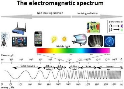
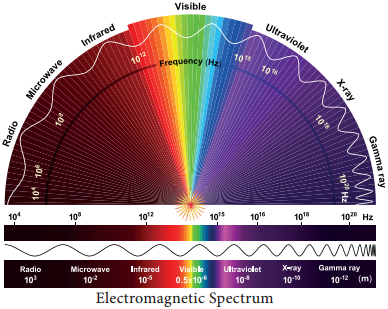
In the total electromagnetic spectrum, visible light is the smallest part. The entire life on earth depends on light and is the driving force for all organisms. Plants have natural potential to utilize solar energy directly. In the given picture electromagnetic radiation spectrum and components of visible spectrum are mentioned. The wavelength of solar radiation which reaches the earth is between 300 to 2600 nm.

The visible spectrum ranges between 390 to 763 nm (3900 å to 7630 å). The colour of the light is determined by the wavelength. Energy of the quantum is inversely proportional to wavelength. Shorter wavelength has more energy than longer wavelength. Electromagnetic spectrum consists of 7 types of radiations such as gamma rays, X rays, U-V rays, Visible light spectrum, infrared rays, electric rays and radio rays (Figure 13. 4).
Spectrum of Electromagnetic Radiation img 1

Properties of Light

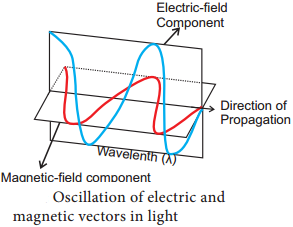
  1. Light is a transverse electromagnetic wave.
  2. It consists of oscillating electric and magnetic fields that are perpendicular to each other and perpendicular to the direction of propagation of the light.
  3. Light moves at a speed of 3 × 10⁸ ms-1
  4. Wavelength is the distance between successive crests of the wave.
  5. Light as a particle is called photon. Each photon contains an amount of energy known as quantum.
  6. The energy of a photon depends on the frequency of the light (Figure 13.5).Spectrum of Electromagnetic Radiation img 2

The entire electromagnetic spectrum, from the lowest to the highest frequency (longest to shortest wavelength), includes all radio waves (e.g., commercial radio and television, microwaves, radar), infrared radiation, visible light, ultraviolet radiation, X-rays, and gamma rays.

The EM spectrum is generally divided into seven regions, in order of decreasing wavelength and increasing energy and frequency. The common designations are: radio waves, microwaves, infrared (IR), visible light, ultraviolet (UV), X-rays and gamma rays.

The electromagnetic spectrum is a continuum of all electromagnetic waves arranged according to frequency and wavelength. The sun, earth, and other bodies radiate electromagnetic energy of varying wavelengths. Electromagnetic energy passes through space at the speed of light in the form of sinusoidal waves.

The characteristics of the electromagnetic spectrum are the propagation features and the amount of information, which signals can carry. In general, signals sent using the higher frequencies have shorter propagation distances but a higher data-carrying capacity.

Radio waves, microwaves, visible light, and x rays are all examples of electromagnetic waves that differ from each other in wavelength.

  • Longer Wavelength
  • Shorter Wavelength

Electromagnetic waves are produced by the motion of electrically charged particles. The different types of waves have different uses and functions in our everyday lives. The most important of these is visible light, which enables us to see. Radio waves have the longest wavelengths of all the electromagnetic waves. They range from around a foot long to several miles long.

“Electromagnetic spectrum” refers to the spectrum of electromagnetic radiation, and electromagnetic radiation is so named because it consists of electric and magnetic fields. In fact, light does affect charges and currents.

These observations enable astronomers to determine certain physical characteristics of objects, such as their temperature, composition and velocity. The electromagnetic spectrum consists of much more than visible light. It includes wavelengths of energy that human eyes can’t perceive.

Cell phones use antennae to transmit and receive radio waves that carry binary information. Every cell tower presides over an area of land, where it receives and transmits radio waves. When a text message is written, it is transmitted as binary code using a particular frequency of radio waves specific to that user.

Almost all of the energy available at Earth’s surface comes from the sun. The sun gets its energy from the process of nuclear fusion. This energy eventually makes its way to the outer regions of the sun and is radiated or emitted away in the form of energy, known as electromagnetic radiation.

Yes, all objects, including human bodies, emit electromagnetic radiation. The wavelength of radiation emitted depends on the temperature of the objects. Such radiation is sometimes called thermal radiation. Most of the radiation emitted by human body is in the infrared region, mainly at the wavelength of 12 micron.
Spectrum of Electromagnetic Radiation img 3

Photosynthetic Pigments

Learninsta presents the core concepts of Biology with high-quality research papers and topical review articles.

Photosynthetic Pigments

A photosynthetic pigment is a pigment that is present in chloroplasts or photosynthetic bacteria which captures the light energy necessary for photosynthesis (Table 13.1).
Photosynthetic Pigments img 1

Chlorophyll

Chlorophyll ‘a’ is the primary pigment which acts as a reaction centre and all other pigments act as accessory pigments and trap solar energy and then transfer it to chlorophyll ‘a’. Chlorophyll molecules have a tadpole like structure. It consists of Mg-Porphyrin head (Hydrophilic Head) and (Lipophilic tail) Phytol tail. The Porphyrin head consists of four pyrrol rings linked together by C-H bridges.

Each pyrrole ring comprises of four carbons and one nitrogen atom. Porphyrin ring has several side groups which alter the properties of the pigment. Different side groups are indicative of various types of chlorophyll. The Phytol tail made up of 20 carbon alcohol is attached to carbon 7 of the Pyrrole ring IV. It has a long propionic acid ester bond. Long lipophilic tail helps in anchoring chlorophyll to the lamellae.

Carotenoids

Carotenoids are yellow to orange pigments, mostly tetraterpens and these pigments absorb light strongly in the blue to violet region of visible spectrum. These pigments protect chlorophyll from photo-oxidative damage. Hence, they are called as shield pigments.

These pigments absorb light and transfer these to chlorophyll. Almost all carotenoid pigments have 40 carbon atoms. Ripening of fruits, floral colours and leaf colour change during autumn is due to Carotenoids (Carotene and Xanthophyll) (Figure 13.2).
Photosynthetic Pigments img 2

(i) Carotenes:

Orange, Red, Yellow and Brownish pigments, hydrocarbons (Lipids) and most of them are tetraterpenes (C40H56). Carotene is the most abundant Carotene in plants and it is a precursor of Vitamin A. Lycopene is the red pigment found in the fruits of tomato, red peppers and roses.

(ii) Xanthophylls:

Yellow (C40H56O2) pigments are like carotenes but contain oxygen. Lutein is responsible for yellow colour change of leaves during autumn season. Examples: Lutein, Violaxanthin and Fucoxanthin.

Phycobilins

They are proteinaceous pigments, soluble in water, and do not contain Mg and Phytol tail. They exist in two forms such as:-

  1. Phycocyanin found in cyanobacteria
  2. Phycoerythrin found in rhodophycean algae (Red algae).