Human Menstrual Cycle

Learninsta presents the core concepts of Biology with high-quality research papers and topical review articles.

Human Menstrual Cycle

The menstrual or ovarian cycle occurs approximately once in every 28/29 days during the reproductive life of the female from menarche (puberty) to menopause except during pregnancy. The cycle of events starting from one menstrual period till the next one is called the menstrual cycle during which cyclic changes occurs in the endometrium every month. Cyclic menstruation is an indicator of normal reproductive phase (Fig. 2.9).
Menstrual Cycle img 1

Menstrual cycle comprises of the following phases

  1. Menstrual phase
  2. Follicular or proliferative phase
  3. Ovulatory phase
  4. Luteal or secretory phase

1. Menstrual phase

The cycle starts with the menstrual phase when menstrual flow occurs and lasts for 3-5 days. Menstrual flow is due to the breakdown of endometrial lining of the uterus, and its blood vessels due to decline in the level of progesterone and oestrogen. Menstruation occurs only if the released ovum is not fertilized. Absence of menstruation may be an indicator of pregnancy. However it could also be due to stress, hormonal disorder and anaemia.

2. Follicular or proliferative phase

The follicular phase extends from the 5th day of the cycle until the time of ovulation. During this phase, the primary follicle in the ovary grows to become a fully mature Graafin follicle and simultaneously, the endometrium regenerates through proliferation. These changes in the ovary and the uterus are induced by the secretion of gonadotropins like FSH and LH, which increase gradually during the follicular phase. It stimulates follicular development and secretion of oestrogen by the follicle cells.

3. Ovulatory phase

Both LH and FSH attain peak level in the middle of the cycle (about the 14th day). Maximum secretion of LH during the mid cycle called LH surge induces the rupture of the Graafin follicle and the release of the ovum (secondary oocyte) from the ovary wall into the peritoneal cavity. Ths process is called as ovulation.

4. Luteal or secretory phase

During luteal phase, the remaining part of the Graafin follicle is transformed into a transitory endocrine gland called corpus luteum. The corpus luteum secretes large amount of progesterone which is essential for the maintenance of the endometrium. If Fertilization takes place, it paves way for the implantation of the fertilized ovum. The uterine wall secretes nutritious fluid in the uterus for the foetus. So, this phase is also called as secretory phase. During pregnancy all events of menstrual cycle stop and there is no menstruation.

In the absence of Fertilization, the corpus luteum degenerates completely and leaves a scar tissue called corpus albicans. It also initiates the disintegration of the endometrium leading to menstruation, marking the next cycle.

Menstrual hygiene

Menstrual hygiene is vital for good health, well-being, dignity, empowerment and productivity of women. The impact of poor menstrual hygiene on girls is increased stress levels, fear and embarrassment during menstruation. This can keep girls inactive during such periods leading to absenteeism from school.

Clean and safe absorbable clothing materials, sanitary napkins, pads, tampons and menstrual cups have been identified as materials used to manage menstruation. Changing sanitary material 4-5 hours as per the requirement, provides comfort, cleanliness and protection from infections.

It also helps in enhancing the quality of life of women during this period. Used sanitary napkins should be wrapped in paper and disposed. It should not be thrown in open areas or drain pipe of toilets. Flushing of sanitary napkins in the drain pipes causes choking of the drainage line leading to water pollution.

Menopause

Menopause is the phase in a women’s life when ovulation and menstruation stops. The average age of menopause is 45-50 years. It indicates the permanent cessation of the primary functions of the ovaries.

Human Reproductive Gametogenesis

Learninsta presents the core concepts of Biology with high-quality research papers and topical review articles.

Human Reproductive Gametogenesis

Gametogenesis is the process of formation of gametes i.e., sperms and ovary from the primary sex organs in all sexually reproducing organisms. Meiosis plays the most significant role in the process of gametogenesis (Fig. 2.5).
Human Reproductive Gametogenesis img 1

Spermatogenesis

Spermatogenesis is the sequence of events in the seminiferous tubules of the testes that produce the male gametes, the sperms. During development, the primordial germ cells migrate into the testes and become immature germ cells called sperm mother cells or spermatogonia in the inner surfaces of the seminiferous tubules (Fig. 2.6 a).
Human Reproductive Gametogenesis img 2

The spermatogonia begin to undergo mitotic division at puberty and continue throughout life. In the first stage of spermatogenesis, the spermatogonia migrate among sertoli cells towards the central lumen of the seminiferous tubule and become modified and enlarged to form primary spermatocytes which are diploid with 23 pairs i.e., 46 chromosomes.

Some of the primary spermatocytes undergo first meiotic division to form two secondary spermatocytes which are haploid with 23 chromosomes each. The secondary spermatocytes undergo second meiotic division to produce four haploid spermatids. The spermatids are transformed into mature spermatozoa (sperms) by the process called spermiogenesis. Sperms are finally released into the cavity of seminiferous tubules by a process called spermiation.

The whole process of spermatogenesis takes about 64 days. At any given time, different regions of the seminiferous tubules contain spermatocytes in different stages of development (Fig. 2.6 b). The sperm production remains nearly constant at a rate of about 200 million sperms per day.
Human Reproductive Gametogenesis img 3

Spermatogenesis starts at the age of puberty and is initiated due to the increase in the release of Gonadotropin Releasing Hormone (GnRH) by the hypothalamus. GnRH acts on the anterior pituitary gland and stimulates the secretion of two gonadotropins namely Follicle Stimulating Hormone (FSH) and Lutenizing Hormone (LH).

FSH stimulates testicular growth and enhances the production of Androgen Binding Protein (ABP) by the sertoli cells and helps in the process of spermiogenesis. LH acts on the Leydig cells and stimulates the synthesis of testosterone which in turn stimulates the process of spermatogenesis.

Structure of human spermatozoan The human sperm is a microscopic, flagellated and motile gamete (Fig. 2.7). The whole body of the sperm is enveloped by plasma membrane and is composed of a head, neck and a tail. The head comprises of two parts namely acrosome and nucleus.

Acrosome is a small cap like pointed structure present at the tip of the nucleus and is formed mainly from the Golgi body of the spermatid. It contains hyaluronidase, a proteolytic enzyme, popularly known as sperm lysin which helps to penetrate the ovum during Fertilization. The nucleus is flat and oval. The neck is very short and is present between the head and the middle piece.

It contains the proximal centriole towards the nucleus which plays a role in the first division of the zygote and the distal centriole gives rise to the axial filament of the sperm. The middle piece possesses mitochondria spirally twisted around the axial filament called mitochondrial spiral or nebenkern. It produces energy in the form of ATP molecules for the movement of sperms. The tail is the longest part of the sperm and is slender and tapering.

It is formed of a central axial filament or axoneme and an outer protoplasmic sheath. The lashing movements of the tail push the sperm forward. The human male ejaculates about 200 to 300 million sperms during coitus. It is estimated that around 60 percent of sperms must have normal shape of which at least 40 per cent must show vigorous motility for normal fertility.
Human Reproductive Gametogenesis img 4

Oogenesis

Oogenesis is the process of development of the female gamete or ovum or egg in the ovaries. During foetal development, certain cells in the germinal epithelium of the foetal ovary divide by mitosis and produce millions of egg mother cells or oogonia.

No more oogonia are formed or added after birth. The oogonial cells start dividing and enter into Prophase I of meiotic division I to form the primary oocytes which are temporarily arrested at this stage. The primary oocytes then get surrounded by a single layer of granulosa cells to form the primordial or primary follicles (Fig. 2.8 a). A large number of follicles degenerate during the period from birth to puberty, so at puberty only
60,000 to 80,000 follicles are lef in each ovary.
Human Reproductive Gametogenesis img 5

The primary follicle gets surrounded by many layers of granulosa cells and a new theca layer to form the secondary follicle. A fluid filed space, the antrum develops in the follicle and gets transformed into a tertiary follicle. The theca layer gets organized into an inner theca interna and an outer theca externa. At this time, the primary oocyte within the tertiary follicle grows in size and completes its first meiotic division and forms the secondary oocyte.

It is an unequal division resulting in the formation of a large haploid secondary oocyte and a first polar body. The first polar body disintegrates. During Fertilization, the secondary oocyte undergoes second meiotic
division and produces a large cell, the ovum and a second polar body.

The second polar body also degenerates. The tertiary follicle eventually becomes a mature follicle or Graafin follicle. If Fertilization does not take place, second meiotic division is never completed and the egg disintegrates. At the end of gametogenesis in females, each primary oocyte gives rise to only one haploid ovum.

Structure of ovum

Human ovum is non-cleidoic, alecithal and microscopic in nature. (Fig. 2.8 (b)). Its cytoplasm called ooplasm contains a large nucleus called the germinal vesicle. The ovum is surrounded by three coverings namely an inner thin transparent vitelline membrane, middle thick zona pellucida and outer thick coat of follicular cells called corona radiata. Between the vitelline membrane and zona pellucida is a narrow perivitelline space.
Human Reproductive Gametogenesis img 6

Human Reproductive System

Learninsta presents the core concepts of Biology with high-quality research papers and topical review articles.

Human Reproductive System

These functions are carried out by the primary and accessory reproductive organs. The primary reproductive organs namely the ovary and testis are responsible for producing the ova and sperms respectively.

Hormones secreted by the pituitary gland and the gonads help in the development of the secondary sexual characteristics, maturation of the reproductive system and regulation of normal functioning of the reproductive system. The accessory organs help in transport and to sustain the gametes and to nurture the developing offspring.

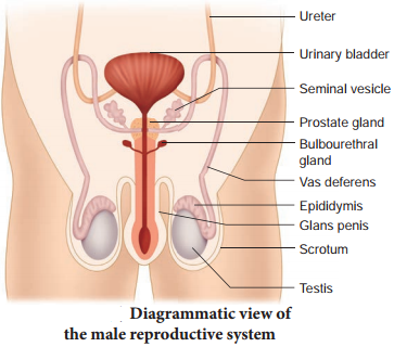
The male reproductive system comprises of a pair of testes, accessory ducts, glands and external genitalia (Fig. 2.1).
Human Reproductive System img 1

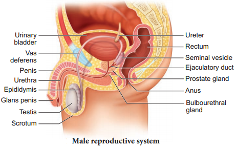
Testes are the primary male sex organs. They are a pair of ovoid bodies lying in the scrotum (Fig. 2.2 a). The scrotum is a sac of skin that hangs outside the abdominal cavity. Since viable sperms cannot be produced at normal body temperature, the scrotum is placed outside the abdominal cavity to provide a temperature 2-3°C lower than the normal internal body temperature. Thus, the scrotum acts as a thermoregulator for spermatogenesis.
Human Reproductive System img 2

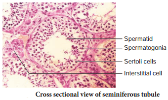
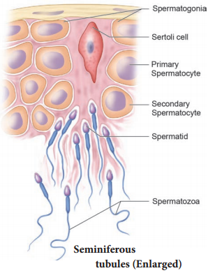
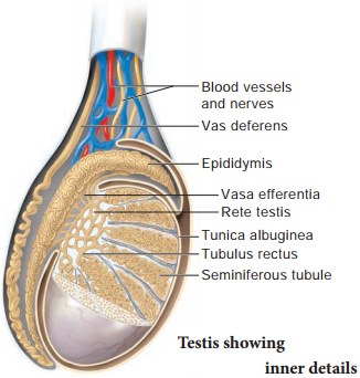
Each testis is covered by an outermost fibrous tunica albuginea and is divided by septa into about 200 – 250 lobules each containing 2-4 highly coiled testicular tubules or seminiferous tubules. These highly convoluted tubules which form 80 percent of the testicular substance are the sites for sperm production.

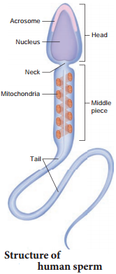
The stratified epithelium of the seminiferous tubule is made of two types of cells namely sertoli cells or nurse cells and spermatogonic cells or male germ cells. Sertoli cells are elongated and pyramidal and provide nourishment to the sperms till maturation. They also secrete inhibin, a hormone which is involved in the negative feedback control of sperm production. Spermatogonic cells divide meiotically and differentiate to produce spermatozoa.

Interstitial cells or Leydig cells are embedded in the soft connective tissue surrounding the seminiferous tubules. These cells are endocrine in nature and secrete androgens namely the testosterone hormone which initiates the process of spermatogenesis. These cells are endocrine in nature and are characteristic features of the testes of mammals. Other immunologically competent cells are also present.

The accessory ducts associated with the male reproductive system include rete testis, vasa efferentia, epididymis and vas deferens (Fig. 2.2 b). The seminiferous tubules of each lobule converge to form a tubulus rectus that conveys the sperms into the rete testis.

The rete testis is a tubular network on the posterior side of the testis. The sperms leave the rete testis and enter the epididymis through the vasa efferentia. The epididymis is a single highly coiled tube that temporarily stores the spermatozoa and they undergo physiological maturation and acquire increased motility and fertilizing capacity.

The epididymis leads to the vas deferens and joins the duct of the seminal vesicle to form the ejaculatory duct which passes through the prostate and opens into the urethra. The urethra is the terminal portion of the male reproductive system and is used to convey both urine and semen at different times. It originates from the urinary bladder and extends through the penis by an external opening called urethral meatus.

The accessory glands of the male reproductive system include the paired seminal vesicles and bulbourethral glands also called Cowper’s gland and a single prostate gland. The seminal vesicles secrete an alkaline flied called seminal plasma containing fructose sugar, ascorbic acid, prostaglandins and a coagulating enzyme called vesiculase which enhances sperm motility. The bulbourethral glands are inferior to the prostate and their secretions also help in the lubrication of the penis.

The prostate encircles the urethra and is just below the urinary bladder and secretes a slightly acidic fluid that contains citrate, several enzymes and prostate specifi antigens. Semen or seminal fluid is a milky white fluid which contains sperms and the seminal plasma (secreted from the seminal vesicles, prostate gland and the bulbourethal glands). The seminal fluid acts as a transport medium, provides nutrients, contains chemicals that protect and activate the sperms and also facilitate their movement.
Human Reproductive System img 3

The penis is the male external genitalia functioning as a copulatory organ. It is made of a special tissue that helps in the erection of penis to facilitate insemination. The enlarged end of the penis called glans penis is covered by a loose fold of skin called foreskin or prepuce.

The female reproductive system is far more complex than the male because in addition to gamete formation, it has to nurture the developing foetus. The female reproductive system consists of a pair of ovaries along with a pair of oviducts, uterus, cervix, vagina and the external genitalia located in the pelvic region (Fig. 2.3 a). These parts along with the mammary glands are integrated structurally and functionally to support the process of ovulation, Fertilization, pregnancy, child birth and child care.

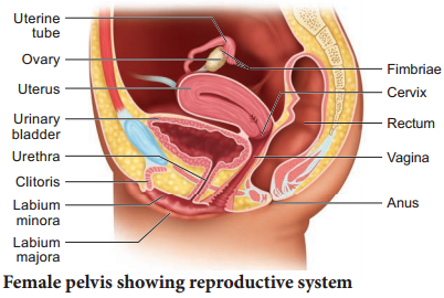
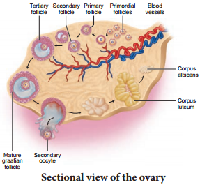
Ovaries are the primary female sex organs that produce the female gamete, ovum. The ovaries are located one on each side of the lower abdomen. The ovary is an elliptical structure about 2-4 cm long. Each ovary is covered by a thin cuboidal epithelium called the germinal epithelium which encloses the ovarian stroma.

The stroma is diffrentiated as the outer cortex and inner medulla. Below the germinal epithelium is a dense connective tissue, the tunica albuginea. The cortex appears dense and granular due to the presence of ovarian follicles in various stages of development. The medulla is a loose connective tissue with abundant blood vessels, lymphatic vessels and nerve fires. The ovary remains attached to the pelvic wall and the uterus by an ovarian ligament called mesovarium.

The fallopian tubes (uterine tubes or oviducts), uterus and vagina constitute the female accessory organs (Fig. 2.3 b). Each fallopian tube extends from the periphery of each ovary to the uterus. The proximal part of the fallopian tube bears a funnel shaped infundibulum.

The edges of the infundibulum have many finger like projections called fimbriae which help in collection of the ovum after ovulation. The infundibulum leads to a wider central portion called ampulla. The last part of the oviduct is the isthmus which is short and thick walled connecting the ampulla and infundibulum to the uterus.
Human Reproductive System img 5

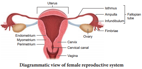
The uterus or womb is a hollow, thick-walled, muscular, highly vascular and inverted pear shaped structure lying in the pelvic cavity between the urinary bladder and rectum. The major portion of the uterus is the body and the rounded region superior to it, is the fundus.

The uterus opens into the vagina through a narrow cervix. The cavity of the cervix called the cervical canal communicates with the vagina through the external orifie and with the uterus through the internal orifie. The cervical canal along with vagina forms the birth canal.

The wall of the uterus has three layers of tissues. The outermost thin membranous serous layer called the perimetrium, the middle thick muscular layer called myometrium and the inner glandular layer called endometrium. The endometrium undergoes cyclic changes during the menstrual cycle while myometrium exhibits strong contractions during parturition.

Vagina is a large fibromuscular tube that extends from the cervix to the exterior. It is the female organ of copulation. The female reproductive structures that lie external to the vagina are called as the external genitalia or vulva comprising of labia majora, labia minora, hymen and clitoris.

The Bartholin’s glands (also called greater vestibular glands) are located posterior to the left and right of the opening of the vagina. They secrete mucus to lubricate the vagina and are homologous to the bulbourethral glands of the male. The Skene’s glands are located on the anterior wall of the vagina and around the lower end of the urethra. They secrete a lubricating fluid and are homologous to the prostate gland of the males.

The external opening of the vagina is partially closed by a thin ring of tissue called the hymen. The hymen is often torn during the first coitus (physical union). However in some women it remains intact. It can be stretched or torn due to a sudden fall or jolt and also during strenuous physical activities such as cycling, horseback riding, etc., and therefore cannot be considered as an indicator of a woman’s virginity.

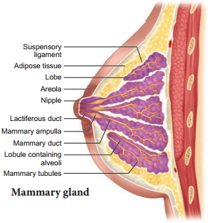
The mammary glands are modified sweat glands present in both sexes. It is rudimentary in the males and functional in the females. A pair of mammary glands is located in the thoracic region. It contains glandular tissue and variable quantities of fat with a median nipple surrounded by a pigmented area called the areola. Several sebaceous glands called the areolar glands are found on the surface and they reduce cracking of the skin of the nipple.

Internally each mammary gland consists of 2-25 lobes, separated by fat and connective tissues (Fig. 2.4). Each lobe is made up of lobules which contain acini or alveoli lined by epithelial cells. Cells of the alveoli secrete milk. The alveoli open into mammary tubules.

The tubules of each lobe join to form a mammary duct. Several mammary ducts join to form a wider mammary ampulla which is connected to the lactiferous duct in the nipple. Under the nipple, each lactiferous duct expands to form the lactiferous sinus which serves as a reservoir of milk. Each lactiferous duct opens separately by a minute pore on the surface of the nipple.
Human Reproductive System img 6

Normal development of the breast begins at puberty and progresses with changes during each menstrual cycle. In non-pregnant women, the glandular structure is largely underdeveloped and the breast size is largely due to amount of fat deposits. The size of the breast does not have an influence on the efficiency of lactation.

Sexual Reproduction | Parthenogenesis (Gr. Parthenos – virgin, Genesisproduce)

Learninsta presents the core concepts of Biology with high-quality research papers and topical review articles.

Sexual Reproduction | Parthenogenesis (Gr. Parthenos – virgin, Genesisproduce)

Sexual reproduction involves the fusion of male and female gametes to form a diploid zygote, which develops into a new organism. It leads to genetic variation. The types of sexual reproduction seen in animals are syngamy (fertilization) and conjugation. In syngamy, the fusion of two haploid gametes takes place to produce a diploid zygote. Depending upon the place where the fertilization takes place, it is of two types.

In external fertilization, the fusion of male and female gametes takes place outside the body of female organisms in the water medium. e.g. sponges, fishes and amphibians. In internal fertilization, the fusion of male and female gametes takes place within the body of female organisms. e.g. reptiles, aves and mammals.

Different kinds of syngamy (fertilization) are prevalent among living organisms.

(a) Autogamy:

The male and female gametes are produced by the same cell or same organism and both the gametes fuse together to form a zygote. e.g. Actinosphaerium and Paramecium.

(b) Exogamy:

The male and female gametes are produced by different parents and they fuse to form a zygote. So it is biparental. e.g. Human – dioecious or unisexual animal.

(c) Hologamy:

Lower organisms, sometimes the entire mature organisms do not form gametes but they themselves behave as gametes and the fusion of such mature individuals is known as hologamy e.g. Trichonympha.

(d) Paedogamy:

It is the sexual union of young individuals produced immediately after the division of the adult parent cell by mitosis.

(e) Merogamy:

The fusion of small sized and morphologically different gametes (merogametes) takes place.

(f) Isogamy:

The fusion of morphological and physiological identical gametes (isogametes) is called isogamy. e.g. Monocystis.

(g) Anisogamy:

The fusion of dissimilar gametes is called anisogamy (Gr. An-without; iso-equal; gam-marriage). Anisogamy occurs in higher animals but it is customary to use the term fertilization instead of anisogamy or syngamy. e.g. higher invertebrates and all vertebrates.

Conjugation is the temporary union of the two individuals of the same species. During their union both individuals, called the conjugants exchange certain amount of nuclear material (DNA) and then get separated. Conjugation is common among ciliates, e.g. Paramecium, Vorticella and bacteria (Prokaryotes).

Phases of life cycle:

Organisms have three phases – Juvenile phase, reproductive phase and senescent phase. Juvenile phase/vegetative phase is the period of growth between the birth of the individual upto reproductive maturity. During reproductive phase/ maturity phase the organisms reproduce and their offsprings
reach maturity period. On the basis of time, breeding animals are of two types: seasonal breeders and continuous breeders.

Seasonal breeders reproduce at particular period of the year such as frogs, lizards, most birds, deers etc., Continuous breeders continue to breed throughout their sexual maturity e.g. honey bees, poultry, rabbit etc., Senescent phase begins at the end of reproductive phase when degeneration sets in the structure and functioning of the body.

Parthenogenesis (Gr. Parthenos – virgin, Genesisproduce)

Development of an egg into a complete individual without fertilization is known as parthenogenesis. It was first discovered by Charles Bonnet in 1745. Parthenogenesis is of two main types namely, Natural Parthenogenesis and Artificial Parthenogenesis. In certain animals, parthenogenesis occurs regularly, constantly and naturally in their life cycle and is known as natural parthenogenesis.

Natural parthenogenesis may be of two types, viz., complete and incomplete. Complete parthenogenesis is the only form of reproduction in certain animals and there is no biparental sexual reproduction. There are no male organisms and so, such individuals are represented by females only.

Incomplete parthenogenesis is found in some animals in which both sexual reproduction and parthenogenesis occurs. e.g. In honeybees; fertilized eggs (zygotes) develop into queen and workers, whereas unfertilized eggs develop into drones (male).

In paedogenetic parthenogenesis (paedogenesis) the larvae produce a new generation of larvae by parthenogenesis. It occurs in the sporocysts and Redia larvae of liver fluke. It is also seen in the larvae of some insects. e.g. Gall fly. In artificial parthenogenesis, the unfertilized egg (ovum) is induced to develop into a complete individual by physical or chemical stimuli. e.g., Annelid and seaurchin eggs.

Asexual Reproduction in Organisms

Learninsta presents the core concepts of Biology with high-quality research papers and topical review articles.

Asexual Reproduction in Organisms

Asexual reproduction is wide spread among different organisms. It is common in members of Protista, Bacteria, Archaea and in multicellular organisms with relatively simple organisation. The offsprings show “uniparental inheritance” without any genetic variation. The different modes of asexual reproduction seen in animals are fission, budding, fragmentation and regeneration.

Fission is the division of the parent body into two or more identical daughter individuals. Five types of fission are seen in animals. They are binary fission, multiple fission, plasmotomy, strobilation and sporulation.

In binary fission, the parent organism divides into two halves and each half forms a daughter individual. The nucleus divides first amitotically or mitotically (karyokinesis), followed by the division of the cytoplasm (cytokinesis). The resultant offsprings are genetically identical to the parent. Depending on the plane of fission, binary fission is of the following types,

  • Simple irregular binary fission
  • Transverse binary fission
  • Longitudinal binary fission
  • Oblique binary fission

Simple irregular binary fission is seen in Amoeba like irregular shaped organisms (Fig. 1.1), where the plane of division is hard to observe. The contractile vacuoles cease to function and disappear. The nucleoli disintegrate and the nucleus divides mitotically. The cell then constricts in the middle, so the cytoplasm divides and forms two daughter cells.
Asexual Reproduction img 1

In transverse binary fission, the plane of the division runs along the transverse axis of the individual. e.g. Paramecium and Planaria. In Paramecium (Fig. 1.2) the macronucleus divides by amitosis and the micronucleus divides by mitosis.
Asexual Reproduction img 2

In longitudinal binary fision, the nucleus and the cytoplasm divides in the longitudinal axis of the organism (Fig 1.3). In flagellates, the flagellum is retained usually by one daughter cell. The basal granule is divided into two and the new basal granule forms a flagellum in the other daughter individual. e.g. Vorticella and Euglena.
Asexual Reproduction img 3

In oblique binary fission the plane of division is oblique. It is seen in dinoflagellates. e.g. Ceratium.

In multiple fission the parent body divides into many similar daughter cells simultaneously. First, the nucleus divides repeatedly without the division of the cytoplasm, later the cytoplasm divides into as many parts as that of nuclei.

Each cytoplasmic part encircles one daughter nucleus. This results in the formation of many smaller individuals from a single parent organism. If multiple fission produces four or many daughter individuals by equal cell division and the young ones do not separate until the process is complete, then this division is called repeated fission. e.g. Vorticella.

During unfavorable conditions (increase or decrease in temperature, scarcity of food) Amoeba withdraws its pseudopodia and secretes a three-layered, protective, chitinous cyst wall around it and becomes inactive (Fig. 1.4). This phenomenon is called encystment.

When conditions become favourable, the encysted Amoeba divides by multiple fission and produces many minute amoebae called pseudopodiospore or amoebulae. The cyst wall absorbs water and breaks off liberating the young pseudopodiospores, each with a fine pseudopodia. They feed and grow rapidly to
lead an independent life.
Asexual Reproduction img 4

In some metazoan animals, a special type of transverse fission called strobilation occurs (Fig. 1.5). In the process of strobilation, several transverse fissions occur simultaneously giving rise to a number of individuals which often do not separate immediately from each other e.g. Aurelia.

Plasmotomy is the division of multinucleated parent into many multinucleate daughter individuals with the
division of nuclei. Nuclear division occurs later to maintain normal number of nuclei. Plasmotomy occurs in Opalina and Pelomyxa (Giant Amoeba).
Asexual Reproduction img 5

During unfavourable conditions Amoeba multiplies bysporulation without encystment. Nucleus breaks into several small fragments or chromatin blocks. Each fragment develops a nuclear membrane, becomes surrounded by cytoplasm and develops a spore-case around it (Fig. 1.6). When conditions become favourable, the parent body disintegrates and the spores are liberated, each hatching into a young amoeba.
Asexual Reproduction img 6

In budding, the parent body produces one or more buds and each bud grows into a young one. The buds separate from the parent to lead a normal life. In sponges, the buds constrict and detach from the parent body and the bud develops into a new sponge (Fig. 1.7).
Asexual Reproduction img 7

When buds are formed on the outer surface of the parent body, it is known as exogenous budding e.g. Hydra. In Hydra when food is plenty, the ectoderm cells increase and form a small elevation on the body surface (Fig. 1.8).

Ectoderm and endoderm are pushed out to form the bud. The bud contains an interior lumen in continuation with parent’s gastro-vascular cavity. The bud enlarges, develops a mouth and a circle of tentacles at its free end. When fully grown, the bud constricts at the base and finally separates from the parent body and leads an independent life.
Asexual Reproduction img 8

In Noctiluca, hundreds of buds are formed inside the cytoplasm and many remain within the body of the parent. This is called endogenous budding. In freshwater sponges and in some marine sponges a regular and peculiar mode of asexual reproduction occurs by internal buds called gemmules is seen (Fig. 1.9).

A completely grown gemmule is a hard ball, consisting of an internal mass of food-laden archaeocytes. During unfavourable conditions, the sponge disintegrates but the gemmule can withstand adverse conditions. When conditions become favourable, the gemmules begin to hatch.
Asexual Reproduction img 9

In fragmentation, the parent body breaks into fragments (pieces) and each of the fragment has the potential to develop into a new individual. Fragmentation or pedal laceration occurs in many genera of sea anemones. Lobes are constricted of from the pedal disc and each of the lobe grows mesenteries and tentacles to form a new sea anemone.

In the tapeworm, Taenia solium the gravid (ripe) proglottids are the oldest at the posterior end of the strobila (Fig. 1.10). The gravid proglottids are regularly cut of either singly or in groups from the posterior end by a process called apolysis. This is very significant since it helps in transferring the developed embryos from the primary host (man) to fid a secondary host (pig).
Asexual Reproduction img 10

Regeneration is regrowth in the injured region. Regeneration was first studied in Hydra by Abraham Trembley in 1740. Regeneration is of two types, morphallaxis and epimorphosis. In morphallaxis the whole body grows from a small fragment e.g. Hydra and Planaria. When Hydra is accidentally cut into several pieces, each piece can regenerate the lost parts and develop into a whole new individual (Fig. 1.11).
Asexual Reproduction img 11

The parts usually retain their original polarity, with oral ends, by developing tentacles and aboral ends, by producing basal discs. Epimorphosis (Fig. 1.12) is the replacement of lost body parts. It is of two types, namely reparative and restorative regeneration. In reparative regeneration, only certain damaged tissue can be regenerated, e.g. human beings whereas in restorative regeneration severed body parts can develop. e.g. star fish, tail of wall lizard.
Asexual Reproduction img 12