Amniocentesis and Its Statutory Ban

Learninsta presents the core concepts of Biology with high-quality research papers and topical review articles.

Amniocentesis and Its Statutory Ban

Due to small family norms and the skewed choice for a male child, female population is decreasing at an alarming rate. Amniocentesis is a prenatal technique used to detect any chromosomal abnormalities in the foetus and it is being often misused to determine the sex of the foetus. Once the sex of the foetus is known, there may be a chance of female foeticide. Hence, a statutory ban on amniocentesis is imposed.

It is used to detect any chromosomal defect in the embryo. However, recently amniocentesis is being used to detect the gender of the foetus which results in several female foeticides. This declines the gender ratio.

Therefore, there is a statutory ban on amniocentesis to avoid female foeticides. The amniotic fluid contains cells from foetus skin and respiratory tract. Sex of the foetus is determined using amniocentesis and then if it turns out to been a female one, foetus is aborted. That is why amniocentesis has been banned in India.

Yes, the ban is necessary because amniocentesis is misused now-a-days. It is used to determine the sex of the foetus and in many cases it led to female foeticide. In such extreme cases that would be incurable, adecision to abort the foetus could be taken.

Amniocentesis is a procedure in which amniotic fluid is removed from the uterus for testing or treatment. Amniotic fluid is the fluid that surrounds and protects a baby during pregnancy. This fluid contains fetal cells and various proteins.

Need For Reproductive Health Problems and Strategies

Learninsta presents the core concepts of Biology with high-quality research papers and topical review articles.

Need For Reproductive Health Problems and Strategies

India is amongst the first few countries in the world to initiate the ‘Family planning programme’ since 1951 and is periodically assessed every decade. These programmes are popularly named as ‘Reproductive and Child Health Care (RCH). Major tasks carried out under these programmes are:

  • Creating awareness and providing medical assistance to build a healthy society.
  • Introducing sex education in schools to provide information about adolescence and adolescence related changes.
  • Educating couples and those in the marriageable age groups about the available birth control methods and family planning norms.
  • Creating awareness about care for pregnant women, post-natal care of mother and child and the importance of breast feeding.
  • Encouraging and supporting governmental and non-governmental agencies to identify new methods and/or to improve upon the existing methods of birth control.

Family planning counselling, pre-natal care, safe delivery, post-natal care, appropriate treatment of infertility, prevention of abortion, treatment of sexually transmitted diseases, responsible parenthood, services against HIV/AIDS, breast cancer should be made available.

Sexually Transmitted Diseases. Ill health of both mother and her baby. Early marriages before attaining the puberty. An increased mortality rate of both mother and Infants. Reproductive health in a society helps to prevent the spread of various sexually transmitted diseases and impart the ability to produce offspring carrying better survival rates. Sex education helps in maintaining the population size and to avoid unwanted pregnancies.

The important conditions to maintain good reproductive health during adolescence are: 1)It is necessary to eat balanced diet during adolescence. 2)It is necessary to maintain personal hygiene during adolescence. 3)It is necessary to take adequate physical exercise during adolescence.

Parturition and Lactation

Learninsta presents the core concepts of Biology with high-quality research papers and topical review articles.

Parturition and Lactation

Parturition is the completion of pregnancy and giving birth to the baby. The series of events that expels the infant from the uterus is collectively called “labour”. Thoughout pregnancy the uterus undergoes periodic episodes of weak and strong contractions. These contractions called Braxter-Hick’s contractions lead to false labour. As the pregnancy progresses, increase in the oestrogen concentration promotes uterine contractions.

These uterine contractions facilitate moulding of the foetus and downward movement of the foetus. The descent of the foetus causes dilation of cervix of the uterus and vaginal canal resulting in a neurohumoral reflex called Foetal ejection reflex or Ferguson reflex.

This initiates the secretion of oxytocin from the neurohypophysis which in turn brings about the powerful contraction of the uterine muscles and leads to the expulsion of the baby through the birth canal. This sequence of events is called as parturition or childbirth.

Relaxin is a hormone secreted by the placenta and also found in the corpus luteum. It promotes parturition by relaxing the pelvic joints and by dilatation of the cervix with continued powerful contractions. The amnion ruptures and the amniotic fluid flows out through the vagina, followed by the foetus. The placenta along with the remains of the umbilical cord called “after birth” is expelled out after delivery.

Lactation is the production of milk by mammary glands. The mammary glands show changes during every menstrual cycle, during pregnancy and lactation. Increased level of oestrogens, progesterone and human Placental Lactogen (hPL) towards the end of pregnancy stimulate the hypothalamus towards prolactin – releasing factors. The anterior pituitary responds by secreting prolactin which plays a major role in lactogenesis.

Oxytocin causes the “Let-Down” reflxthe actual ejection of milk from the alveoli of the mammary glands. During lactation, oxytocin also stimulates the recently emptied uterus to contract, helping it to return to pre – pregnancy size.

The mammary glands secrete a yellowish fluid called colostrum during the initial few days after parturition. It has less lactose than milk and almost no fat, but it contains more proteins, vitamin A and minerals. Colostrum is also rich in IgA antibodies.

This helps to protect the infant’s digestive tract against bacterial infection. Breast milk is the ideal food for infants as it contains all the constituents in suitable concentration and is easily digestible. It is fully sufficient till about 6 months of age and all infants must be breast fed by the mother to ensure the growth of a healthy baby.
Parturition And Lactation img 1

Maintenance Of Pregnancy and Embryonic Development

Learninsta presents the core concepts of Biology with high-quality research papers and topical review articles.

Maintenance Of Pregnancy and Embryonic Development

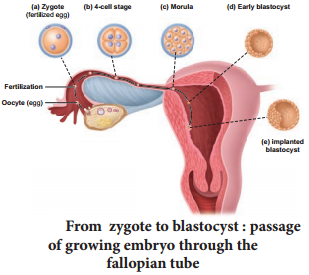
The inner cell mass in the blastula is differentiated into epiblast and hypoblast immediately after implantation. The hypoblast is the embryonic endoderm and the epiblast is the ectoderm. The cells remaining in between the epiblast and the endoderm form the mesoderm.

Thus the transformation of the blastocyst into a gastrula with the primary germ layers by the movement of the blastomeres is called gastrulation. Each germ layer gives rise to specific tissues, organs and organ systems during organogenesis.

The extra embryonic membranes namely the amnion, yolk sac, allantois and chorion protect the embryo from dessication, mechanical shock and help in the absorption of nutrients and exchange of gases (Fig. 2.12). The amnion is a double layered translucent membrane filled with the amniotic fluid.

It provides a buoyant environment to protect the developing embryo from injury, regulates the temperature of the foetus and provides a medium in which the foetus can move. The yolk sac forms a part of the gut and is the source of the earliest blood cells and blood vessels.
Maintenance Of Pregnancy And Embryonic Development img 1

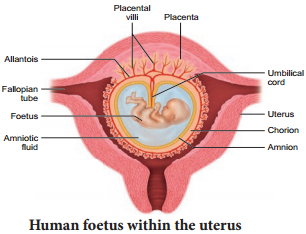
The allantois forms a small out pocketing of embryonic tissue at the caudal end of the yolk, sac. It is the structural base for the umbilical cord that links the embryo to the placenta and ultimately it becomes part of the urinary bladder. The chorion is the outermost membrane which encloses the embryo and all other membranes and also helps in the formation of the placenta.

The trophoblast cells in the blastocyst send out several finger like projections called chorionic villi carrying foetal blood and are surrounded by sinuses that contain maternal blood. The chorionic villi and the uterine tissues form the disc-shaped placenta.

Placenta is a temporary endocrine organ formed during pregnancy and it connects the foetus to the uterine wall through the umbilical cord. It is the organ by which the nutritive, respiratory and excretory functions are fulfilled.

The embryo’s heart develops during the fourth week of pregnancy and circulates blood through the umbilical cord and placenta as well as through its own tissues. The primary germ layers serve as the primitive tissues from which all body organs develop. The ectoderm gives rise to the central nervous system (brain and spinal cord), peripheral nervous system, epidermis and its derivatives and mammary glands.

The connective tissue, cartilage and bone, muscles, organs of urinogenital system (kidney, ureter and gonads) arise from the mesoderm. The endodermal derivatives are epithelium of gastrointestinal and respiratory tract, liver, pancreas, thyroid and parathyroids.

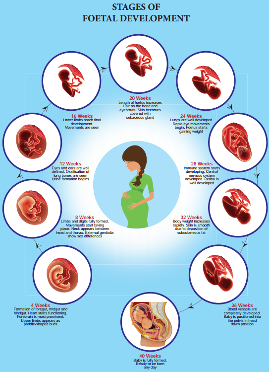
Human pregnancy lasts for about 280 days or 40 weeks and is called the gestation period. It can be divided for convenience into three trimesters of three months each. The first trimester is the main period of organogenesis, the body organs namely the heart, limbs, lungs, liver and external genital organs are well developed.

By the end of the second trimester, the face is well formed with features, eyelids and eyelashes, eyes blink, body is covered with fie hair, muscle tissue develops and bones become harder. The foetus is fully developed and is ready for delivery by the end of nine months (third trimester).

During pregnancy, the placenta acts as a temporary endocrine gland and produces large quantities of human Chorionic Gonadotropin (hCG), human Chorionic Somatomammotropin (hCS) or human Placental Lactogen (hPL), oestrogens and progesterone which are essential for a normal pregnancy.

A hormone called relaxin is also secreted during the later phase of pregnancy which helps in relaxation of the pelvic ligaments at the time of parturition. It should be noted that hCG, hPL and relaxin are produced only during pregnancy. In addition, during pregnancy the level of other hormones like oestrogen and progesterone, cortisol, prolactin, thyroxine, etc., is increased several folds in the maternal blood. These hormones are essential for supporting foetal growth.

Fertilization and Implantation

Learninsta presents the core concepts of Biology with high-quality research papers and topical review articles.

Fertilization and Implantation

Fertilization occurs when a haploid sperm fuses with a haploid ovum to form a fertilized egg or diploid zygote. The sperms deposited in the female reproductive tract undergo capacitation, which is a biochemical event that enables the sperm to penetrate and fertilise the egg. Fertilization occurs only if the ovum and sperms are transported simultaneously to the ampullary isthmic junction of the fallopian tube.

Before a sperm can enter the egg, it must penetrate the multiple layers of granulosa (follicular) cells which are around the ovum forming the corona radiata (Fig. 2.10). The follicular cells are held together by an adhesive cementing substance called hyaluronic acid. The acrosomal membrane disintegrates releasing the proteolytic enzyme, hyaluronidase during sperm entry through the corona radiata and zona pellucida.

This is called acrosomal reaction. Once Fertilization is accomplished, cortical granules from the cytoplasm of the ovum form a barrier called the Fertilization membrane around the ovum preventing further penetration of other sperms. This polyspermy is prevented.

The first cleavage produces two identical cells called blastomeres. These produce 4 cells, then 8 and so on. After 72 hours of Fertilization, a loose collection of cells forms a berry shaped cluster of 16 or more cells called the morula (Fig. 2.11).

Under the influence of progesterone, smooth muscles of the fallopian tube relax and the dividing embryo takes 4-5 days to move through the fallopian tube into the uterine cavity and finally gets implanted in the uterine wall.

At this point the embryo consists of a fluid filled hollow ball of about 100 cells, called the blastocyst. The blastocyst is composed of a single layer of large flattened cells called trophoblast and a small cluster of 20-30 rounded cells called the inner cell mass. The inner cell mass of the blastocyst develops into the embryo and becomes embedded in the endometrium of the uterus.
Fertilization And Implantation img 1

This process is called implantation and it results in pregnancy. If the fertilized ovum is implanted outside the uterus it results in ectopic pregnancy. About 95 percent of ectopic pregnancies occur in the fallopian tube. The growth of the embryo may cause internal bleeding, infection and in some cases even death due to rupture of the fallopian tube.
Fertilization And Implantation img 2