Genetic Control of Rh Factor

Learninsta presents the core concepts of Biology with high-quality research papers and topical review articles.

Genetic Control of Rh Factor

Fisher and Race Hypothesis:

Rh factor involves three different pairs of alleles located on three different closely linked loci on the chromosome pair. This system is more commonly in use today, and uses the ‘Cde’ nomenclature.
Genetic Control Of Rh Factor img 1

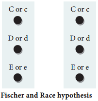
In the above Fig. 4.1, three pairs of Rh alleles (Cc, Dd and Ee) occur at 3 different loci on homologous chromosome pair. The possible genotypes will be one C or c, one D or d, one E or e from each chromosome. For e.g. CDE/cde; CdE/cDe; cde/cde; CDe/CdE etc., All genotypes carrying a dominant ‘D’ allele will produce Rh positive phenotype and double recessive genotype ‘dd’ will give rise to Rh negative phenotype.

Wiener Hypothesis

Wiener proposed the existence of eight alleles (R1, R2, RO, Rz, r, r1, r11, ry) at a single Rh locus. All genotypes carrying a dominant ‘R allele’ (R1, R2, RO, Rz) will produce Rh positive phenotype and double recessive genotypes (rr, r1r1, r11r11, ry ry) will give rise to Rh negative phenotype.

Incompatibility of Rh Factor – Erythroblastosis foetalis

Rh incompatability has great signifiance in child birth. If a woman is Rh negative and the man is Rh positive, the foetus may be Rh positive having inherited the factor from its father. The Rh negative mother becomes sensitized by carrying Rh positive foetus within her body. Due to damage of blood vessels, during child birth, the mother’s immune system recognizes the Rh antigens and gets sensitized. The sensitized mother produces Rh antibodies.

The antibodies are IgG type which are small and can cross placenta and enter the foetal circulation. By the time the mother gets sensitized and produce anti ‘D’ antibodies, the child is delivered.

Usually no effects are associated with exposure of the mother to Rh positive antigen during the first child birth, subsequent Rh positive children carried by the same mother, may be exposed to antibodies produced by the mother against Rh antigen, which are carried across the placenta into the foetal blood circulation. This causes haemolysis of foetal RBCs resulting in haemolytic jaundice and anaemia. This condition is known as Erythoblastosis foetalis or Haemolytic disease of the new born (HDN).

Prevention of Erythroblastosis Foetalis

If the mother is Rh negative and foetus is Rh positive, anti D antibodies should be administered to the mother at 28th and 34th week of gestation as a prophylactic measure. If the Rh negative mother delivers Rh positive child then anti D antibodies should be administered to the mother soon after delivery. This develops passive immunity and prevents the formation of anti D antibodies in the mothers blood by destroying the Rh foetal RBC before the mother’s immune system is sensitized. This has to be done whenever the woman attains pregnancy.

Human Blood Groups | ABO Blood Groups and Its Types

Learninsta presents the core concepts of Biology with high-quality research papers and topical review articles.

Human Blood Groups | ABO Blood Groups and Its Types

Multiple allelism occurs in humans, particularly in the inheritance of different types of blood groups. The blood group inheritance in human can be understood by learning about antigens and antibodies. The composition of blood, different types of blood groups (ABO) the blood antigens and antibodies were discussed in chapter 7 of class XI.

ABO blood types

Multiple allele inheritance of ABO blood groups Blood differs chemically from person to person. When two different incompatible blood types are mixed, agglutination (clumping together) of erythrocytes (RBC) occurs.

The basis of these chemical differences is due to the presence of antigens (surface antigens) on the membrane of RBC and epithelial cells. Karl Landsteiner discovered two kinds of antigens called antigen ‘A’ and antigen ‘B’ on the surface of RBC’s of human blood.

Based on the presence or absence of these antigens three kinds of blood groups, type ‘A’, type ‘B’, and type ‘O’ (universal donor)were recognized. The fourth and the rarest blood group ‘AB’ (universal recipient) was discovered in 1902 by two of Landsteiner’s students Von De Castelle and Sturli.

Bernstein in 1925 discovered that the inheritance of different blood groups in human beings is determined by a number of multiple allelic series. The three autosomal alleles located on chromosome 9 are concerned with the determination of blood group in any person.

The gene controlling blood type has been labeled as ‘L’ (after the name of the discoverer, Landsteiner) or I (from isoagglutination). The I gene exists in three allelic forms, IA, IB and IO. IA specifies A antigen. IB allele determines B antigen and IO allele specifies no antigen. Individuals who possess these antigens in their fluids such as the saliva are called secretors.

Each allele (IA and IB) produces a transferase enzyme. IA allele produces N-acetyl galactose transferase and can add N-acetyl galactosamine (NAG) and IB allele encodes for the enzyme galactose transferase that adds galactose to the precursor (i.e., H substances) In the case of IO/IO allele no terminal transferase enzyme is produced and therefore called “null” allele and hence cannot add NAG or galactose to the precursor.

From the phenotypic combinations it is evident that the alleles IA and IB are dominant to IO, but co-dominant to each other (IA = IB). Their dominance hierarchy can be given as (IA = IB > IO). A child receives one of three alleles from each parent, giving rise to six possible genotypes and four possible blood types (phenotypes). The genotypes are IAIA, IA IO, IBIB, IBIO, IAIB and IO IO.
Human Blood Groups img 1
Genetic basis of the human ABO blood groups

Rhesus or Rh Factor

The Rh factor or Rh antigen is found on the surface of erythrocytes. It was discovered in 1940 by Karl Landsteiner and Alexander Wiener in the blood of rhesus monkey, Macaca rhesus and later in human beings. The term ‘Rh factor’ refers to “immunogenic D antigen of the Rh blood group system. An individual having D antigen are Rh D positive (Rh+) and those without D antigen are Rh D negative (Rh)”.

Rhesus factor in the blood is inherited as a dominant trait. Naturally occurring Anti D antibodies are absent in the plasma of any normal individual. However if an Rh (Rh negative) person is exposed to Rh+ (Rh positive) blood cells (erythrocytes) for the first time, anti D antibodies are formed in the blood of that individual. On the other hand, when an Rh positive person receives Rh negative blood no effect is seen.

Principles of Inheritance and Variation Multiple Alleles

Learninsta presents the core concepts of Biology with high-quality research papers and topical review articles.

Principles of Inheritance and Variation Multiple Alleles

The genetic segregations in Mendelian inheritance reveal that all genes have two alternative forms – dominant and recessive alleles e.g. tall versus dwarf (T and t). The former is the normal allele or wild allele and the latter the mutant allele.

A gene can mutate several times producing several alternative forms. When three or more alleles of a gene that control a particular trait occupy the same locus on the homologous chromosome of an organism, they are called multiple alleles and their inheritance is called multiple allelism.

Multiple alleles exist in a population when there are many variations of a gene present. In organisms with two copies of every gene, also known as diploid organisms, each organism has the ability to express two alleles at the same time. They can be the same allele, which is called a homozygous genotype.

Multiple alleles refer to the occurrence of three or more than three alleles for a particular gene. Alleles are different or contrasting forms of a gene. For example, for the gene encoding for height, one allele can be for tallness, whereas the other can be for dwarfness.

Mendel implied that only two alleles, one dominant and one recessive, could exist for a given gene. The variant may be recessive or dominant to the wild-type allele. An example of multiple alleles is the ABO blood-type system in humans.

Polygenic Inheritance: Human skin color is a good example of polygenic (multiple gene) inheritance. A genotype with all “dominant” capital genes (AABBCC) has the maximum amount of melanin and very dark skin. A genotype with all “recessive” small case genes (aabbcc) has the lowest amount of melanin and very light skin.

Multiple alleles exist in a population when there are many variations of a gene present. In both haploid and diploid organisms, new alleles are created by spontaneous mutations. These mutations can arise in a variety of ways, but the effect is a different sequence of nucleic acid bases in the DNA.

Multiple alleles are present at the same locus of the chromosomes. A classical example of multiple alleles is found in ABO blood group system of humans. Despite the multiple alleles of any gene, an individual possess and can have only two alleles at a time.
Multiple Alleles img 1

Detection of Foetal Disorders During Early Pregnancy

Learninsta presents the core concepts of Biology with high-quality research papers and topical review articles.

Detection of Foetal Disorders During Early Pregnancy

Ultrasound scanning Ultrasound has no known risks other than mild discomfort due to pressure from the transducer on the abdomen or vagina. No radiation is used during this procedure. Ultrasonography is usually performed in the first trimester for dating, determination of the number of foetuses, and for assessment of early pregnancy complications.

Amniocentesis

Amniocentesis involves taking a small sample of the amniotic fluid that surrounds the foetus to diagnose for chromosomal abnormalities (Fig. 3.1).
Detection Of Foetal Disorders During Early Pregnancy img 1

Amniocentesis is generally performed in a pregnant woman between the 15th and 20th weeks of pregnancy by inserting a long, thin needle through the abdomen into the amniotic sac to withdraw a small sample of amniotic fluid. The amniotic fluid contains cells shed from the foetus.

Chorionic villus sampling (CVS)

CVS is a prenatal test that involves taking a sample of the placental tissue to test for chromosomal abnormalities.

Foetoscope

Foetoscope is used to monitor the foetal heart rate and other functions during late pregnancy and labour. The average foetal heart rate is between 120 and 160 beats per minute. An abnormal foetal heart rate or pattern may mean that the foetus is not getting enough oxygen and it indicates other problems.

A hand-held doppler device is often used during prenatal visits to count the foetal heart rate. During labour, continuous electronic foetal monitoring is often used.

Assisted Reproductive Technology (Art)

Learninsta presents the core concepts of Biology with high-quality research papers and topical review articles.

Assisted Reproductive Technology (Art)

A collection of procedures, which includes the handling of gametes and/or embryos outside the body to achieve pregnancy is known as Assisted Reproductive Technology. It increases the chance of pregnancy in infertile couples. ART includes intra-uterine insemination (IUI), in vitro fertilization, (IVF) Embryo transfer (ET), Zygote intra-fallopian transfer (ZIFT), Gamete intrafallopian transfer (GIFT), Intra-cytoplasmic sperm injection (ICSI), Preimplantation genetic diagnosis, oocyte and sperm donation and surrogacy.

Intra-uterine insemination (IUI)

This is a procedure to treat infertile men with low sperm count. The semen is collected either from the husband or from a healthy donor and is introduced into the uterus through the vagina by a catheter after stimulating the ovaries to produce more ova. The sperms swim towards the fallopian tubes to fertilize the egg, resulting in normal pregnancy.

In vitro fertilization (IVF) or Test tube baby

In this technique, sperm and eggs are allowed to unite outside the body in a laboratory. One or more fertilized eggs may be transferred into the woman’s uterus, where they may implant in the uterine lining and develop. Excess embryos may be cryopreserved (frozen) for future use. Initially, IVF was used to treat women with blocked, damaged, or absent fallopian tubes. Today, IVF is used to treat many causes of infertility.

The basic steps in an IVF treatment cycle are ovarian stimulation, egg retrieval, fertilization, embryo culture, and embryo transfer. Egg retrieval is done by minor surgery under general anesthesia, using ultrasound guide aftr 34 to 37 hours of hCG (human chorionic gonadotropin) injection.

The eggs are prepared and stripped from the surrounding cells. At the same time, sperm preparation is done using a special media. After preparing the sperms, the eggs are brought together.

10,000-1,00,000 motile sperms are needed for each egg. Then the zygote is allowed to divide to form 8 celled blastomere and then transferred into the uterus for a successful pregnancy. The transfer of an embryo with more than 8 blastomeres stage into uterus is called Embryo transfer technique.

Zygote intra-fallopian transfer (ZIFT)

As in IVF, the zygote upto 8 blastomere stage is transferred to the fallopian tube by laparoscopy. The zygote continues its natural divisions and migrates towards the uterus where it gets implanted.

Intra uterine transfer (IUT)

Embryo with more than 8 blastomeres is inserted into uterus to complete its further development.

Gamete intra-fallopian transfer (GIFT)

Transfer of an ovum collected from a donor into the fallopian tube. In this the eggs are collected from the ovaries and placed with the sperms in one of the fallopian tubes. The zygote travels toward the uterus and gets implanted in the inner lining of the uterus.

Intra-cytoplasmic sperm injection (ICSI)

In this method only one sperm is injected into the focal point of the egg to fertilize. The sperm is carefully injected into the cytoplasm of the egg. Fertilization occurs in 75 – 85% of eggs injected with the sperms. The zygote is allowed to divide to form an 8 celled blastomere and then transferred to the uterus to develop a protective pregnancy.

Surrogacy

Surrogacy is a method of assisted reproduction or agreement whereby a woman agrees to carry a pregnancy for another person, who will become the newborn child’s parent after birth. Though in vitro fertilization (IVF), embryos are created in a lab and are transferred into the surrogate mother’s uterus.

Male infertility

Azoospermia is defined as the absence of spermatozoa in the ejaculate semen on atleast two occasions and is observed approximately in 1% of the population.

Micro-testicular sperm extraction (TESE)

Microsurgical sperm retrieval from the testicle involves a small midline incision in the scrotum, through which one or both testicles can be seen. Under the microscope, the seminiferous tubules are dilated and small amount of testicular tissue in areas of active sperm production are removed and improved for sperm yield compared to traditional biopsy techniques.