Mendelian Disorders | Thalassemia | Phenylketonuria | Albinism | Huntington’s Chorea

Learninsta presents the core concepts of Biology with high-quality research papers and topical review articles.

Mendelian Disorders | Thalassemia | Phenylketonuria | Albinism | Huntington’s Chorea

Alteration or mutation in a single gene causes Mendelian disorders. These disorders are transmitted to the offsprings on the same line as the Mendelian pattern of inheritance. Some examples for Mendelian disorders are Thlassemia, albinism, phenylketonuria, sickle cell anaemia, Huntington’s chorea, etc., These disorders may be dominant or recessive and autosomal or sex linked.

Thlassemia

Thlassemia is an autosomal recessive disorder. It is caused by gene mutation resulting in excessive destruction of RBC’s due to the formation of abnormal haemoglobin molecules. Normally haemoglobin is composed of four polypeptide chains, two alpha and two beta globin chains. Thlassemia patients have defects in either the alpha or beta globin chain causing the production of abnormal haemoglobin molecules resulting in anaemia.

Thlassemia is classified into alpha and beta based on which chain of haemoglobin molecule is affected. It is controlled by two closely linked genes HBA1 and HBA2 on chromosome 16. Mutation or deletion of one or more of the four alpha gene alleles causes Alpha Thlassemia.

In Beta Thlassemia, production of beta globin chain is affected. It is controlled by a single gene (HBB) on chromosome 11. It is the most common type of Thlassemia and is also known as Cooley’s anaemia. In this disorder the alpha chain production is increased and damages the membranes of RBC.

Phenylketonuria

It is an inborn error of Phenylalanine metabolism caused due to a pair of autosomal recessive genes. It is caused due to mutation in the gene PAH (phenylalanine hydroxylase gene) located on chromosome 12 for the hepatic enzyme “phenylalanine hydroxylase” This enzyme is essential for the conversion of phenylalanine to tyrosine.

Affected individual lacks this enzyme, so phenylalanine accumulates and gets converted to phenylpyruvic acid and other derivatives. It is characterized by severe mental retardation, light pigmentation of skin and hair. Phenylpyruvic acid is excreted in the urine.
Mendelian Disorders img 1

Albinism

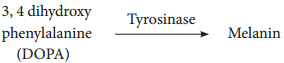
Albinism is an inborn error of metabolism, caused due to an autosomal recessive gene. Melanin pigment is responsible for skin colour. Absence of melanin results in a condition called albinism. A person with the recessive allele lacks the tyrosinase enzyme system, which is required for the conversion of dihydroxyphenyl alanine (DOPA) into melanin pigment inside the melanocytes. In an albino, melanocytes are present in normal numbers in their skin, hair, iris, etc., but lack melanin pigment.
Mendelian Disorders img 2

Huntington’s chorea

It is inherited as an autosomal dominant lethal gene in man. It is characterized by involuntary jerking of the body and progressive degeneration of the nervous system, accompanied by gradual mental and physical deterioration. The patients with this disease usually die between the age of 35 and 40.

Pedigree Analysis – Genetic Disorders

Learninsta presents the core concepts of Biology with high-quality research papers and topical review articles.

Pedigree Analysis – Genetic Disorders

Pedigree is a “family tree”, drawn with standard genetic symbols, showing the inheritance pathway for specific phenotypic characters.(Fig. 4.6). Pedigree analysis is the study of traits as they have appeared in a given family line for several past generations.
Pedigree Analysis img 1

Genetic Disorders

A genetic disorder is a disease or syndrome that is caused by an abnormality in an individual DNA. Abnormalities can range from a small mutation in a single gene to the addition or subtraction of an entire chromosome or even a set of chromosomes. Genetic disorders are of two types namely, Mendelian disorders and chromosomal disorders.

A pedigree shows relationships between family members and indicates which individuals have certain genetic pathogenic variants, traits, and diseases within a family as well as vital status. A pedigree shows relationships between family members and patterns of inheritance for certain traits and diseases.

Pedigrees are drawn using standard symbols and formatting. Males are represented by squares and females by circles. Individuals who are deceased have a slash through the symbol representing them. Symbols for individuals affected by a particular disorder are shaded.

It can be simply called as a “family tree” Pedigrees use a standardized set of symbols, squares represent males and circles represent females.

The modes of inheritance are autosomal dominant , autosomal recessive, and X-linked. To simplify the discussion of these different forms, the trait used in the following text will be a hereditary disease.

In this family pedigree, black squares indicate the presence of a particular trait in a male, and white squares represent males without the trait. White circles are females. A trait in one generation can be inherited, but not outwardly apparent before two more generations (compare black squares).

There are five basic modes of inheritance for single-gene diseases: autosomal dominant, autosomal recessive, X-linked dominant, X-linked recessive, and mitochondrial. Genetic heterogeneity is a common phenomenon with both single-gene diseases and complex multi-factorial diseases.

The study of an inherited trait in a group of related individuals to determine the pattern and characteristics of the trait, including its mode of inheritance, age of onset, and phenotypic variability.

Karyotyping Preparation and Its Applications

Learninsta presents the core concepts of Biology with high-quality research papers and topical review articles.

Karyotyping Preparation and Its Applications

Karyotyping is a technique through which a complete set of chromosomes is separated from a cell and the chromosomes are arranged in pairs. An idiogram refers to a diagrammatic representation of chromosomes.

Preparation of Karyotype

Tjio and Levan (1960) described a simple method of culturing lymphocytes from the human blood. Mitosis is induced followed by addition of colchicine to arrest cell division at metaphase stage and the suitable spread of metaphase chromosomes is photographed. The individual chromosomes are cut from the photograph and are arranged in an orderly fashion in homologous pairs. This arrangement is called a karyotype. Chromosome banding permits structural definitions and diffrentiation of chromosomes.

Applications of Karyotyping:

  • It helps in gender identification.
  • It is used to detect the chromosomal aberrations like deletion, duplication, translocation, nondisjunction of chromosomes.
  • It helps to identify the abnormalities of chromosomes like aneuploidy.
  • It is also used in predicting the evolutionary relationships between species.
  • Genetic diseases in human beings can be detected by this technique.
    Karyotyping Preparation and Its Applications img 1

Human Karyotype

Depending upon the position of the centromere and relative length of two arms, human chromosomes are of three types: Metacentric, sub metacentric and acrocentric. The photograph of chromosomes are arranged in the order of descending length in groups from A to G (Fig. 4.5).

Sex Linked Inheritance

Learninsta presents the core concepts of Biology with high-quality research papers and topical review articles.

Sex Linked Inheritance

The inheritance of a trait that is determined by a gene located on one of the sex chromosomes is called sex linked inheritance. Genes present on the differential region of X or Y chromosomes are called sex linked genes. The genes present in the differential region of “X” chromosome are called X linked genes. The X-linked genes have no corresponding alleles in the Y chromosome. The genes present in the differential region of Y chromosome are called Y – linked or holandric genes.

The Y linked genes have no corresponding allele in X chromosome. The Y linked genes inherit along with Y chromosome and they phenotypically express only in the male sex. Sex linked inherited traits are more common in males than females because, males are hemizygous and therefore express the trait when they inherit one mutant allele. The X – linked and Y – linked genes in the differential region (non-homologus
region) do not undergo pairing or crossing over during meiosis. The inheritance of X or Y linked genes is called sex-linked inheritance.

Inheritance of X – linked genes

Red-green colour blindness or daltonism, haemophilia and Duchenne’s muscular dystrophy are examples of X-linked gene inheritance in humans.

1. Haemophilia

Haemophilia is commonly known as bleeder’s disease, which is more common in men than women. This hereditary disease was first reported by John Cotto in 1803. Haemophilia is caused by a recessive X-linked gene.

A person with a recessive gene for haemophilia lacks a normal clotting substance (thromboplastin) in blood, hence minor injuries cause continuous bleeding, leading to death. The females are carriers of the disease and would transmit the disease to 50% of their sons even if the male parent is normal. Haemophilia follows the characteristic criss – cross pattern of inheritance.

2. Colour blindness

In human beings a dominant X – linked gene is necessary for the formation of colour sensitive cells, the cones. The recessive form of this gene is incapable of producing colour sensitive cone cells. Homozygous recessive females (XcXc) and hemizygous recessive males (XcY) are unable to distinguish red and green colour. The inheritance of colour blindness can be studied in the following two types of marriages.

(i) Marriage between colour blind man and normal visioned woman

A marriage between a colour blind man and a normal visioned woman will produce normal visioned male and female individuals in F1 generation but the females are carriers. The marriage between a F1 normal visioned carrier woman and a normal visioned male will produce one normal visioned female, one carrier female, one normal visioned male and one colour blind male in F2 generation.

Colour blind trait is inherited from the male parent to his grandson through carrier daughter, which is an example of criss-cross pattern of inheritance (Fig. 4.3).
Sex Linked Inheritance img 1

(ii) Marriage between normal visioned man and colour blind woman

If a colour blind woman (XcXc) marries a normal visioned male (XcXc), all F1 sons will be colourblind and daughters will be normal visioned but are carriers. Marriage between F1 carrier female with a colour blind male will produce normal visioned carrier daughter, colourblind daughter, normal visioned son and a colourblind son in the F2 generation (Fig. 4.4).
Sex Linked Inheritance img 2

Inheritance of Y-linked genes

Genes in the non-homologous region of the Y-chromosome are inherited directly from male to male. In humans, the Y-linked or holandric genes for hypertrichosis (excessive development of hairs on pinna of the ear) are transmitted directly from father to son, because males inherit the Y chromosome from the father. Female inherits only X chromosome from the father and are not affected.

Sex Determination – An Overview

Learninsta presents the core concepts of Biology with high-quality research papers and topical review articles.

Sex Determination – An Overview

Sex determination is the method by which the distinction between male and female is established in a species. Sex chromosomes determine the sex of the individual in dioecious or unisexual organisms. The chromosomes other than the sex chromosomes of an individual are called autosomes.

Sex chromosomes may be similar (homomorphic) in one sex and dissimilar (heteromorphic) in the other. Individuals having homomorphic sex chromosomes produce only one type of gametes (homogametic) whereas heteromorphic individuals produce two types of gametes (heterogametic).

Chromosomal basis of sex determination

Heterogametic Sex Determination:

In heterogametic sex determination one of the sexes produces similar gametes and the other sex produces dissimilar gametes. The sex of the offspring is determined at the time of fertilization.

Heterogametic Males

In this method of sex determination the males are heterogametic producing dissimilar gametes while females are homogametic producing similar gametes. It is of two kinds XX-XO type (e.g. Bugs, cockroaches and grasshoppers) and XX-XY type (e.g. Human beings and Drosophila).

Heterogametic Females

In this method of sex determination the females are heterogametic producing dissimilar gametes while males are homogametic producing similar gametes. To avoid confusion with the XX-XO and XX-XY types of sex determination, the alphabets ‘Z’ and ‘W’ are used here instead of X and Y respectively. Heterogametic females are of two types, ZO-ZZ type (eg. Moths, butterflies and domestic chickens) and ZW-ZZ type (eg. Gypsy moth, fihes, reptiles and birds).

Sex determination in human beings

Genes determining sex in human beings are located on two sex chromosomes, called allosomes. In mammals, sex determination is associated with chromosomal differences between the two sexes, typically XX females and XY males. 23 pairs of human chromosomes include 22 pairs of autosomes (44A) and one pair of sex chromosomes (XX or XY).

Females are homogametic producing only one type of gamete (egg), each containing one X chromosome while the males are heterogametic producing two types of sperms with X and Y chromosomes. An independently evolved XX: XY system of sex chromosomes also exist in Drosophila (Fig. 4.2).
Sex Determination img 1

The Y Chromosome and Male Development

Current analysis of Y chromosomes has revealed numerous genes and regions with potential genetic function; some genes with or without homologous counterparts are seen on the X. Present at both ends of the Y chromosome are the pseudoautosomal regions (PARs) that are similar with regions on the X chromosome which synapse and recombine during meiosis.

The remaining 95% of the Y chromosome is referred as the Non – combining Region of the Y (NRY). The NRY is divided equally into functional genes (euchromatic) and non functional genes (heterochromatic). Within the euchromatin regions, is a gene called Sex determining region Y (SRY). In humans, absence of Y chromosome inevitably leads to female development and this SRY gene is absent in X chromosome. The gene product of SRY is the testes determining factor (TDF) present in the adult male testis.

Dosage compensation Barr Body

In 1949, Barr and Bertram first observed a condensed body in the nerve cells of female cat which was absent in the male. This condensed body was called sex chromatin by them and was later referred as Barr body. In the XY chromosomal system of sex determination, males have only one X chromosome, whereas females have two. A question arises: how does the organism compensate for this dosage differences between the sexes? In mammals the necessary dosage compensation is accomplished by the inactivation of one of the X chromosome in females so that both males and females have only one functional X chromosome per cell.

Mary Lyon suggested that Barr bodies represented an inactive chromosome, which in females becomes tightly coiled into a heterochromatin, a condensed and visible form of chromatin (Lyon’s hypothesis). The number of Barr bodies observed in cell was one less than the number of X-Chromosome. XO females have no Barr body, whereas XXY males have one Barr body.

Haplodiploidy in Honeybees

In hymenopteran insects such as honeybees, ants and wasps a mechanism of sex determination called haplodiploidy mechanism of sex determination is common. In this system, the sex of the offspring is determined by the number of sets of chromosomes it receives.

Fertilized eggs develop into females (Queen or Worker) and unfertilized eggs develop into males (drones) by parthenogenesis. It means that the males have half the number of chromosomes (haploid) and the females have double the number (diploid), hence the name haplodiplody for this system of sex determination.

This mode of sex determination facilitates the evolution of sociality in which only one diploid female becomes a queen and lays the eggs for the colony. All other females which are diploid having developed from fertilized eggs help to raise the queen’s eggs and so contribute to the queen’s reproductive success and indirectly to their own, a phenomenon known as Kin Selection. The queen constructs their social environment by releasing a hormone that suppresses fertility of the workers.