Chemistry Of Nucleic Acids

Learninsta presents the core concepts of Biology with high-quality research papers and topical review articles.

Chemistry Of Nucleic Acids

Having identified the genetic material as the nucleic acid DNA (or RNA), we proceed to examine the chemical structure of these molecules. Generally nucleic acids are a long chain or polymer of repeating subunits called nucleotides. Each nucleotide subunit is composed of three parts: a nitrogenous base, a five carbon sugar (pentose) and a phosphate group.

Pentose sugar

There are two types of nucleic acids depending on the type of pentose sugar. Those containing deoxyribose sugar are called Deoxyribo Nucleic Acid (DNA) and those with ribose sugar are known as Ribonucleic Acid (RNA). DNA is found in the nucleus of eukaryotes and nucleoid of prokaryotes. The only difference between these two sugars is that there is one oxygen atom less in deoxyribose.

Nitrogenous bases

The bases are nitrogen containing molecules having the chemical properties of a base (a substance that accepts H+ ion or proton in solution). DNA and RNA both have four bases (two purines and two pyrimidines) in their nucleotide chain. Two of the bases, Adenine (A) and Guanine (G) have double carbon – nitrogen ring structures and are called purines. The bases, Thymine (T), Cytosine (C) and Uracil (U) have single ring structure and these are called pyrimidines. Thymine is unique for DNA, while Uracil is unique for RNA.

The phosphate functional group

It is derived from phosphoric acid (H3PO4), has three active OH groups of which two are involved in strand formation. The phosphate functional group (PO4) gives DNA and RNA the property of an acid (a substance
that releases an H+ ion or proton in solution) at physiological pH, hence the name nucleic acid.

The bonds that are formed from phosphates are esters. The oxygen atom of the phosphate group is negatively charged after the formation of the phosphodiester bonds. This negatively charged phosphate ensures the retention of nucleic acid within the cell or nuclear membrane.

Nucleoside and nucleotide

The nitrogenous base is chemically linked to one molecule of sugar (at the 1-carbon of the sugar) forming a nucleoside. When a phosphate group is attached to the 5′ carbon of the same sugar, the nucleoside becomes a nucleotide. The nucleotides are joined (polymerized) by condensation reaction to form a polynucleotide chain.

The hydroxyl group on the 3′ carbon of a sugar of one nucleotide forms an ester with the phosphate of another nucleotide. The chemical bonds that link the sugar components of adjacent nucleotides are called phosphodiester bond (5′ → 3′), indicating the polarity of the strand.

The ends of the DNA or RNA are distinct. The two ends are designated by the symbols 5′ and 3′. The symbol 5′ refers to carbon in the sugar to which a phosphate (PO4) functional group is attached. The symbol 3′ refers to carbon in the sugar to which hydroxyl (OH) functional group is attached. In RNA, every nucleotide residue has an additional OH group at 2′ position in the ribose. Understanding the 5′ → 3′ direction of a nucleic acid is critical for understanding the aspects of replication and transcription.

Based on the X – ray diffraction analysis of Maurice Wilkins and Rosalind Franklin, the double helix model for DNA was proposed by James Watson and Francis Crick in 1953. The highlight was the base pairing between the two strands of the polynucleotide chain. This proposition was based on the observations of Erwin Chargaff that Adenine pairs with Thymine (A = T) with two hydrogen bonds and Guanine pairs with Cytosine (G ≡ C) with three hydrogen bonds.

The ratios between Adenine with Thymine and Guanine with Cytosine are constant and equal. The base pairing confers a unique property to the polynucleotide chain. They are said to be complementary to each other, that is, if the sequence of bases in one strand (template) is known, then the sequence in the other strand can be predicted. The salient features of DNA structure has already been dealt in class XI.
DNA Is The Genetic Material

Learninsta presents the core concepts of Biology with high-quality research papers and topical review articles.

DNA Is The Genetic Material

Many biologists despite the earlier experiments of Griffith, Avery and others, still believed that protein, not DNA, was the hereditary material in a cell. As eukaryotic chromosomes consist of roughly equal amounts of protein and DNA, it was said that only a protein had sufficient chemical diversity and complexity to encode the information required for genetic material. In 1952, however, the results of the Hershey-Chase experiment finally provided convincing evidence that DNA is the genetic material.

Hershey and Chase experiment on T2 bacteriophage

Alfred Hershey and Martha Chase (1952) conducted experiments on bacteriophages that infect bacteria. Phage T2 is a virus that infects the bacterium Escherichia coli. When phages (virus) are added to bacteria, they adsorb to the outer surface, some material enters the bacterium, and then later each bacterium lyses to release a large number of progeny phage.

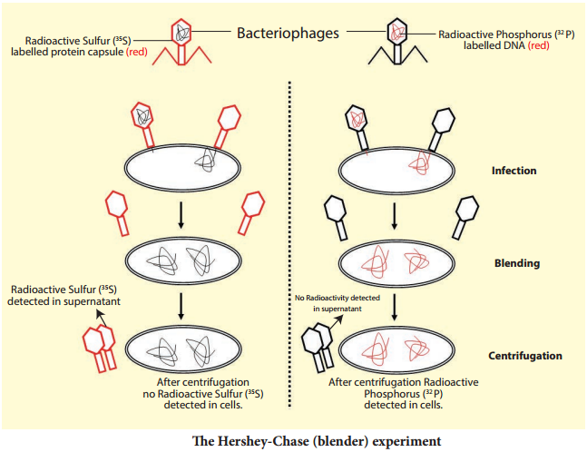
Hershey and Chase wanted to observe whether it was DNA or protein that entered the bacteria. All nucleic acids contain phosphorus, and proteins contain sulphur (in the amino acid cysteine and methionine). Hershey and Chase designed an experiment using radioactive isotopes of Sulphur (35S) and phosphorus (32P) to keep separate track of the viral protein and nucleic acids during the infection process.

The phages were allowed to infect bacteria in culture medium which containing the radioactive isotopes 35S or 32P. The bacteriophage that grew in the presence of 35S had labelled proteins and bacteriophages grown in the presence of 32P had labelled DNA.

The differential labelling thus enabled them to identify DNA and proteins of the phage. Hershey and Chalse mixed the labelled phages with unlabeled E coli and allowed bacteriophages to attack and inject their genetic material. Soon after infection (before lysis of bacteria), the bacterial cells were gently agitated in a blender to loosen the adhering phase particles.

It was observed that only 32P was found associated with bacterial cells and 32S was in the surrounding medium and not in the bacterial cells. When phage progeny was studied for radioactivity, it was found that it carried only 32P and not 35S (Fig. 5.2). These results clearly indicate that only DNA and not protein coat entered the bacterial cells. Hershey and Chase thus conclusively proved that it was DNA, not protein, which carries the hereditary information from virus to bacteria.

In Search Of The Genetic Material

Learninsta presents the core concepts of Biology with high-quality research papers and topical review articles.

In Search Of The Genetic Material

As early as 1848, Wilhelm Hofmeister, a German botanist, had observed that cell nuclei organize themselves into small, rod like bodies during mitosis called chromosomes. In 1869, Friedrich Miescher, a Swiss physician, isolated a substance from the cell nuclei and called it as nuclein. It was renamed as nucleic acid by Altman (1889), and is now known as DNA. By 1920, it became clear that chromosomes are made up of proteins and DNA.

Many experiments were carried out to study the actual carriers of genetic information. Griffith’s experiment proved that DNA is the genetic material which has been dealt in class XI. Bacterial transformation experiments provided the first proof that DNA is the genetic material. However, he could not understand the cause of bacterial transformation, and the biochemical nature of genetic material was not defined from his experiments.

Later, Oswald Avery, Colin Macleod and Maclyn McCarty in 1944 repeated Griffith’s experiments in an ‘in vitro’ system in order to identify the nature of the transforming substance responsible for converting a nonvirulent strain into virulent strain. They observed that the DNA, RNA and proteins isolated from the heat-killed S-strain when added to R-strain changed their surface character from rough to smooth and also made them pathogenic (Fig. 5.1).

But when the extract was treated with DNase (an enzyme which destroys DNA) the transforming ability was lost. RNase (an enzyme which destroys RNA) and proteases (an enzyme which destroys protein) did not affect the transformation.

Digestion with DNase inhibited transformation suggesting that the DNA caused the transformation. These experiments suggested that DNA and not proteins is the genetic material. The phenomenon, by which DNA isolated from one type of cell (S – strain), when introduced into another type (R – strain), is able to retain some of the properties of the S – strain is referred to as transformation.
In Search Of The Genetic Material img 1
In Search Of The Genetic Material img 2

Gene As The Functional Unit Of Inheritance

Learninsta presents the core concepts of Biology with high-quality research papers and topical review articles.

Gene As The Functional Unit Of Inheritance

A gene is a basic physical and functional unit of heredity. The concept of the gene was first explained by Gregor Mendel in 1860’s. He never used the term ‘gene’. He called it ‘factor’. In 1909, the Danish biologist Wilhelm Johannsen, coined the term ‘gene’, that was referred to discrete determiners of inherited characteristics.

According to the classical concept of gene introduced by Sutton in 1902, genes have been defined as discrete particles that follow Mendelian rules of inheritance, occupy a definite locus in the chromosome and are responsible for the expression of specific phenotypic character. They show the following properties:

  • Number of genes in each organism is more than the number of chromosomes; hence several genes are located on the same chromosome.
  • The genes are arranged in a single linear order like beads on a string.
  • Each gene occupies a specific position called locus.
  • Genes may exist in several alternate forms called alleles.
  • Genes may undergo sudden change in positions and composition called mutations.
  • Genes are capable of self-duplication producing their own copies.
    Gene As The Functional Unit Of Inheritance img 1

Chromosomal Abnormalities

Learninsta presents the core concepts of Biology with high-quality research papers and topical review articles.

Chromosomal Abnormalities

Each human diploid (2n) body cell has 46 chromosomes (23 pairs). Chromosomal disorders are caused by errors in the number or structure of chromosomes. Chromosomal anomalies usually occur when there is an error in cell division. Failure of chromatids to segregate during cell division resulting in the gain or loss of one or more chromosomes is called aneuploidy.

It is caused by nondisjunction of chromosomes. Group of signs and symptoms that occur together and characterize a particular abnormality is called a syndrome. In humans, Down’s syndrome, Turner’s syndrome, Klinefelter’s syndrome, Patau’s syndrome are some of the examples of chromosomal disorders.

a. Autosomal aneuploidy in human beings

Several autosomal aneuploidies have been reported in human beings. eg. Down’s syndrome (21-Trisomy), Patau’s syndrome (13-Trisomy).

1. Down’s Syndrome/Trisomy – 21

Trisomic condition of chromosome – 21 results in Down’s syndrome. It is characterized by severe mental retardation, defective development of the central nervous system, increased separation between the eyes, flttened nose, ears are malformed, mouth is constantly open and the tongue protrudes.

2. Patau’s Syndrome/Trisomy-13

Trisomic condition of chromosome 13 results in Patau’s syndrome. Meiotic non disjunction is thought to be the cause for this chromosomal abnormality. It is characterized by multiple and severe body malformations as well as profound mental deficiency. Small head with small eyes, clef palate, malformation of the brain and internal organs are some of the symptoms of this syndrome.

b. Allosomal abnormalities in human beings

Mitotic or meiotic non-disjunction of sex chromosomes causes allosomal abnormalities. Several sex chromosomal abnormalities have been detected. Eg. Klinefelter’s syndrome and Turner’s syndrome.

1. Klinefelter’s Syndrome (XXY Males)

This genetic disorder is due to the presence of an additional copy of the X chromosome resulting in a karyotype of 47,XXY. Persons with this syndrome have 47 chromosomes (44AA+XXY). They are usually sterile males, tall, obese, with long limbs, high pitched voice, under developed genitalia and have feeble breast (gynaecomastia) development.

2. Turner’s Syndrome (XO Females)

This genetic disorder is due to the loss of a X chromosome resulting in a karyotype of 45, X. Persons with this syndrome have 45 chromosomes (44 autosomes and one X chromosome) (44AA+XO) and are sterile females. Low stature, webbed neck, under developed breast, rudimentary gonads lack of menstrual cycle during puberty, are the main symptoms of this syndrome.