DNA Finger Printing Technique

Learninsta presents the core concepts of Biology with high-quality research papers and topical review articles.

DNA Finger Printing Technique

The DNA fingerprinting technique was first developed by Alec Jeffreys in 1985 (Recipient of the Royal Society’s Copley Medal in 2014). Each of us have the same chemical structure of DNA. But there are millions of differences in the DNA sequence of base pairs. This makes the uniqueness among us so that each of us except identical twins is different from each other genetically.

The DNA of a person and finger prints are unique. There are 23 pairs of human chromosomes with 1.5 million pairs of genes. It is a well known fact that genes are segments of DNA which differ in the sequence of their nucleotides.

Not all segments of DNA code for proteins, some DNA segments have a regulatory function, while others are intervening sequences (introns) and still others are repeated DNA sequences. In DNA fingerprinting, short repetitive nucleotide sequences are specific for a person. These nucleotide sequences are called as variable number tandem repeats (VNTR).The VNTRs of two persons generally show variations and are useful as genetic markers.

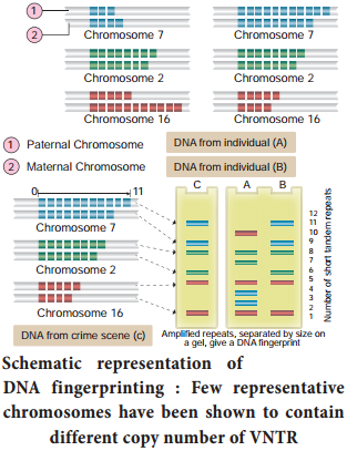
DNA figer printing involves identifying diffrences in some specific regions in DNA sequence called repetitive DNA, because in these sequences, a small stretch of DNA is repeated many times. These repetitive DNA are separated from bulk genomic DNA as different peaks during density gradient centrifugation. The bulk DNA forms a major peak and the other small peaks are referred to as satellite DNA.

Depending on base composition (A : T rich or G : C rich), length of segment and number of repetitive units, the satellite DNA is classified into many sub categories such as micro-satellites, minisatellites, etc., These sequences do not code for any proteins, but they form a large portion of human genome.

These sequences show high degree of polymorphism and form the basis of DNA figerprinting (Fig. 5.15). DNA isolated from blood, hair, skin cells, or other genetic evidences lef at the scene of a crime can be compared through VNTR patterns, with the DNA of a criminal suspect to determine guilt or innocence. VNTR patterns are also useful in establishing the identity of a homicide victim, either from DNA found as evidence or from the body itself.
DNA Finger Printing Technique img 1

The Steps in DNA Fingerprinting technique is depicted in Fig. 5.16.

1. Extraction of DNA

The process of DNA fingerprinting starts with obtaining a sample of DNA from blood, semen, vaginal fluids, hair roots, teeth, bones, etc.,

2. Polymerase chain reaction (PCR)

In many situations, there is only a small amount of DNA available for DNA figerprinting. If needed many copies of the DNA can be produced by PCR (DNA amplifiation).

3. Fragmenting DNA

DNA is treated with restriction enzymes which cut the DNA into smaller fragments at specific sites.
DNA Finger Printing Technique img 2

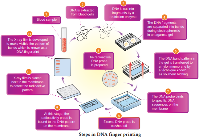
4. Separation of DNA by electrophoresis

During electrophoresis in an agarose gel, the DNA fragments are separated into bands of different sizes. The bands of separated DNA are sieved out of the gel using a nylon membrane (treated with chemicals that allow for it to break the hydrogen bonds of DNA so there are single strands).

5. Denaturing DNA

The DNA on gels is denatured by using alkaline chemicals or by heating.

6. Blotting

The DNA band pattern in the gel is transferred to a thin nylon membrane placed over the ‘size fractionated DNA strand’ by Southern blotting.

7. Using probes to identify specific DNA

A radioactive probe (DNA labeled with a radioactive substance) is added to the DNA bands. The probe attaches by base pairing to those restriction fragments that are complementary to its sequence. The probes can also be prepared by using either ‘florescent substance’ or ‘radioactive isotopes’.

8. Hybridization with probe

After the probe hybridizes and the excess probe washed off a photographic film is placed on the membrane containing ‘DNA hybrids’.

9. Exposure on fim to make a genetic/DNA Fingerprint

The radioactive label exposes the film to form an image (image of bands) corresponding to specific DNA bands. The thick and thin dark bands form a pattern of bars which constitutes a genetic fingerprint.

Application of DNA figer printing

Forensic analysis:

1. It can be used in the identification of a person involved in criminal activities, for settling paternity or maternity disputes, and in determining relationships for immigration purposes.

2. Pedigree analysis – inheritance pattern of genes through generations and for detecting inherited diseases.

3. Conservation of wild life – protection of endangered species. By maintaining DNA records for identification of tissues of the dead endangered organisms.

4. Anthropological studies – It is useful in determining the origin and migration of human populations and genetic diversities.

Human Genome Project (HGP)

Learninsta presents the core concepts of Biology with high-quality research papers and topical review articles.

Human Genome Project (HGP)

The international human genome project was launched in the year 1990. It was a mega project and took 13 years to complete. The human genome is about 25 times larger than the genome of any organism sequenced to date and is the first vertebrate genome to be completed. Human genome is said to have approximately 3 × 10⁹ bp. HGP was closely associated with the rapid development of a new area in biology called bioinformatics.

Goals and methodologies of Human Genome Project

The main goals of Human Genome Project are as follows

  • Identify all the genes (approximately 30000) in human DNA.
  • Determine the sequence of the three billion chemical base pairs that makeup the human DNA.
  • To store this information in databases.
  • Improve tools for data analysis.
  • Transfer related technologies to other sectors, such as industries.
  • Address the ethical, legal and social issues (ELSI) that may arise from the project.

The methodologies of the Human Genome Project involved two major approaches. One approach was focused on identifying all the genes that are expressed as RNA (ESTS – Expressed Sequence Tags). The other approach was sequence annotation. Here, sequencing the whole set of genome was taken, that contains all the coding and non-coding sequences and later assigning different regions in the sequences with functions.

For sequencing, the total DNA from a cell is isolated and converted into random fragments of relatively smaller sizes and cloned in suitable hosts using specialized vectors. This cloning results in amplification of pieces of DNA fragments so that it could subsequently be sequenced with ease.

Bacteria and yeast are two commonly used hosts and these vectors are called as BAC (Bacterial Artificial Chromosomes) and YAC (Yeast Artificial Chromosomes). The fragments are sequenced using automated DNA sequencers (developed by Frederick Sanger).

The sequences are then arranged based on few overlapping regions, using specialized computer based programs. These sequences were subsequently annotated and are assigned to each chromosome. The genetic and physical maps on the genome are assigned using information on polymorphism of restriction endonuclease recognition sites and some repetitive DNA sequences, called microsatellites.

The latest method of sequencing even longer fragments is by a method called Shotgun sequencing using super computers, which has replaced the traditional sequencing methods.

Salient features of Human Genome Project:

  • The human genome contains 3 billion nucleotide bases.
  • An average gene consists of 3000 bases, the largest known human gene being dystrophin with 2.4 million bases.
  • Genes are distributed over 24 chromosomes. Chromosome 19 has the highest gene density. Chromosome 13 and Y chromosome have lowest gene densities.
  • The chromosomal organization of human genes shows diversity.
  • There may be 35000-40000 genes in the genome and almost 99.9 nucleotide bases are exactly the same in all people.
  • Functions for over 50 percent of the discovered genes are unknown.
  • Less than 2 percent of the genome codes for proteins.
  • Repeated sequences make up very large portion of the human genome. Repetitive sequences have no direct coding functions but they shed light on chromosome structure, dynamics and evolution (genetic diversity).
  • Chromosome 1 has 2968 genes whereas chromosome ’Y’ has 231 genes.
  • Scientists have identified about 1.4 million locations where single base DNA differences (SNPs – Single nucleotidepolymorphism – pronounce as ‘snips’) occur in humans.
  • Identification of ‘SNIPS’ is helpful in finding chromosomal locations for disease associated sequences and tracing human history.

Applications and future challenges

The mapping of human chromosomes is possible to examine a person’s DNA and to identify genetic abnormalities. This is extremely useful in diagnosing diseases and to provide genetic counselling to those planning to have children.

This kind of information would also create possibilities for new gene therapies. Besides providing clues to understand human biology, learning about non-human organisms, DNA sequences can lead to an understanding of their natural capabilities that can be applied towards solving challenges in healthcare, agriculture, energy production and environmental remediation.

A new era of molecular medicine, characterized by looking into the most fundamental causes of disease than treating the symptoms will be an important advantage.

  • Once genetic sequence becomes easier to determine, some people may attempt to use this information for profit or for political power.
  • Insurance companies may refuse to insure people at ‘genetic risk’ and this would save the companies the expense of future medical bills incurred by ‘less than perfect’ people.
  • Another fear is that attempts are being made to “breed out” certain genes of people from the human population in order to create a ‘perfect race’.

Regulation Of Gene Expression

Learninsta presents the core concepts of Biology with high-quality research papers and topical review articles.

Regulation Of Gene Expression

We have previously established how DNA is organized into genes, how genes store genetic information, and how this information is expressed. We now consider the most fundamental issues in molecular genetics. How is genetic expression regulated? Evidence in support of the idea that genes can be turned on and of is very convincing. Regulation of gene expression has been extensively studied in prokaryotes, especially in E. coli.

Gene expression can be controlled or regulated at transcriptional or post transcriptional or translational level. Here, we are going to discuss regulation of gene expression at transcriptional level. Usually, small extracellular or intracellular metabolites trigger initiation or inhibition of gene expression. The clusters of gene with related functions are called operons. They usually transcribe single mRNA molecules. In E.coli, nearly 260 genes are grouped into 75 different operons.

Structure of the operon:

Each operon is a unit of gene expression and regulation and consists of one or more structural genes and an adjacent operator gene that controls transcriptional activity of the structural gene.

  • The structural gene codes for proteins, rRNA and tRNA required by the cell.
  • Promoters are the signal sequences in DNA that initiate RNA synthesis. RNA polymerase binds to the promoter prior to the initiation of transcription.
  • The operators are present between the promoters and structural genes. The repressor protein binds to the operator region of the operon.

The Lac (Lactose) operon:

The metabolism of lactose in E.coli requires three enzymes – permease, β-galactosidase (β-gal) and transacetylase. The enzyme permease is needed for entry of lactose into the cell, β-galactosidase brings about hydrolysis of lactose to glucose and galactose, while transacetylase transfers acetyl group from acetyl Co A to β-galactosidase.

The lac operon consists of one regulator gene (‘i’ gene refers to inhibitor) promoter sites (p), and operator site (o). Besides these, it has three structural genes namely lac z, y and lac a. The lac ‘z’ gene codes for β-galactosidase, lac ‘y’ gene codes for permease and ‘a’ gene codes for transacetylase.

Jacob and Monod proposed the classical model of Lac operon to explain gene expression and regulation in E.coli. In lac operon, a polycistronic structural gene is regulated by a common promoter and regulatory gene. When the cell is using its normal energy source as glucose, the ‘i’ gene transcribes a repressor mRNA and after its translation, a repressor protein is produced.

It binds to the operator region of the operon and prevents translation, as a result, β-galactosidase is not produced. In the absence of preferred carbon source such as glucose, if lactose is available as an energy source for the bacteria then lactose enters the cell as a result of permease enzyme. Lactose acts as an inducer and interacts with the repressor to inactivate it.

The repressor protein binds to the operator of the operon and prevents RNA polymerase from transcribing the operon. In the presence of inducer, such as lactose or allolactose, the repressor is inactivated by interaction with the inducer.

This allows RNA polymerase to bind to the promotor site and transcribe the operon to produce lac mRNA which enables formation of all the required enzymes needed for lactose metabolism (Fig. 5.14). This regulation of lac operon by the repressor is an example of negative control of transcription initiation. Lac operon is also under the control of positive regulation as well.
Regulation Of Gene Expression img 1

Rna – The Adapter Molecule

Learninsta presents the core concepts of Biology with high-quality research papers and topical review articles.

Rna – The Adapter Molecule

The transfer RNA, (tRNA) molecule of a cell acts as a vehicle that picks up the amino acids scattered through the cytoplasm and also reads specific codes of mRNA molecules. Hence it is called an adapter molecule. This term was postulated by Francis Crick.

The two dimensional clover leaf model of tRNA was proposed by Robert Holley. The secondary structure of tRNA depicted in Fig. 5.11 looks like a clover leaf. In actual structure, the tRNA is a compact molecule which looks like an inverted L. The clover leaf model of tRNA shows the presence of three arms namely DHU arm, middle arm and TΨC arm. These arms have loops such as amino acyl binding loop, anticodon loop and ribosomal binding loop at their ends.
Rna - The Adapter Molecule img 1

In addition it also shows a small lump called variable loop or extra arm. The amino acid is attached to one end (amino acid acceptor end) and the other end consists of three anticodon nucleotides. The anticodon pairs with a codon in mRNA ensuring that the correct amino acid is incorporated into the growing polypeptide chain.

Four different regions of double-stranded RNA are formed during the folding process. Modified bases are especially common in tRNA. Wobbling between anticodon and codon allows some tRNA molecules to read more than one codon.

The process of addition of amino acid to tRNA is known as aminoacylation or charging and the resultant product is called aminoacyl – tRNA (charged tRNA). Without aminoacylation tRNA is known as uncharged tRNA (Fig. 5.12).

If two such tRNAs are brought together peptide bond formation is favoured energetically. Numbers of amino acids are joined by peptide bonds to form a polypeptide chain. This aminoacylation is catalyzed by an enzyme aminoacyl – tRNA synthetase. This is an endothermic reaction and is associated with ATP hydrolysis. 20 different aminoacyl – tRNA synthetases are known. The power to recognize codon on the mRNA lies in the tRNA and not in the attached amino acid molecule.
Rna - The Adapter Molecule img 2

Translation and Its Mechansim

Learninsta presents the core concepts of Biology with high-quality research papers and topical review articles.

Translation and Its Mechansim

Translation refers to the process of polymerization of amino acids to form poly peptide chain. The decoding process is carried out by ribosomes that bind mRNA and charged tRNA molecules. The mRNA is translated, starting at the 5′ end.

After binding to mRNA, the ribosomes move along it, adding new amino acids to the growing polypeptide chain each time it reads a codon. Each codon is read by an anticodon on the corresponding tRNA. Hence the order and sequence of amino acids are defined by the sequence of bases in the mRNA.

Mechanism of Translation

The cellular factory responsible for synthesizing protein is the ribosome. The ribosome consists of structural RNAs and about 80 different proteins. In inactive state, it exists as two subunits; large subunit and small subunit.

When the subunit encounters an mRNA, the process of translation of the mRNA to protein begins. The prokaryotic ribosome (70 S) consists of two subunits, the larger subunit (50 S) and smaller subunit (30 S). The ribosomes of eukaryotes (80 S) are larger, consisting of 60 S and 40 S sub units. ‘S’ denotes the sedimentation coefficient which is expressed as Svedberg unit (S).

One of the alternative ways of dividing up a sequence of bases in DNA or RNA into codons is called reading frame. Any sequence of DNA or RNA, beginning with a start codon and which can be translated into a protein is known as an Open Reading Frame (ORF). A translational unit in mRNA is the sequence of RNA that is flanked by the start codon (AUG) and the stop codon and codes for polypeptides.

mRNA also have some additional sequences that are not translated and are referred to as Untranslated Regions (UTR). UTRs are present at both 5′ end (before start codon) and at 3′ end (after stop codon). The start codon (AUG) begins the coding sequence and is read by a special tRNA that carries methionine (met).

The initiator tRNA charged with methionine binds to the AUG start codon. In prokaryotes, N – formyl methionine (f met) is attached to the initiator tRNA whereas in eukaryotes unmodified methionine is used. The 5′ end of the mRNA of prokaryotes has a special sequence which precedes the initial AUG start codon of mRNA.

This ribosome binding site is called the Shine – Dalgarno sequence or S-D sequence. This sequences base-pairs with a region of the 16Sr RNA of the small ribosomal subunit facilitating initiation. The subunits of the ribosomes (30 S and 50 S) are usually dissociated from each other when not involved in translation (Fig. 5.13 a).
Translation and Its Mechansim img 1

Initiation of translation in E. coli begins with the formation of an initiation complex, consisting of the 30S subunits of the ribosome, a messenger RNA and the charged N-formyl methionine tRNA (fmet – t RNA fmet), three proteinaceous initiation factors (IF1, IF2, IF3), GTP (Guanine Tri Phosphate) and Mg2+.

The components that form the initiation complex interact in a series of steps. IF3 binds to the 30S and allows the 30S subunit to bind to mRNA. Another initiation protein (IF2) then enhances the binding of charged formyl methionine tRNA to the small subunit in response to the AUG triplet. This step ‘sets’ the reading frame so that all subsequent groups of three ribonucleotides are translated accurately.

The assembly of ribosomal subunits, mRNA and tRNA represent the initiation complex. Once initiation complex has been assembled, IF3 is released and allows the initiation complex to combine with the 50S ribosomal subunit to form the complete ribosome (70S). In this process a molecule of GTP is hydrolyzed providing the required energy and the initiation factors (IF1 and IF2 and GDP) are released (Fig. 5.13 b).
Translation and Its Mechansim img 2

Elongation is the second phase of translation. Once both subunits of the ribosomes are assembled with the mRNA, binding sites for two charged tRNA molecules are formed. The sites in the ribosome are referred to as the aminoacyl site (A site), the peptidyl site (P site) and the exit site (E site). The charged initiator tRNA binds to the P site.

The next step in prokaryotic translation is to position the second tRNA at the ‘A’ site of the ribosome to form hydrogen bonds between its anticodon and the second codon on the mRNA (step1). This step requires the correct transfer RNA, another GTP and two proteins called elongation factors (EF-Ts and EF-Tu).

Once the charged tRNA molecule is positioned at the A site, the enzyme peptidyl transferase catalyses the formation of peptide bonds that link the two amino acids together (step 2). At the same time, the covalent bond between the amino acid and tRNA occupying the P site is hydrolyzed (broken).

The product of this reaction is a dipeptide which is attached to the 3′ end of tRNA still residing in the A site. For elongation to be repeated, the tRNA attached to the P site, which is now uncharged is released from the large subunit. The uncharged tRNA moves through the ‘E’ site on the ribosome.

The entire mRNA-tRNA-aa1-aa2 complex shift in the direction of the ‘P’ site by a distance of three nucleotides (step 3). This step requires several elongation factors (EFs) and the energy derived from hydrolysis of GTP. This results in the third triplet of mRNA to accept another charged tRNA into the A site (step 4).

The sequence of elongation is repeated over and over (step 5 and step 6). An additional amino acid is added to the growing polypeptide, each time mRNA advances through the ribosome. Once a polypeptide chain is assembled, it emerges out from the base of the large subunit (Fig. 5.13 c).
Translation and Its Mechansim img 3

Termination is the third phase of translation. Termination of protein synthesis occurs when one of the three stop codons appears in the ‘A’ site of the ribosome. The terminal codon signals the action of GTP – dependent release factor, which cleaves the polypeptide chain from the terminal tRNA releasing it from the translational complex (step 1). The tRNA is then released from the ribosome, which then dissociates into its subunits (step 2) (Fig. 5.13 d).
Translation and Its Mechansim img 4