Transgenic Animals

Learninsta presents the core concepts of Biology with high-quality research papers and topical review articles.

Transgenic Animals

In early days selective breeding methods were carried out to improve the genetic characteristics of live stock and other domestic animals. With the advent of modern biotechnology it is possible to carry out manipulations at the genetic level to get the desired traits in animals. Transgenesis is the process of introduction of extra (foreign/exogenous) DNA into the genome of the animals to create and maintain stable heritable characters.

The foreign DNA that is introduced is called the transgene and the animals that are produced by DNA manipulations are called transgenic animals or the genetically engineered or genetically modified organisms.

The various steps involved in the production of transgenic organisms are

  • Identification and separation of desired gene.
  • Selection of a vector (generally a virus) or direct transmission.
  • Combining the desired gene with the vector.
  • Introduction of transferred vector into cells, tissues, embryo or mature individual.
  • Demonstration of integration and expression of foreign gene in transgenic tissue or animals.
    Transgenic animals such as mice, rat, rabbit, pig, cow, goat, sheep and fish have been produced (Fig. 9.7).

Transgenic Animals img 1

Uses Of Transgenesis

  • Transgenesis is a powerful tool to study gene expression and developmental processes in higher organisms.
  • Transgenesis helps in the improvement of genetic characters in animals.
  • Transgenic animals serve as good models for understanding human diseases which help in the investigation of new treatments for diseases.
  • Transgenic models exist for many human diseases such as cancer, Alzheimer’s, cystic fibrosis, rheumatoid arthritis and sickle cell anemia.
  • Transgenic animals are used to produce proteins which are important for medical and pharmaceutical applications.
  • Transgenic mice are used for testing the safety of vaccines.
  • Transgenic animals are used for testing toxicity in animals that carry genes which make them sensitive to toxic substances than non-transgenic animals exposed to toxic substances and their effects are studied.
  • Transgenesis is important for improving the quality and quantity of milk, meat, eggs and wool production in addition to testing drug resistance.

Molecular Diagnostics

Learninsta presents the core concepts of Biology with high-quality research papers and topical review articles.

Molecular Diagnostics

Early diagnosis of infectious diseases or inherent genetic defects is essential for appropriate treatment. Early detection of the disease is not possible using conventional diagnostic methods like microscopic examinations, serum analysis and urine analysis. These laboratory techniques are indirect and not always specific. Scientists are continuously searching for specific, sensitive and simple diagnostic techniques for diagnosis of diseases.

Recombinant DNA technology, Polymerase Chain Reactions (PCR) and Enzyme Linked Immunosorbent Assay (ELISA) are some of the techniques that are reliable and help in early diagnosis. Presence of pathogens like virus, bacteria, etc., is detected only when the pathogen produces symptoms in the patient. By the time the symptoms appear concentration of pathogen becomes very high in the body. However very low concentration of a bacteria or a virus, even when the symptoms of the disease does not appear, can be detected by amplification of their nucleic acid.

ELISA [Enzyme Linked Immunosorbent Assay]

ELISA is a biochemical procedure discovered by Eva Engvall and Peter Perlmanin (1971) to detect the presence of specific antibodies or antigens in a sample of serum, urine, etc., It is a very important diagnostic tool to determine if a person is HIV positive or negative.

ELISA is a tool for determining serum antibody concentrations (such as the antibodies produced in a person infected by pathogens such as HIV) and also for detecting the presence of specific antigens and hormones such as human chorionic gonadotropins.

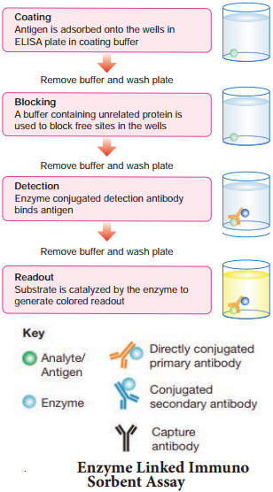
During diagnosis the sample suspected to contain the antigen is immobilized on the surface of an ELISA plate (Fig. 9.5). The antibody specific to this antigen is added and allowed to react with the immobilized antigen. The anti-antibody is linked to an appropriate enzyme like peroxidase.

The unreacted anti-antbody is washed away and the substrate of the enzyme (hydrogen peroxidase) is added with certain reagents such as 4-chloronaphthol. The activity of the enzyme yields a coloured product indicating the presence of the antigen. The intensity of the colour is directly proportional to the amount of the antigen. ELISA is highly sensitive and can detect antigens in the range of a nanogram.
Molecular Diagnostics image 1

There are four kinds of ELISA namely, Direct ELISA, Indirect ELISA, sandwich ELISA and competitive ELISA. It is a highly sensitive and specific method used for diagnosis. ELISA possesses the added advantages of not requiring radioisotopes or a radiation counting apparatus. PCR (Polymerase Chain Reaction) The polymerase chain reaction (PCR) is an invitro amplification technique used for synthesising multiple identical copies (billions) of DNA of interest. The technique was developed by Kary Mullis (Nobel laureate, 1993) in the year 1983.

Denaturation, renaturation or primer annealing and synthesis or primer extension, are the three steps involved in PCR (Fig. 9.6). The double stranded DNA of interest is denatured to separate into two individual strands by high temperature .

This is called denaturation. Each strand is allowed to hybridize with a primer (renaturation or primer annealing). The primer template is used to synthesize DNA by using Taq – DNA polymerase.
Molecular Diagnostics image 2

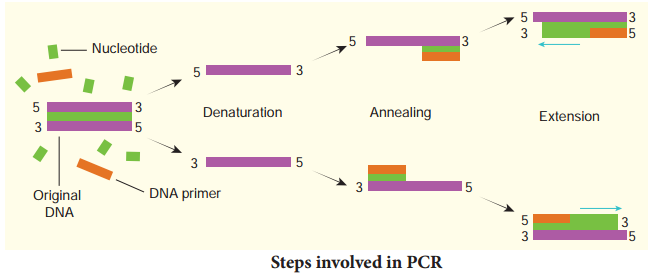
During denaturation the reaction mixture is heated to 95° C for a short time to denature the target DNA into single strands that will act as a template for DNA synthesis. Annealing is done by rapid cooling of the mixture, allowing the primers to bind to the sequences on each of the two strands flanking the target DNA. During primer extension or synthesis the temperature of the mixture is increased to 75° C for a sufficient
period of time to allow Taq DNA polymerase to extend each primer by copying the single stranded template.

At the end of incubation both single template strands will be made partially double stranded. The new strand of each double stranded DNA extends to a variable distance downstream. These steps are repeated again and again to generate multiple forms of the desired DNA. This process is also called DNA amplification.

The PCR technique can also be used for amplifications of RNA in which case it is referred to as reverse transcription PCR (RT-PCR). In this process the RNA molecules (mRNA) must be converted to complementary DNA by the enzyme reverse transcriptase. The CDNA then serves as the template for PCR.

PCR In Clinical Diagnosis

The specificity and sensitivity of PCR is useful for the diagnosis of inherited disorders (genetic diseases), viral diseases, bacterial diseases, etc., The diagnosis and treatment of a particular disease often requires identifying a particular pathogen. Traditional methods of identification involve culturing these organisms from clinical specimens and performing metabolic and other tests to identify them.

The concept behind PCR based diagnosis of infectious diseases is simple – if the pathogen is present in a clinical specimen its DNA will be present.

Its DNA has unique sequences that can be detected by PCR, often using the clinical specimen (for example, blood, stool, spinal fluid, or sputum) in the PCR mixture. PCR is also employed in the prenatal diagnosis of inherited diseases by using chorionic villi samples or cells from amniocentesis. Diseases like sickle cell anemia, β-thalassemia and phenylketonuria can be detected by PCR in these samples.

CDNA from PCR is a valuable tool for diagnosis and monitoring retroviral infections e.g., Tuberculosis by Mycobacterium tuberculosis. Several virally induced cancers, like cervical cancer caused by Papilloma virus can be detected by PCR. Sex of human beings and live stocks, embryos fertilized invitro can be determined by PCR by using primers and DNA probes specific for sex chromosomes. PCR technique is also used to detect sex-linked disorders in fertilized embryos.

Applications of PCR

The differences in the genomes of two different organisms can be studied by PCR. PCR is very important in the study of evolutions, more specifically phylogenetics. As a technique which can amplify even minute quantities of DNA from any source, like hair, mummified tissues, bones or any fossilized materials.

PCR technique can also be used in the field of forensic medicine. A single molecule of DNA from blood stains, hair, semen of an individual is adequate for amplification by PCR. The amplified DNA is used to develop DNA fingerprint which is used as an important tool in forensic science. Thus, PCR is very useful for identification of criminals. PCR is also used in amplification of specific DNA segment to be used in gene therapy.

Stem Cell Therapy

Learninsta presents the core concepts of Biology with high-quality research papers and topical review articles.

Stem Cell Therapy

Stem cells are undifferentiated cells found in most of the multi cellular animals. These cells maintain their undifferentiated state even after undergoing numerous mitotic divisions.

Stem cell research has the potential to revolutionize the future of medicine with the ability to regenerate damaged and diseased organs. Stem cells are capable of self renewal and exhibit ‘cellular potency’. Stem cells can differentiate into all types of cells that are derived from any of the three germ layers ectoderm, endoderm and mesoderm.

In mammals there are two main types of stem cells – embryonic stem cells (ES cells) and adult stem cells. ES cells are pluripotent and can produce the three primary germ layers ectoderm, mesoderm and endoderm. Embryonic stem cells are multipotent stem cells that can differentiate into a number of types of cells (Fig. 9.4). ES cells are isolated from the epiblast tissue of the inner cell mass of a blastocyst.
Stem Cell Therapy img 1

When stimulated ES can develop into more than 200 cells types of the adult body. ES cells are immortal i.e., they can proliferate in a sterile culture medium and maintain their undifferentiated state. Adult stem cells are found in various tissues of children as well as adults. An adult stem cell or somatic stem cell can divide and create another cell similar to it. Most of the adult stem cells are multipotent and can act as a repair system of the body, replenishing adult tissues. The red bone marrow is a rich source of adult stem cells.

The most important and potential application of human stem cells is the generation of cells and tissues that could be used for cell based therapies. Human stem cells could be used to test new drugs.

Stem Cell Banks

Stem cell banking is the extraction, processing and storage of stem cells, so that they may be used for treatment in the future, when required. Amniotic cell bank is a facility that stores stem cells derived from amniotic fluid for future use.

Stem cells are stored in banks specifically for use by the individual from whom such cells have been collected and the banking costs are paid. Cord Blood Banking is the extraction of stem cells from the umbilical cord during childbirth. While the umbilical cord and cord blood are the most popular sources of stem cells, the placenta, amniotic sac and amniotic fluid are also rich sources in terms of both quantity and quality.

Biotechnology Of Gene Therapy

Learninsta presents the core concepts of Biology with high-quality research papers and topical review articles.

Biotechnology Of Gene Therapy

If a person is born with a hereditary disease, can a corrective therapy be given for such disease? Yes, this can be done by a process known as gene therapy. This process involves the transfer of a normal gene into a person’s cells that carries one or more mutant alleles. Expression of normal gene in the person results in a functional gene product whose action produces a normal phenotype.

Delivery of the normal gene is accomplished by using a vector. The main thrust of gene therapy has been directed at correcting single gene mutations as in cystic firosis and haemophilia. At present most genetic diseases have no effective treatment and so gene therapy could offer hope for many people.

There are two strategies involved in gene therapy namely; Gene augmentation therapy which involves insertion of DNA into the genome to replace the missing gene product and Gene inhibition therapy which involves insertion of the anti sense gene which inhibits the expression of the dominant gene (Fig. 9.3). The two approaches to achieve gene therapy are somatic cell and germ line gene therapy.
Biotechnology Of Gene Therapy img 1

Somatic cell therapy involves the insertion of a fully functional and expressible gene into a target somatic cell to correct a genetic disease permanently whereas Germline gene therapy involves the introduction of DNA into germ cells which is passed on to the successive generations. Gene therapy involves isolation of a specific gene and making its copies and inserting them into target cells to make the desired proteins.

It is absolutely essential for gene therapists to ensure that the gene is harmless to the patient and it is appropriately expressed and that they body’s immune system does not react to the foreign proteins produced by the new genes.

Applications In Medicine

Learninsta presents the core concepts of Biology with high-quality research papers and topical review articles.

Applications In Medicine

Recombinant Human Insulin

The Human insulin is synthesized by the β cells of Islets of Langerhans in the pancreas. It is formed of 51 aminoacids which are arranged in two polypeptide chains, A and B. The polypeptide chain A has 21 amino acids while the polypeptide chain B has 30 amino acids. Both A and B chains are attached together by disulphide bonds.

Insulin controls the levels of glucose in blood. It facilitates the cellular uptake and utilization of glucose for the release of energy. Deficiency of insulin leads to diabetes mellitus which is characterized by increased blood glucose concentration and a complex of symptoms which may lead to death, if untreated. A continuous program of insulin dependence is required to treat this deficiency.

In the early years, insulin isolated and purified from the pancreas of pigs and cows was used to treat diabetic patients. Due to minor differences in the structure of the animal insulin as compared to human insulin, it resulted in the occurrence of allergic reactions in some diabetic patients.

Production of insulin by recombinant DNA technology started in the late 1970s. This technique involved the insertion of human insulin gene on the plasmids of E.coli. The polypeptide chains are synthesized as a precursor called pre-pro insulin, which contains A and B segments linked by a third chain (C) and preceded by a leader sequence.

The leader sequence is removed after translation and the C chain is excised, leaving the A and B polypeptide chains (Fig. 9.1). Insulin was the first ever pharmaceutical product of recombinant DNA technology administered to humans. The approval to use recombinant insulin for diabetes mellitus was given in 1982. In 1986 human insulin was marketed under the trade name Humulin.
Applications In Medicine img 1

Interferons

Interferons are proteinaceous, antiviral, species specific substances produced by mammalian cells when infected with viruses. Interferons were discovered by Alick Isaacs and Jean Lindemann in 1957. Based on the structure of interferons they are classifid as α, β and γ interferons.

They stimulate the cellular DNA to produce antiviral enzymes which inhibit viral replication and protect the cells. Interferons could be isolated from blood, but the amount of blood required for isolation of interferons is enormous and not practical. To overcome this issue interferons could be produced by rDNA technology.

The yeast Saccharomyces cerevisiae is more suitable for production of recombinant interferons than E.coli, since E.coli does not possess the machinery for glycosylation of proteins. Interferons are used for the treatment of various diseases like cancer, AIDS, multiple sclerosis, hepatitis C and herpes zoster. In spite of the therapeutic applications interferons are not within the reach of the common man due to high cost for its production.

Recombinant Vaccines

Recombinant DNA technology has been used to produce new generation vaccines. The limitations of traditional vaccine production could be overcome by this approach. The recombinant vaccines are generally of uniform quality and produce less side effects as compared to the vaccines produced by conventional methods. Different types of recombinant vaccines include subunit recombinant vaccines, attenuated recombinant vaccines and DNA vaccines.

Subunit recombinant vaccines

Vaccines that use components of a pathogenic organism rather than the whole organism are called subunit vaccines; recombinant DNA technology is very suited for developing new subunit vaccines. It includes components like proteins, peptides and DNAs of pathogenic organisms. The advantages of these vaccines include their purity in preparation, stability and safe use.

Attenuated recombinant vaccines

This includes genetically modified pathogenic organisms (bacteria or viruses) that are made nonpathogenic and are used as vaccines. It is now possible to genetically engineer the organisms (bacteria or viruses) and use them as live vaccines and such vaccines are referred to as attenuated recombinant vaccines.

DNA Vaccines

Genetic immunisation by using DNA vaccines is a novel approach that came into being in 1990. The immune response of the body is stimulated by a DNA molecule. A DNA vaccine consists of a gene encoding an antigenic protein, inserted onto a plasmid, and then incorporated into the cells in a target animal. DNA instructs the cells to make antigenic molecules which are displayed on its surfaces.

This would evoke an antibody response to the free floating antigen secreted by the cells. The DNA vaccine cannot cause the disease as it contains only copies of a few of its genes. DNA vaccines are relatively easy and inexpensive to design and produce. Vaccines produced by these new techniques have definite advantages like producing target proteins, long lasting immunity and trigger immune response only against specific pathogens with less toxic effects.

Recombinant hepatitis B vaccine as a subunit vaccine is produced by cloning hepatitis B surface antigen (HbsAg) gene in the yeast, Saccharomyces cerevisiae (Fig. 9.2).
Applications In Medicine img 2