Neural Tissue Definition and its Uses

Learninsta presents the core concepts of Biology with high-quality research papers and topical review articles.

Neural Tissue Definition and its Uses

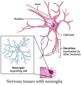
Nervous tissue exerts the greatest control over the body’s responsiveness to changing conditions. Neurons, the unit of neural system are excitable cells (Figure 3.8).
Neural Tissue img 1

The neuroglial cells which constitute the rest of the neural system protect and support the neurons. Neuroglia makes up more than one-half of the volume of neural tissue in our body. When a neuron is suitably stimulated, an electrical disturbance is generated which swifty travels along its plasma membrane. Arrival of the disturbance at the neuron’s endings, or output zone, triggers events that may cause stimulation or inhibition of adjacent neurons and other cells (You will study in detail in Chapter 10)

Neurons, or nerves, transmit electrical impulses, while neuroglia do not; neuroglia have many other functions including supporting and protecting neurons.

Integration and communication are the two major functions of nervous tissue. Nervous tissue contains two categories of cells – neurons and neuroglia. Neurons are highly specialized nerve cells that generate and conduct nerve impulses.

Nervous tissue contains two major cell types, neurons and glial cells. Neurons are the cells responsible for communication through electrical signals.

Nervous tissue is made up of different types of neurons, all of which have an axon. Bundles of axons make up the nerves in the PNS and tracts in the CNS. Functions of the nervous system are sensory input, integration, control of muscles and glands, homeostasis, and mental activity.

The function of muscle tissue (smooth, skeletal, and cardiac) is to contract, while nervous tissue is responsible for communication.

Brain, spinal cord and nerves constitute nervous tissue. Tendon is a fibrous connective tissue connecting bones to muscles. Nervous tissue is absent in tendon. These are made up of collagen.

Neurons, also known as nerve cells, send and receive signals from your brain. While neurons have a lot in common with other types of cells, they’re structurally and functionally unique. Specialized projections called axons allow neurons to transmit electrical and chemical signals to other cells.

Although the nervous system is very complex, there are only two main types of cells in nerve tissue. The actual nerve cell is the neuron. It is the “conducting” cell that transmits impulses and the structural unit of the nervous system. The other type of cell is neuroglia, or glial, cell.

Muscle Tissue Function and Its Types

Learninsta presents the core concepts of Biology with high-quality research papers and topical review articles.

Muscle Tissue Function and Its Types

Each muscle is made of many long, cylindrical fires arranged in parallel arrays. These fibres are composed of numerous fine firils, called myofirils. Muscle fires contract (shorten) in response to stimulation, then relax (lengthen) and return to their uncontracted state in a coordinated fashion. In general muscles play an active role in all the movements of the body.

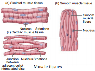
Muscles are of three types, skeletal, smooth and cardiac. Skeletal muscle tissue is closely attached to skeletal bones. In a typical muscle such as the biceps, the striated (striped) skeletal muscle fires are bundled together in a parallel fashion. A sheath of tough connective tissue encloses several bundles of muscle fires (You will learn more about this in Chapter 9).

The smooth muscle fires taper at both ends (fusiform) and do not show striations (Figure 3.7). Cell junctions hold them together and they are bundled together in a connective tissue sheath. The walls of internal organs such as the blood vessels, stomach and intestine contain this type of muscle tissue. Smooth muscles are ‘involuntary’ as their functions cannot be directly controlled. Unlike the smooth muscles, skeletal muscles can be controlled by merely thinking.

Cardiac muscle tissue is a contractile tissue present only in the heart. Cell junctions fuse the plasma membranes of cardiac muscle cells and make them stick together. Communication junctions (intercalated discs) at some fusion points allow the cells to contract as a unit, i.e., when one cell receives a signal to contract, its neighbours are also stimulated to contract.
Muscle Tissue img 1

An Overview Of Connective Tissue

Learninsta presents the core concepts of Biology with high-quality research papers and topical review articles.

An Overview Of Connective Tissue

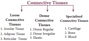
Connective tissue develops from the mesoderm and is widely distributed in the body. There are three main classes namely Loose connective tissue, Dense connective tissue and Specialized connective tissue. Major functions of connective tissues are binding, support, protection, insulation and transportation.
An Overview Of Connective Tissue img 1

Components of Connective Tissue

All connective tissues consist of three main components namely fires, ground substance and cells. The ‘Fibres’ of connective tissue provide support. Three types of fires are found in the connective tissue matrix. They are collagen, elastic and reticular fires.

Connective tissues are of three types namely, Loose connective tissues (Areolar, Adipose and Reticular) and Dense connective tissues (dense regular, dense irregular and elastic) and Specialized connective tissues (cartilage, bone and blood).

Loose Connective Tissues

In this tissue the cells and fires are loosely arranged in a semi fluid ground substances. For example the Areolar connective tissue beneath the skin acts as a support framework for epithelium and acts as a reservoir of water and salts for the surrounding body tissues, hence apply called tissue fluid. It contains firoblasts, macrophages, and mast cells (Figure 3.5).
An Overview Of Connective Tissue img 2

Adipose tissue is similar to areolar tissue in structure and function and located beneath the skin. Adipocytes commonly called adipose or fat cells predominate and account for 90% of this tissue mass. The cells of this tissue store fats and the excess nutrients which are not utilised immediately are converted to fats and are stored in tissues. Adipose tissue is richly vascularised indicating its high metabolic activity. While fasting, these cells maintain life by producing and supplying energy as fuel.

Adipose tissues are also found in subcutaneous tissue, surrounding the kidneys, eyeball, heart, etc. Adipose tissue is called ‘white fat’ or white adipose tissue. The adipose tissue which contains abundant mitochondria is called ‘Brown fat’ or Brown adipose tissue. White fat stores nutrients whereas brown fat is used to heat the blood stream to warm the body. Brown fat produces heat by nonshivering thermogenesis in neonates.

Reticular connective tissue resembles areolar connective tissue, but, the matrix is filled with firoblasts called reticular cells. It forms an internal framework (stroma) that supports the blood cells (largely lymphocytes) in the lymph nodes, spleen and bone marrow. Dense connective tissues (connective tissue proper) Fibres and firoblasts are compactly packed in the dense connective tissues. Orientation of fires show a regular or irregular pattern and is called dense regular and dense irregular tissues.

Dense regular connective tissues primarily contain collagen fires in rows between many parallel bundles of tissues and a few elastic fires. The major cell type is firoblast. It attaches muscles and bones and withstands great tensile stress when pulling force is applied in one direction. This connective tissue is present in tendons, that attach skeletal muscles to bones and ligaments attach one bone to another. Dense irregular connective tissues have bundles of thick collagen fires and firoblasts which are arranged irregularly.

The major cell type is the firoblast. It is able to withstand tension exerted in many directions and provides structural strength. Some elastic fires are also present. It is found in the skin as the leathery dermis and forms firous capsules of organs such as kidneys, bones, cartilages, muscles, nerves and joints.

Elastic connective tissue contains high proportion of elastic fires. It allows recoil of tissues following stretching. It maintains the pulsatile flow of blood through the arteries and the passive recoil of lungs following inspiration. It is found in the walls of large arteries; ligaments associated with vertebral column and within the walls of the bronchial tubes.

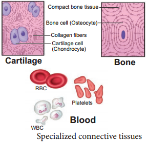
Specialised connective tissues are classified as cartilage, bones and blood. The intercellular material of cartilage is solid and pliable and resists compression. Cells of this tissue (chondrocytes) are enclosed in small cavities within the matrix secreted by them (Figure 3.6). Most of the cartilages in vertebrate embryos are replaced by bones in adults. Cartilage is present in the tip of nose, outer ear joints, ear pinna, between adjacent bones of the vertebral column, limbs and hands in adults.
An Overview Of Connective Tissue img 3

Bones have a hard and non-pliable ground substance rich in calcium salts and collagen fires which gives strength to the bones. It is the main tissue that provides structural frame to the body. Bones support and protect softer tissues and organs.

The bone cells (osteocytes) are present in the spaces called lacunae. Limb bones, such as the long bones of the legs, serve weightbearing functions. They also interact with skeletal muscles attached to them to bring about movements.

The bone marrow in some bones is the site of production of blood cells. Blood is the fluid connective tissue containing plasma, red blood cells (RBC), white blood cells (WBC) and platelets. It functions as the transport medium for the cardiovascular system, carrying nutrients, wastes, respiratory gases throughout the body. You will learn more about blood in Chapter 7.

Organisation of Epithelial Tissue

Learninsta presents the core concepts of Biology with high-quality research papers and topical review articles.

Organisation of Epithelial Tissue

Epithelial tissue is a sheet of cells that covers the body surface or lines the body cavity. It occurs in the body as a covering, as a lining epithelium and as glandular, epithelium. The functions of epithelium includes protection, absorption, filtration, excretion, secretion and sensory reception.

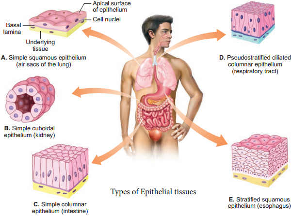
Based on the structural modification of the cells, the epithelial tissues are classified into simple epithelium and compound epithelium or stratified epithelium. Simple epithelium is composed of a single layer of cells. They are found in the organs of absorption, secretion and filtration. Simple epithelial tissue is further classified into squamous epithelium, cuboidal epithelium, columnar epithelium, ciliated epithelium and pseudostratifid epithelium (Figure 3.2).

The squamous epithelium is made of a single thin layer of flattened cells with irregular boundaries. They are found in the kidney glomeruli, air sacs of lungs, lining of heart, blood vessels and lymphatic vessels and are involved in functions like forming a diffusion boundary and filtration in sites where protection is not important.

The cuboidal epithelium is made of a single layer of cube like cells. This tissue is commonly found in the kidney tubules, ducts and secretory portions of small glands and surface of the ovary. Its main functions are secretion and absorption.

The columnar epithelium is composed of single layer of tall cells with round to oval nuclei at the base. It lines the digestive tract from the stomach to the rectum. The two modifications of this lining are the presence of microvilli on the apical surface of the absorptive cells and Goblet cell which secretes the protective lubricating mucus. The functions of this epithelium include absorption, secretion of mucus, enzymes and other substances.

If the columnar cells bear cilia on their free surfaces they are called ciliated epithelium. This ciliated type propels mucus by ciliary actions and it lines the small bronchioles, fallopian tubes and uterus. Nonciliated type lines most of the digestive tract, gall bladder and secretory ducts of glands.

Pseudo-stratified epithelial cells are columnar, but unequal in size. Although the epithelium is single layered yet it appears to be multi-layered because the nuclei lie at different levels in different cells. Hence, it is also called pseudostratified epithelium and its functions are protection, secretion and absorption. Ciliated forms line the trachea and the upper respiratory tract. The non ciliated forms, line the epididymis, large ducts of a glands and tracts of male urethra.
Epithelial Tissue img 1

Glandular Epithelium

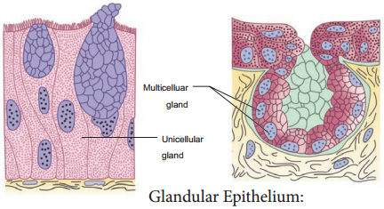
Some of the cuboidal or columnar cells get specialized for secretion and are called glandular epithelium (Figure 3.3). They are mainly of two types: unicellular, consisting of isolated glandular cells (goblet cells of the alimentary canal), and multicellular, consisting of cluster of cells (salivary gland). On the basis of the mode of pouring of their secretions, glands are divided into two categories namely exocrine and endocrine
glands. Exocrine glands secrete mucus, saliva, earwax, oil, milk, digestive enzymes and other cell products.

These products are released through ducts or tubes. In contrast endocrine glands do not have ducts. Their secretions called hormones are secreted directly into the fluid bathing the gland. The exocrine glands are classified as unicellular and multicelluar glands.

The multicelluar glands are further classified based on the structure as simple and compound glands, based on their secretory units as tubular, alveolar (Acinus) and tubulo alveolar. Based on the mode of secretion exocrine glands are classified as merocrine, holocrine and apocrine.
Epithelial Tissue img 2

Compound epithelium is made of more than one layer (multi-layered) of cells and thus has a limited role in secretion and absorption(Figure 3.4). The compound epithelia may be stratified and transitional. Their main function is to provide protection against chemical and mechanical stresses. They cover the dry surface of the skin, the moist surface of buccal cavity, pharynx, inner lining of ducts of salivary glands and of pancreatic ducts.

There are four types of compound epithelium namely, stratified squamous epithelium, cuboidal epithelium, columnar epithelium and transitional epithelium. Stratified squamous epithelium is of two types called keratinized type which forms the dry epidermis of the skin and the non keratinized type forms the moist lining of the oesophagus, mouth, conjunctiva of the eyes and vagina.

Stratified cuboidal epithelium mostly found in the ducts of sweat glands and mammary glands. Stratified columnar epithelium has limited distribution in the body, found around the lumen of the pharynx, male urethra and lining of some glandular ducts.

Transitional Epithelium is found lining the ureters, urinary bladder and part of the urethra. This epithelium allows stretching and is protective in function. All cells of the epithelium are held together with little intercellular material. In most of the animal tissues, specialized junctions provide both structural and functional links between its individual cells.

Three types of cell junctions are found in the epithelium and other tissues. These are called as tight, adhering and gap junctions. Tight junctions help to stop substances from leaking across a tissue. Adhering junctions perform cementing to keep neighbouring cells together. Gap junctions facilitate the cells to communicate with each other by connecting the cytoplasm of adjoining cells, for rapid transfer of ions, small molecules and sometimes big molecules.
Tissue Level of Organisation – Animal Tissues

Learninsta presents the core concepts of Biology with high-quality research papers and topical review articles.

Tissue Level of Organisation – Animal Tissues

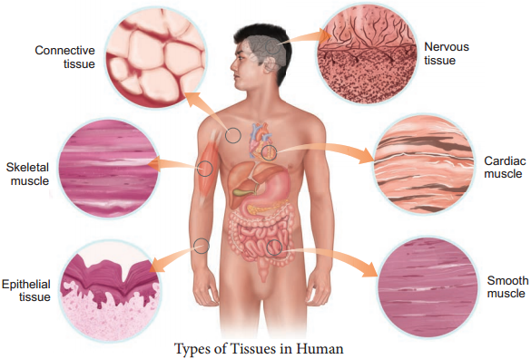
Animal tissues are classified according to the size, shape and function of the cells. There are four primary (basic) tissue types that interweave to form the ‘fabric’ of the body. They are, the epithelial tissue (covering), the connective tissue (support), the muscle tissue (movement) and the nervous tissue (control) (Figure 3.1).
Animal Tissues img 1

There are four types of animal tissues: epithelial tissue, connective tissue, muscle tissue and nervous tissue. Key Outcomes: Understand the differentiation of animal tissues and the relationship between structure and function of the various tissues. Know the location of the various tissues within the animal body.

Epithelial Tissue:
They cover the body, organs, blood vessels and all body cavities.

Muscular Tissue:
Smooth, Skeletal, and Cardiac Muscles.

Connective Tissue:
Connective tissues are made up of fibrous cells.

Nervous Tissue:
Neuron Structure.

Organs are composed of tissues, which are in turn composed of cells. Plants have three tissue types: ground, dermal, and vascular. Animals have four: epithelial, connective, muscle, and bone.

Supports an animal’s body – Connective tissue(supportive) Binds different tissues together – Fibrous connective tissue.

Connective tissue assists in support and protection of organs and limbs and depending on the location in the body it may join or separate organs or parts of the body. Muscle tissue enables various forms of movement, both voluntary and involuntary.

What are Parenchyma tissues? Parenchyma is a type of simple permanent tissue that makes a major part of ground tissues in plants, where other tissues like vascular tissues are embedded. They are non-vascular and composed of simple, living and undifferentiated cells, which are modified to perform various functions.

The nervous tissue is composed of two cell types: neurons and glia. The main function of nervous tissue is the processing of information coming from the external and internal environments, and then triggers a response.

Animal tissues are grouped into four basic types: connective, muscle, nervous, and epithelial. Collections of tissues joined in units to serve a common function compose organs.

Types of Tissues

  1. Simple squamous
  2. Simple cuboidal
  3. Cardiac muscle
  4. Skeletal muscle
  5. Bone
  6. Dense fibrous tissue
  7. Nerve
  8. Cartilage