Regulation of Kidney Function

Learninsta presents the core concepts of Biology with high-quality research papers and topical review articles.

Regulation of Kidney Function

ADH and Diabetes Insipidus

The functioning of kidneys is efficiently monitored and regulated by hormonal feedback control mechanism involving the hypothalamus, juxta glomerular apparatus and to a certain extent the heart. Osmoreceptors in the hypothalamus are activated by changes in the blood volume, body fluid volume and ionic concentration.

When there is excessive loss of fluid from the body or when there is an increase in the blood pressure, the osmoreceptors of the hypothalamus respond by stimulating the neurohypophysis to secrete the antidiuretic hormone (ADH) or vasopressin (a positive feedback). ADH facilitates reabsorption of water by increasing the number of aquaporins on the cell surface membrane of the distal convoluted tubule and collecting duct.

This increase in aquaporins causes the movement of water from the lumen into the interstitial cells, thereby preventing excess loss of water by diuresis. When you drink excess amounts of your favourite juice, osmoreceptors of the hypothalamus is no longer stimulated and the release of ADH is suppressed from the neurohypophysis (negative feedback) and the aquaporins of the collecting ducts move into the cytoplasm.

This makes the collecting ducts impermeable to water and the excess fluid flows down the collecting duct without any water loss. Hence dilute urine is produced to maintain the blood volume. Vasopressin secretion is controlled by positive and negative feedback mechanism.

Defects in ADH receptors or inability to secrete ADH leads to a condition called diabetes insipidus, characterized by excessive thirst and excretion of large quantities of dilute urine resulting in dehydration and fall in blood pressure.

Renin Angiotensin

Juxta glomerular apparatus (JGA) is a specialized tissue in the afferent arteriole of the nephron that consists of macula densa and granular cells. The macula densa cells sense distal tubular flow and affect afferent arteriole diameter, whereas the granular cells secrete an enzyme called renin. A fall in glomerular blood flow, glomerular blood pressure and glomerular filtration rate, can atctivate JG cells to release renin which converts a plasma protein, angiotensinogen (synthesized in the liver) to angiotensin I.

Angiotensin converting enzyme (ACE) converts angiotensin I to angiotensin II. Angiotensin II stimulates Na+ reabsorption in the proximal convoluted tubule by vasoconstriction of the blood vessels and increases the glomerular blood pressure.

Angiotensin II acts at different sites such as heart, kidney, brain, adrenal cortex and blood vessels. It stimulates adrenal cortex to secrete aldosterone that causes reabsorption of Na+, K+ excretion and absorption of water from the distal convoluted tubule and collecting duct.

This increases the glomerular blood pressure and glomerular filtration rate. This complex mechanism is generally known as Renin-AngiotensinAldosterone System (RAAS). Figure 8.9 shows the schematic representation of the various hormones in the regulation of body fluid concentration.
Regulation of Kidney Function img 1

Atrial Natriuretic Factor

Excessive stretch of cardiac atrial cells cause an increase in blood flow to the atria of the heart and release Atrial Natriuretic Peptide or factor (ANF) travels to the kidney where it increases Na+ excretion and increases the blood flow to the glomerulus, acting on the afferent glomerular arterioles as a vasodilator or on efferent arterioles as a vasoconstrictor.

It decreases aldosterone release from the adrenal cortex and also decreases release of renin, thereby decreasing angiotensin II. ANF acts antagonistically to the renin – angiotensin system, aldosterone and vasopressin.

Mechanism of Urine Formation in Human

Learninsta presents the core concepts of Biology with high-quality research papers and topical review articles.

Mechanism of Urine Formation in Human

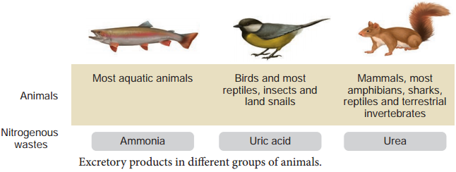
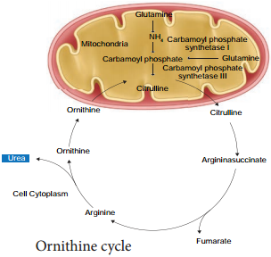
The nitrogenous waste formed as a result of breakdown of amino acids is converted to urea in the liver by the Ornithine cycle or urea cycle (Figure 8.7).
Mechanism of Urine Formation in Human img 1

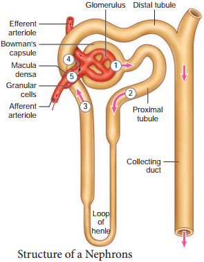
Urine formation involves three main processes namely, glomerular fitration, tubular reabsorption and tubular secretion.

(i) Glomerular Filtration

Blood enters the kidney from the renal artery, into the glomerulus. Blood is composed of large quantities of water, colloidal proteins, sugars, salts and nitrogenous end product. The first step in urine formation is the filtration of blood that takes place in the glomerulus.

This is called glomerular filtration which is a passive process. The fluid that leaves the glomerular capillaries and enters the Bowman’s capsule is called the glomerular filtrate.

The glomerular membrane has a large surface area and is more permeable to water and small molecules present in the blood plasma. Blood enters the glomerulus faster with greater force through the afferent arteriole and leaves the glomerulus through the efferent arterioles, much slower.

This force is because of the difference in sizes between the afferent and efferent arteriole (afferent arteriole is wider than efferent arteriole) and glomerular hydrostatic pressure which is around 55mm Hg.

Kidneys produce about 180L of glomerular filtrate in 24 hours. The molecules such as water, glucose, amino acids and nitrogenous substances pass freely from the blood into the glomerulus. Molecules larger than 5nm are barred from entering the tubule.

Glomerular pressure is the chief force that pushes water and solutes out of the blood and across the filtration membrane. The glomerular blood pressure (approximately 55 mmHg) is much higher than in other capillary beds. The two opposing forces are contributed by the plasma proteins in the capillaries.

These includes, colloidal osmotic pressure (30 mmHg) and the capsular hydrostatic pressure (15 mmHg) due to the fluids in the glomerular capsule. The net filtration pressure of 10 mmHg is responsible for the renal filtration.

Net filtration Pressure = Glomerular
hydrostatic pressure – (Colloidal osmotic pressure + Capsular hydrostatic pressure).
Net filtration pressure = 55 mmHg – (30 mmHg + 15 mmHg) = 10mmHg

The effective glomerular pressure of 10 mmHg results in ultrafiltration. Glomerular filtration rate (GFR) is the volume of filtrate formed min-1 in all nephrons (glomerulus) of both the kidneys. In adults the GFR is approximately 120-125mL/min. Blood from the glomerulus is passed out through the efferent arteriole.

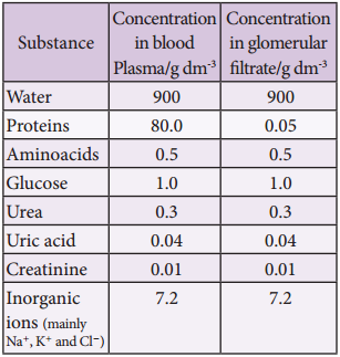
The smooth muscle of the efferent arteriole contract resulting in vasoconstriction. Table 8.1 shows the relative concentrations of substances in the blood plasma and the glomerular filtrate. The glomerular filtrate is similar to blood plasma except that there are no plasma proteins.

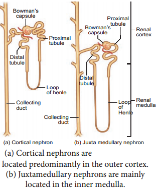
In cortical nephrons, blood from efferent arteriole flows into peritubular capillary beds and enters the venous system carrying with it recovered solutes and water from the interstitial fluid that surrounds the tubule.

Table 8.1 Concentration of substances in the blood plasma and in the glomerular filtrate
Mechanism of Urine Formation in Human img 2

(ii) Tubular Reabsorption

This involves movement of the filtrate back into the circulation. The volume of filtrate formed per day is around 170-180 L and the urine released is around 1.5 L per day, i.e., nearly 99% of the glomerular filtrate that has to be reabsorbed by the renal tubules as it contains certain substances needed by the body.

This process is called selective reabsorption. Reabsorption takes place by the tubular epithelial cells in different segments of the nephron either by active transport or passive transport, diffusion and osmosis.

Proximal Convoluted Tubule (PCT):

Glucose, lactate, amino acids, Na+ and water in the filtrate is reabsorbed in the PCT. Sodium is reabsorbed by active transport through sodium-potassium (Na+K+) pump in the PCT. Small amounts of urea and uric acid are also reabsorbed.

Descending Limb

Of Henle’s loop is permeable to water due the presence of aquaporins, but not permeable to salts. Water is lost in the descending limb, hence Na+ and Cl gets concentrated in the filtrate.

Ascending Limb of Henle’s Loop

Is impermeable to water but permeable to solutes such as Na+, Cl and K+.

The distal convoluted tubule recovers water and secretes potassium into the tubule. Na+, Cl and water remains in the filtrate of the DCT. Most of the reabsorption from this point is dependent on the body’s need and is regulated by hormones. Reabsorption of bicarbonate (HCO3) takes place to regulate the blood pH. Homeostasis of K+ and Na+ in the blood is also regulated in this region.

Collecting Duct

Is permeable to water, secretes K+ (potassium ions are actively transported into the tubule) and reabsorbs Na+ to produce concentrated urine. The change in permeability to water is due to the presence of number of waterpermeable channels called aquaporins.

Tubular Secretion:

Substances such as H+, K+, NH4+, creatinine and organic acids move into the filtrate from the peritubular capillaries into the tubular fluid. Most of the water is absorbed in the proximal convoluted tubule and Na+ is exchanged for water in the loop of Henle. Hypotonic fluid enters the distal convoluted tubule and substances such as urea and salts pass from peritubular blood into the cells of DCT.

The urine excreted contains both filtered and secreted substances. Once it enters the collecting duct, water is absorbed and concentrated hypertonic urine is formed. For every H+ secreted into the tubular filtrate, a Na+ is absorbed by the tubular cell.

The H+ secreted combines with HCO3, HPO3 and NH3and gets fixed as H2CO4+, H2PO4+ and NH4+ respectively. Since H+ gets fixed in the fluid, reabsorption of H+ is prevented.

Formation of Concentrated Urine

Formation of concentrated urine is accomplished by kidneys using counter current mechanisms. The major function of Henle’s loop is to concentrate Na+ and Cl. There is low osmolarity near the cortex and high osmolarity towards the medulla.

This osmolarity in the medulla is due to the presence of the solute transporters and is maintained by the arrangement of the loop of Henle, collecting duct and vasa recta. This arrangement allows movement of solutes from the filtrate to the interstitial fluid. At the transition between the proximal convoluted tubule and the descending loop of Henle the osmolarity of the interstitial fluid is similar to that of the blood – about 300mOsm.

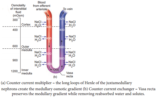
Ascending and Descending Limbs of Henle, Create a Counter Current Multiplier

(Interaction between flow of filtrate through the limbs of Henle’s and JMN) by active transport. Figure 8.8 (a) shows the counter current multiplier created by the long loops of Henle of the JM nephrons which creates medullary osmotic gradient.
Mechanism of Urine Formation in Human img 3

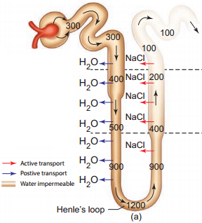
As the fluid enters the descending limb, water moves from the lumen into the interstitial fluid and the osmolarity of interstitial fluid decreases. To counteract this dilution the region of the ascending limb actively pumps solutes from the lumen into the interstitial fluid and the osmolarity increases to about 1200mOsm in medulla. This mismatch between water and salts creates osmotic gradient in the medulla. The osmotic gradient is also due to the permeability of the collecting duct to urea.

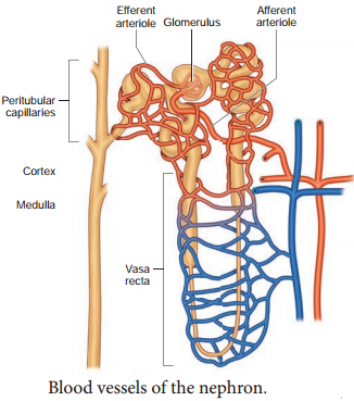
The vasa recta, maintains the medullary osmotic gradient via counter current exchanger (the flow of blood through the ascending and descending vasa recta blood vessels) by passive transport. Figure 8.8 (b) shows counter current exchanger where the vasa recta preserves the medullary gradient while removing reabsorbed water and solutes.

This system does not produce an osmotic gradient, but protects the medulla by removal of excess salts from the interstitial fluid and removing reabsorbed water. The vasa recta leave the kidney at the junction between the cortex and medulla. The interstitial fluid at this point is iso-osmotic to the blood.

When the blood leaves the efferent arteriole and enters vasa recta the osmolarity in the medulla increases (1200mOsm) and results in passive uptake of solutes and loss of water in descending vasa recta. As the blood enters the cortex, the osmolarity in the blood decreases (300mOsm) and the blood loses solutes and gains water.

At the final stage in collecting duct to form concentrated urine (hypertonic). Human kidneys can produce urine nearly four times concentrated than the initial filtrate formed.
Mechanism of Urine Formation in Human img 4

Human Excretory System – Structure of Kidney, Nephron

Learninsta presents the core concepts of Biology with high-quality research papers and topical review articles.

Human Excretory System – Structure of Kidney, Nephron

Structure of kidney

Excretory system in human consists of a pair of kidneys, a pair of ureters, urinary bladder and urethra (Figure. 8.2). Kidneys are reddish brown, bean shaped structures that lie in the superior lumbar region between the levels of the last thoracic and third lumber vertebra close to the dorsal inner wall of the abdominal cavity.

The right kidney is placed slightly lower than the left kidney. Each kidney weighs an average of 120-170 grams. The outer layer of the kidney is covered by three layers of supportive tissues namely, renal fascia, perirenal fat capsule and fibrous capsule.
Human Excretory System - Structure of Kidney, Nephron img 1

The longitudinal section of kidney (Figure. 8.3) shows, an outer cortex, inner medulla and pelvis. The medulla is divided into a few conical tissue masses called medullary pyramids or renal pyramids. The part of cortex that extends in between the medullary pyramids is the renal columns of Bertini.
Human Excretory System - Structure of Kidney, Nephron img 2

The centre of the inner concave surface of the kidney has a notch called the renal hilum, through which ureter, blood vessels and nerves innervate. Inner to the hilum is a broad funnel shaped space called the renal pelvis with projection called calyces.

The renal pelvis is continuous with the ureter once it leaves the hilum. The walls of the calyces, pelvis and ureter have smooth muscles which contracts rhythmically. The calyces collect the urine and empties into the ureter, which is stored in the urinary bladder temporarily. The urinary bladder opens into the urethra through which urine is expelled out.

Structure of a Nephron

Each kidney has nearly one million complex tubular structures called nephron (Figure 8.4). Each nephron consists of a filtering corpuscle called renal corpuscle (malpighian body) and a renal tubule. The renal tubule opens into a longer tubule called the collecting duct. The renal tubule begins with a double walled cup shaped structure called the Bowman’s capsule, which encloses a ball of capillaries that delivers fluid to the tubules, called the glomerulus.

The Bowman’s capsule and the glomerulus together constitute the renal corpuscle. The endothelium of glomerulus has many pores (fenestrae). The external parietal layer of the Bowman’s capsule is made up of simple squamous epithelium and the visceral layer is made of epithelial cells called podocytes. The podocytes end in foot processes which cling to the basement membrane of the glomerulus. The openings between the foot processes are called filtration slits.
Human Excretory System - Structure of Kidney, Nephron img 3

The renal tubule continues further to form the proximal convoluted tubule [PCT] followed by a U-shaped loop of Henle (Henle’s loop) that has a thin descending and a thick ascending limb. The ascending limb continues as a highly coiled tubular region called the distal convoluted tubule [DCT].

The DCT of many nephrons open into a straight tube called collecting duct. The collecting duct runs through the medullary pyramids in the region of the pelvis. Several collecting ducts fuse to form papillary duct that delivers urine into the calyces, which opens into the renal pelvis.

In the renal tubules, PCT and DCT of the nephron are situated in the cortical region of the kidney whereas the loop of Henle is in the medullary region. In majority of nephrons, the loop of Henle is too short and extends only very little into the medulla and are called cortical nephrons. Some nephrons have very long loop of Henle that run deep into the medulla and are called juxta medullary nephrons (JMN) (Figure 8.5 a and b)
Human Excretory System - Structure of Kidney, Nephron img 4

The capillary bed of the nephrons. First capillary bed of the nephron is the glomerulus and the other is the peritubular capillaries. The glomerular capillary bed is different from other capillary beds in that it is supplied by the afferent and drained by the efferent arteriole.

The efferent arteriole that comes out of the glomerulus forms a fine capillary network around the renal tubule called the peritubular capillaries. The efferent arteriole serving the juxta medullary nephron forms bundles of long straight vessel called vasa recta and runs parallel to the loop of Henle. Vasa recta is absent or reduced in cortical nephrons (Figure 8.6).
Human Excretory System - Structure of Kidney, Nephron img 5

Models of Excretion Definition and Its Explanation

Learninsta presents the core concepts of Biology with high-quality research papers and topical review articles.

Models of Excretion Definition and Its Explanation

Excretory system helps in collecting nitrogenous waste and expelling it into the external environment. Animals have evolved different strategies to get rid of these nitrogenous wastes. Ammonia produced during amino acid breakdown is toxic hence must be excreted either as ammonia, urea or uric acid.

The type of nitrogenous end product an animal excretes depends upon the habitat of the animal. Ammonia requires large amount of water for its elimination, whereas uric acid, being the least toxic can be removed with the minimum loss of water, and urea can be stored in the body for considerable periods of time, as it is less toxic and less soluble in water than ammonia.

Animals that excrete most of its nitrogen in the form of ammonia are called ammonoteles. Many fishes, aquatic amphibians and aquatic insects are ammonotelic. In bony fishes, ammonia diffuses out across the body surface or through gill surface as ammonium ions.

Reptiles, birds, land snails and insects excrete uric acid crystals, with a minimum loss of water and are called uricoteles. In terrestrial animals, less toxic urea and uric acid are produced to conserve water. Mammals and terrestrial amphibians mainly excrete urea and are called ureoteles. Earthworms while in soil are ureoteles and when in water are ammonoteles. Figure 8.1 shows the excretory products in different groups of animals.
Models of Excretion img 1

The animal kingdom presents a wide variety of excretory structures. Most invertebrates have a simple tubular structure in the form of primitive kidneys called protonephridia and metanephridia. Vertebrates have complex tubular organs called kidneys.

Protonephridia are excretory structures with specialized cells in the form of flame cells (cilia) in Platyhelminthes (example tapeworm) and Solenocytes (flagella) in Amphioxus. Nematodes have rennette cells, Metanephridia are the tubular excretory structures in annelids and molluscs.

Malpighian tubules are the excretory structures in most insects. Antennal glands or green glands perform excretory function in crustaceans like prawns. Vertebrate kidney differs among taxa in relation to the environmental conditions.

Nephron is the structural and functional unit of kidneys. Reptiles have reduced glomerulus or lack glomerulus and Henle’s loop and hence produce very little hypotonic urine, whereas mammalian kidneys produce concentrated (hyperosmotic) urine due to the presence of long Henle’s loop.

The Loop of Henle of the nephron has evolved to form hypertonic urine. Aglomerular kidneys of marine fishes produce little urine that is isoosmotic to the body fluid. Amphibians and fresh water fish lack Henle’s loop hence produce dilute urine (hypoosmotic).

Cardio Pulmonary Resuscitation (CPR)

Learninsta presents the core concepts of Biology with high-quality research papers and topical review articles.

Cardio Pulmonary Resuscitation (CPR)

In 1956, James Elam and Peter Safar were the first to use mouth to mouth resuscitation. CPR is a life saving procedure that is done at the time of emergency conditions such as when a person’s breath or heart beat has stopped abruptly in case of drowning, electric shock or heart attack.

CPR includes rescue of breath, which is achieved by mouth to mouth breathing, to deliver oxygen to the victim’s lungs by external chest compressions which helps to circulate blood to the vital organs.

CPR must be performed within 4 to 6 minutes after cessation of breath to prevent brain damage or death. Along with CPR, defibrillation is also done. Defibrillation means a brief electric shock is given to the heart to recover the function of the heart.

Cardiopulmonary resuscitation (CPR) is an emergency procedure that combines chest compressions often with artificial ventilation in an effort to manually preserve intact brain function until further measures are taken to restore spontaneous blood circulation and breathing in a person who is in cardiac arrest.

5 Steps for Performing CPR

  • Check the patient’s responsiveness.
  • Shake the unresponsive person by the shoulders and speak loudly to them in an attempt to rouse them.
  • Check their breathing and pulse.
  • Administer chest compressions.
  • Recheck breathing and pulse.

After every 30 chest compressions at a rate of 100 to 120 a minute, give 2 breaths. Continue with cycles of 30 chest compressions and 2 rescue breaths until they begin to recover or emergency help arrives.

Cardiopulmonary resuscitation (CPR) is a lifesaving technique. It aims to keep blood and oxygen flowing through the body when a person’s heart and breathing have stopped. CPR can be performed by any trained person. It involves external chest compressions and rescue breathing.

Types of CPR

High-Frequency Chest Compressions. This technique involves imitating hear beats by giving more chest compressions at intervals of time in high frequency. Open-Chest CPR. Open chest CPR is a procedure in which the heart is retrieved through thoracotomy. Interposed Abdominal Compression CPR.

How is CPR Performed? There are two commonly known versions of CPR: For healthcare providers and those trained: conventional CPR using chest compressions and mouth-to-mouth breathing at a ratio of 30:2 compressions-to-breaths.

CPR stands for cardiopulmonary resuscitation. It is an emergency life-saving procedure that is done when someone’s breathing or heartbeat has stopped.

The three basic parts of CPR are easily remembered as “CAB”: C for compressions, A for airway, and B for breathing. C is for compressions. Chest compressions can help the flow of blood to the heart, brain, and other organs.
Cardio Pulmonary Resuscitation (CPR) img 1