Types of Movement in Human Body

Learninsta presents the core concepts of Biology with high-quality research papers and topical review articles.

Types of Movement in Human Body

The different types of movements that occur in the cells of our body are amoeboid, ciliary, flagellar and muscular movement. Amoeboid movement – Cells such as macrophages exhibit amoeboid movement for engulfing pathogens by pseudopodia formed by the streaming movement of the cytoplasm.

Ciliary Movement:

This type of movement occurs in the respiratory passages and genital tracts which are lined by ciliated epithelial cells.

Flagellar Movement:

This type of movement occurs in the cells which are having flagella or whip-like motile organelle. The sperm cells show flagellar movement.

Muscular Movement:

The movement of hands, legs, jaws, tongue are caused by the contraction and relaxation of the muscle which is termed as the muscular movement.

The different types of movement that are permitted at each joint are described below.

  • Flexion – bending a joint.
  • Extension – straightening a joint.
  • Abduction – movement away from the midline of the body.
  • Adduction – movement towards the midline of the body.
  • Circumduction – this is where the limb moves in a circle.

In the world of mechanics, there are four basic types of motion. These four are rotary, oscillating, linear and reciprocating.

Flexibility is extending and contracting the muscle tissues, joints, and ligaments into a greater range of motion accepted by the nervous system.

Mobility is neuromuscular active control of the range of motion within the muscle tissue, joints, and ligaments.

  • Strength
  • Power
  • Endurance
  • Stability

Body movement involves a complex cascade transforming neural signals to depolarization of myofibres, binding of individual myosin and actin filaments in the sarcomeres leading to myofibre contraction, and myofibre cross-linking transmitting force throughout muscle groups and into the skeletal system via their tendinous.

Muscles move body parts by contracting and then relaxing. Muscles can pull bones, but they can’t push them back to the original position. So they work in pairs of flexors and extensors. The flexor contracts to bend a limb at a joint.
Process of Haemodialysis and its Kidney Transplantation

Learninsta presents the core concepts of Biology with high-quality research papers and topical review articles.

Process of Haemodialysis and its Kidney Transplantation

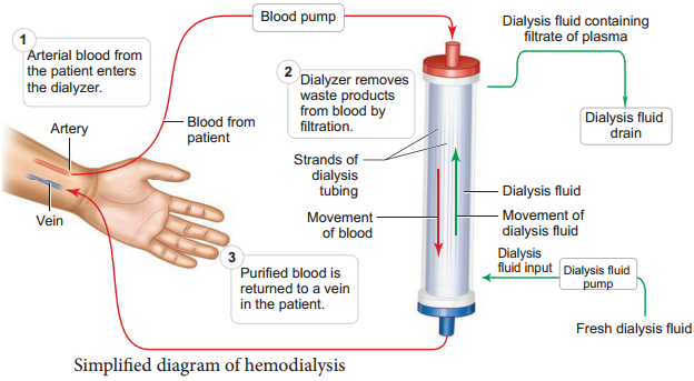
Malfunctioning of the kidneys can lead to accumalation of urea and other toxic substances, leading to kidney failure. In such patients toxic urea can be removed from the blood by a process called haemodialysis. A dialyzing machine or an artificial kidney is connected to the patient’s body.

A dialyzing machine consists of a long cellulose tube surrounded by the dialysing fluid in a water bath. The patient’s blood is drawn from a conveinent artery and pumped into the dialysing unit after adding an anticoagulant like heparin.

The tiny pores in the dialysis tube allows small molecules such as glucose, salts and urea to enter into the water bath, whereas blood cells and protein molecules do not enter these pores. This stage is similar to the filtration process in the glomerulus.

The dialysing liquid in the water bath consists of solution of salt and sugar in correct proportion in order to prevent loss of glucose and essential salts from the blood. The cleared blood is then pumped back to the body through a vein Figure 8.10.
Disorders Related to the Excretory System img 1

Kidney Transplantation

It is the ultimate method for correction of acute renal failures. This involves transfer of healthy kidney from one person (donor) to another person with kidney failure. The donated kidney may be taken from a healthy person who is declared brain dead or from sibling or close relatives to minimise the chances of rejection by the immune system of the host. Immunosuppressive drugs are usually administered to the patient to avoid tissue rejection.

Haemodialysis is a way of replacing some of the functions of your kidney, if your kidneys have failed, by using a machine to filter and clean your blood. Blood is pumped out of your body to the machine where it is passed through a series of tiny tubes, in an ‘artificial kidney’ or ‘dialyser’.

In hemodialysis, blood is removed from the body and filtered through a man-made membrane called a dialyzer, or artificial kidney, and then the filtered blood is returned to the body. The average person has about 10 to 12 pints of blood; during dialysis only one pint (about two cups) is outside of the body at a time.

There are three different types of dialysis. Hemodialysis. Hemodialysis is the most common type of dialysis. Peritoneal dialysis. Peritoneal dialysis involves surgery to implant a peritoneal dialysis (PD) catheter into your abdomen. Continuous renal replacement therapy (CRRT).

When your kidneys fail, dialysis keeps your body in balance by: removing waste, salt and extra water to prevent them from building up in the body. keeping a safe level of certain chemicals in your blood, such as potassium, sodium and bicarbonate helping to control blood pressure.

Hemodialysis is a procedure where a dialysis machine and a special filter called an artificial kidney, or a dialyzer, are used to clean your blood. To get your blood into the dialyzer, the doctor needs to make an access, or entrance, into your blood vessels. This is done with minor surgery, usually to your arm.

The Benefits of Hemodialysis Include:

  • Nurses perform treatments for the patient.
  • Regular contact with other hemodialysis patients and staff.
  • Patients usually only have three treatments per week; giving them four days off.
  • No equipment or supplies have to be kept at home.
  • In an emergency, medical help is available quickly.

Disorders Related to the Excretory System

Learninsta presents the core concepts of Biology with high-quality research papers and topical review articles.

Disorders Related to the Excretory System

Urinary Tract Infection

Female’s urethra is very short and its external opening is close to the anal opening, hence improper toilet habits can easily carry faecal bacteria into the urethra. The urethral mucosa is continuous with the urinary tract and the inflammation of the urethra (urethritis) can ascend the tract to cause bladder inflammation (cystitis) or even renal inflammation (pyelitis or pyelonephritis).

Symptoms include dysuria (painful urination), urinary urgency, fever and sometimes cloudy or blood tinged urine. When the kidneys are inflammed, back pain and severe headache often occur. Most urinary tract infections can be treated by antibiotics.

Renal Failure (Kidney Failure)

Failure of the kidneys to excrete wastes may lead to accumulation of urea with marked reduction in the urine output. Renal failure are of two types, Acute and chronic renal failure. In acute renal failure the kidney stops its function abruptly, but there are chances for recovery of kidney functions. In chronic renal failure there is a progressive loss of function of the nephrons which gradually decreases the function of kidneys.

Uremia

Uremia is characterized by increase in urea and other non-protein nitrogenous substances like uric acid and creatinine in blood. Normal urea level in human blood is about 17-30mg/100mL of blood. The urea concentration rises as 10 times of normal levels during chronic renal failure.

Renal Calculi

Kidney stone or calculi, also called renal stone or nephrolithiasis, is the formation of hard stone like masses in the renal tubules of renal pelvis. It is mainly due to the accumulation of soluble crystals of salts of sodium oxalates and certain phosphates.

This result in severe pain called “renal colic pain” and can cause scars in the kidneys. Renal stones can be removed by techniques like pyleothotomy or lithotripsy.

Glomerulonephritis

It is also called Bright’s disease and is characterized by inflammation of the glomeruli of both kidneys and is usually due to poststreptococcal infection that occurs in children. Symptoms are haematuria, proteinuria, salt and water retention, oligouria, hypertension and pulmonary oedema.

Role of Other Organs in Excretion

Learninsta presents the core concepts of Biology with high-quality research papers and topical review articles.

Role of Other Organs in Excretion

Apart from kidneys, organs such as lungs, liver and skin help to remove wastes. Our lungs remove large quantities of carbon dioxide (18 L/day) and significant quantities of water every day. Liver secretes bile containing substances like, bilirubin and biliverdin, cholesterol, steroid hormones, vitamins and drugs which are excreted out along with the digestive wastes.

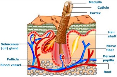
Sweat and sebaceous glands in the skin eliminate certain wastes through their secretions. Sweat produced by the sweat glands primarily helps to cool the body and secondarily excretes Na+ and Cl, small quantities of urea and lactate.

Sebaceous glands eliminate certain substances like sterols, hydrocarbons and waxes through sebum that provides a protective oily covering for the skin. Small quantities of nitrogenous wastes are also excreted through saliva.

Kidneys play a major role in the process of excretion in humans. Kidneys help in the elimination of wastes from the body in the form of urine. Apart from kidneys, organs like lungs, liver, skin and sebaceous glands help in excretion.

The excretory system in humans consists mainly of the kidneys and bladder. The kidneys filter urea and other waste products from the blood, which are then added to the urine within the bladder. Other organs, such as the liver, process toxins but put their wastes back into the blood.

Excretory Organs. Organs of excretion include the skin, liver, large intestine, lungs, and kidneys (see the figure below). Together, these organs make up the excretory system. They all excrete wastes, but they don’t work together in the same way that organs do in most other body systems. The appendix is a vestigial organ that has no role to play in excretion.

The liver regulates most chemical levels in the blood and excretes a product called bile. This helps carry away waste products from the liver. Production of bile, which helps carry away waste and break down fats in the small intestine during digestion. Production of certain proteins for blood plasma.

These chemical reactions produce waste products such as carbon dioxide, water, salts, urea and uric acid. Accumulation of these wastes beyond a level inside the body is harmful to the body. The excretory organs remove these wastes. This process of removal of metabolic waste from the body is known as excretion.

Humans have two kidneys and each kidney is supplied with blood from the renal artery. The kidneys remove from the blood the nitrogenous wastes such as urea, as well as salts and excess water, and excrete them in the form of urine.

Role of Liver in Excretion:

Liver converts the amino acids present in blood into ammonia and pyruvic acid. Pyruvic acid gets oxidized to release energy and ammonia gets converted into urea. Kidney helps in the filtration of the urea and urea gets excreted in the form of urine.

Certain waste and harmful substances are formed during the functioning of body cells. When these toxic materials are not removed from the body, they get mixed with blood and can damage the cells of the body. The removal of such poisonous waste materials is therefore necessary.

Skin has an important role in excretion in man . So Skin is important to clean our body by the process of excretion. Lungs release Carbon dioxide (CO2) which helps in the process of respiration and purifies blood. Intestine helps in excretion of food digested in stomach and in duodenum.

Excretion, the process by which animals rid themselves of waste products and of the nitrogenous by-products of metabolism. Through excretion organisms control osmotic pressure the balance between inorganic ions and water and maintain acid-base balance.
Role of Other Organs in Excretion img 1

The Physiology of Micturition Definition and its Uses

Learninsta presents the core concepts of Biology with high-quality research papers and topical review articles.

The Physiology of Micturition Definition and its Uses

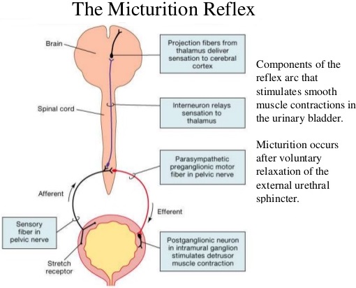
The process of release of urine from the bladder is called micturition or urination. Urine formed by the nephrons is ultimately carried to the urinary bladder where it is stored till it receives a voluntary signal from the central nervous system. The stretch receptors present in the urinary bladder are stimulated when it gets filled with urine.

Stretching of the urinary bladder stimulates the CNS via the sensory neurons of the parasympathetic nervous system and brings about contraction of the bladder. Simultaneously, somatic motor neurons induce the sphincters to close. Smooth muscles contracts resulting in the opening of the internal sphincters passively and relaxing the external sphincter.

When the stimulatory and inhibitory controls exceed the threshold, the sphincter opens and the urine is expelled out. An adult human on an average excretes 1 to 1.5 L of urine per day. The urine formed is a yellow coloured watery fluid which is slightly acidic in nature (pH 6.0), Changes in diet may cause pH to vary between 4.5 to 8.0 and has a characteristic odour. The yellow colour of the urine is due to the presence of a pigment, urochrome.

On an average, 25-30 gms of urea is excreted per day. Various metabolic disorders can affect the composition of urine. Analysis of urine helps in clinical diagnosis of various metabolic disorders and the malfunctioning of the kidneys. For example the presence of glucose (glucosuria) and ketone bodies (ketonuria) in the urine are indications of diabetes mellitus.

The exact cause of micturition syncope isn’t fully understood. But it may be related to opening (vasodilation) of the blood vessels that occurs when getting up and standing at the toilet or that occurs at the rapid emptying of a full bladder. This is thought to result in a sudden drop in blood pressure.

Micturition involves the simultaneous coordinated contraction of the bladder detrusor muscle, which is controlled by parasympathetic (cholinergic) nerves, and the relaxation of the bladder neck and sphincter, which are controlled by sympathetic (α-adrenergic) nerves.

Micturition syncope causes more than 8 percent of all episodes of fainting. People who experience it are more prone to fainting under other circumstances, too. Micturition syncope occurs more often in men. It often happens after using the bathroom in the middle of the night or first thing in the morning.

The pons is a major relay center between the brain and the bladder. The mechanical process of urination is coordinated by the pons in the area known as the pontine micturition center (PMC). The conscious sensations associated with bladder activity are transmitted to the pons from the cerebral cortex.

Introduction. Micturition is the process of eliminating water and electrolytes from the urinary system, commonly known as urinating. It has two discrete phases: the storage/continence phase, when urine is stored in the bladder; and the voiding phase, where urine is released through the urethra.

Micturition is the process by which the urine from the urinary bladder is excreted. This reflex stimulates the urge to pass out urine. To discharge urine, the urethral sphincter relaxes and the smooth muscles of the bladder contract. This forces the urine out from the bladder.
Micturition img 1