Mechanism of Muscle Contraction

Learninsta presents the core concepts of Biology with high-quality research papers and topical review articles.

Mechanism of Muscle Contraction

Sliding Filament Theory:

In 1954, Andrew F. Huxley and Rolf Niedergerke proposed the sliding-filament theory to explain muscle contraction. According to this theory, overlapping actin and myosin filaments of fixed length slide past one another in an energy requiring process, resulting in muscle contraction. The contraction of muscle fire is a remarkable process that helps in creating a force to move or to resist a load.

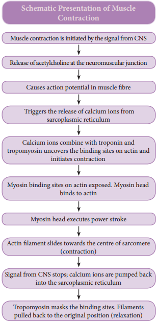
The force which is created by the contracting muscle is called muscle tension. The load is a weight or force that opposes contraction of a muscle. Contraction is the creation of tension in the muscle which is an active process and relaxation is the release of tension created by contraction. Muscle contraction is initiated by a nerve impulse sent by the central nervous system (CNS) through a motor neuron.

The junction between the motor neuron and the sarcolemma of the muscle fire is called the neuromuscular junction or motor end plate. When nerve impulse reaches a neuromuscular junction, acetylcholine is released. It initiates the opening of multiple gated channels in sarcolemma. The action potential travels along the T-tubules and triggers the release of calcium ions from the sarcoplasmic reticulum.

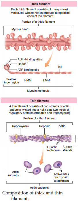
The released calcium ions bind to troponin on thin filaments. The tropomyosin uncovers the myosin-binding sites on thin filaments. Now the active sites are exposed to the heads of myosin to form a cross-bridge (Figure 9.3).

During cross-bridge formation acting and myosin form a protein complex called actomyosin. Utilizing the energy released from hydrolysis of ATP, the myosin head rotates until it forms a 90° angle with the long axis of the filament. In this position myosin binds to an actin and activates a contraction – relaxation cycle which is followed by a power stroke.
Mechanism of Muscle Contraction img 1

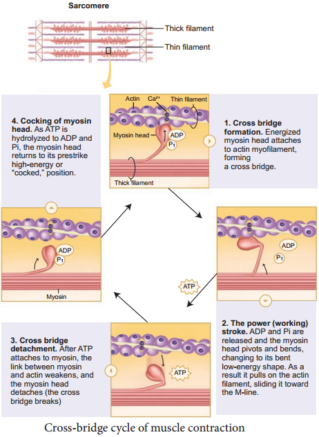
The power stroke (cross-bridge tilting) begins after the myosin head and hinge region tilt from a 90° angle to a 45° angle. The crossbridge transforms into strong, high-force bond which allows the myosin head to swivel. When the myosin head swivels it pulls the attached acting filament towards the centre of the A-band.

The myosin returns back to its relaxed state and releases ADP and phosphate ion. A new ATP molecule then binds to the head of the myosin and the cross-bridge is broken. At the end of each power stroke, each myosin head detaches from actin, then swivels back and binds to a new actin molecule to start another contraction cycle.

This movement is similar to the motion of an oar on a boat. At the end of each power stroke, each myosin head detaches from actin, then swivels back and binds to a new actin molecule to start another contraction cycle. The power stroke repeats many times until a muscle fire contracts.

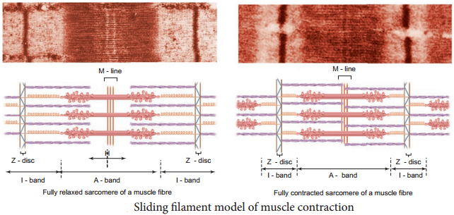
The myosin heads bind, push and release actin molecules over and over as the thin filaments move toward the centre of the sarcomere. The repeated formation of cross-bridge cycles cause the sliding of the filaments only but there is no change in the lengths of either the thick or thin filaments.

The Z – discs attached to the actin filaments are also pulled inwards from both the sides, causing the shortening of the sarcomere (i.e. contraction). This process continues as long as the muscle receives the stimuli and with a steady flow of calcium ions.

When motor impulse stops, the calcium ions are pumped back into the sarcoplasmic reticulum which result in the masking of the active sites of the actin filaments. The myosin head fails to bind with the active sites of actin and these changes cause the return of Z – discs back to their original position, i.e. relaxation. (Figure 9.4)
Mechanism of Muscle Contraction img 2
Mechanism of Muscle Contraction img 3

Types of Skeletal Muscle Contraction

Learninsta presents the core concepts of Biology with high-quality research papers and topical review articles.

Types of Skeletal Muscle Contraction

There are two primary types of muscle contractions. They are isotonic contraction and isometric contraction. The types of contractions depend on the changes in the length and tension of the muscle fibres at the time of its contraction.

Isotonic contraction (iso – same, tonweight/resistance) In isotonic contraction the length of the muscle changes but the tension remains constant. Here, the force produced is unchanged. Example: lifting dumb bells and weightlifting. Isometric contraction (iso – same, metric – distance)

In isometric contraction the length of the muscle does not change but the tension of the muscle changes. Here, the force produced is changed. Example: pushing against a wall, holding a heavy bag.

Types of Skeletal Muscle Fibres

The muscle fibres can be classified on the basis of their rate of shortening, either fast or slow and the way in which they produce the ATP needed for contraction, either oxidative or glycolytic. Fibres containing myosin with high ATPase activity are classified as fast fibres and with lower ATPase activity are classified as slow fibres.

Fibres that contain numerous mitochondria and have a high capacity for oxidative phosphorylation are classified as oxidative fibres. Such fibres depend on blood flow to deliver oxygen and nutrients to the muscles. The oxidative fibres are termed as red muscle fibres.

Fibres that contain few mitochondria but possess a high concentration of glycolytic enzymes and large stores of glycogen are called glycolytic fibres. The lack of myoglobin gives pale colour to the fibres, so they are termed as white muscle fibres.

Skeletal muscle fibres are further classified into three types based on the above classification. They are slow – oxidative fibres, fast – oxidative fibres and fast – glycolytic fibres.

1. Slow – Oxidative Fibres

Have low rates of myosin ATP hydrolysis but have the ability to make large amounts of ATP. These fibres are used for prolonged, regular activity such as long distance swimming. Long – distance runners have a high proportion of these fibres in their leg muscles.

2. Fast – Oxidative Fibres

Have high myosin ATPase activity and can make large amounts of ATP. They are particularly suited for rapid actions.

3. Fast – Glycolytic Fibres

Have myosin ATPase activity but cannot make as much ATP as oxidative fires, because their source of ATP is glycolysis. These fibres are best suited for rapid, intense actions, such as short sprint at maximum speed.

Isometric:
A muscular contraction in which the length of the muscle does not change.

Isotonic:
A muscular contraction in which the length of the muscle changes.

Eccentric:
An isotonic contraction where the muscle lengthens.

Concentric:
An isotonic contraction where the muscle shortens. Types of Contractions. There are three types of muscle contraction: concentric, isometric, and eccentric.

Isometric:
Of or involving muscular contraction against resistance in which the length of the muscle remains the same.

Isotonic:
Of or involving muscular contraction against resistance in which the length of the muscle changes.

The process of muscular contraction occurs over a number of key steps, including: Depolarisation and calcium ion release.

Actin and myosin cross-bridge formation.
Sliding mechanism of actin and myosin filaments.
Sarcomere shortening (muscle contraction)

Isotonic contractions – These occur when a muscle contracts and changes length and there are two types:
Isotonic concentric contraction – This involves the muscle shortening.
Isotonic eccentric contraction – This involves the muscle lengthening whilst it is under tension.

The first step in the process of contraction is for Ca++ to bind to troponin so that tropomyosin can slide away from the binding sites on the actin strands. This allows the myosin heads to bind to these exposed binding sites and form cross-bridges.

Abstract. Skeletal muscle is one of the most dynamic and plastic tissues of the human body. In humans, skeletal muscle comprises approximately 40% of total body weight and contains 50-75% of all body proteins.

Skeletal muscles only pull in one direction. For this reason they always come in pairs. When one muscle in a pair contracts, to bend a joint for example, its counterpart then contracts and pulls in the opposite direction to straighten the joint out again.

This is a table of skeletal muscles of the human anatomy. There are around 650 skeletal muscles within the typical human body. Almost every muscle constitutes one part of a pair of identical bilateral muscles, found on both sides, resulting in approximately 320 pairs of muscles, as presented in this article.
Types of Skeletal Muscle Contraction img 1

Structure of Contractile Proteins

Learninsta presents the core concepts of Biology with high-quality research papers and topical review articles.

Structure of Contractile Proteins

Contraction of the muscle depends on the presence of contractile proteins (Figure 9.2) such as actin and myosin in the myofilaments. The thick filaments are composed of the protein myosin which are bundled together whose heads produce at opposite ends of the filament.

Each myosin molecule is made up of a monomer called meromyosin. The meromyosin has two regions, a globular head with a short arm and a tail. The short arm constitutes the heavy meromyosin (HMM).

The tail portion forms the light meromyosin (LMM). The head bears an actin-binding site and an ATP – binding site. It also contains ATP case enzyme that split ATP to generate energy for the contraction of muscle. The thin filaments are composed of two interwined actin molecules. Actin has polypeptide subunits called globular actin or G-actin and filamentous form or F-actin.

Each thin filament is made of two F-actins helically wound to each other. Each F-actin is a polymer of monomeric G-actins. It also contains a binding site for myosin. The thin filaments also contain several regulatory proteins like tropomyosin and troponin which help in regulating the contraction of muscles along with actin and myosin.

Structure of Contractile Proteins img 1

Structure of Contractile Proteins. Each actin (thin) filament is made of two ‘F’ (filamentous) actins helically wound to each other. Each ‘F’ actin is a polymer of monomeric ‘G’ (Globular) actins. Two filaments of another protein, tropomyosin also run close to the ‘F’ actins throughout its length.

The contractile proteins are myosin, the principal component of thick myofilaments, and actin, which is the principal component of thin myofilaments.

Contractile fibers are intracellular protein filament-based structures that are primarily composed of actin, myosin and tropomyosin.

Contractile proteins are proteins that mediate sliding of contractile fibres (contraction) of a cell’s cytoskeleton, and of cardiac and skeletal muscle.

Thick filaments contain myosin, thin filaments contain actin , troponin and tropomyosin. Scientists think that muscles contract by the two types of filament sliding over each other so that they overlap more.

Contractile function is a fundamental part of the CMR examination. Contractile function imaging is used for global and regional wall motion assessment and has been demonstrated to be highly accurate and reproducible for LV and right ventricular (RV) volume, ejection fraction, and mass measurements. Non-contractile (inert) tissues – joint capsules, ligaments, nerves and their sheaths, bursae, and cartilages.

Fiber-tracheids are intermediate forms between tracheids and libriform fibers. Tracheids are not fibers, as their major function is conducting water and the cell shape is not typical of a fiber, though they have relatively thick cell walls.

ATP is a nucleotide that consists of three main structures: the nitrogenous base, adenine; the sugar, ribose; and a chain of three phosphate groups bound to ribose. The phosphate tail of ATP is the actual power source which the cell taps.

Any metabolic process that requires oxygen to occur is referred to as aerobic. Humans, most other multicellular organisms, and some microorganisms require oxygen for the efficient capture of the chemical energy from food and its transformation into the cellular energy form known as ATP.

Skeletal Muscle (Voluntary Muscle) Definition and its Uses

Learninsta presents the core concepts of Biology with high-quality research papers and topical review articles.

Skeletal Muscle (Voluntary Muscle) Definition and its Uses

Skeletal muscle is attached to the bone by a bundle of collagen fibres known as tendon. Each muscle is made up of bundles of muscle fibres called fascicle. Each muscle fibre contains hundreds to thousands of rod-like structures called myofibrils that run parallel to its length.

The connective tissue covering the whole muscle is the epimysium, the covering around each fascicle is the perimysium and the muscle fibre is surrounded by the endomysium. They control the voluntary actions such as walking, running, swimming, writing hence termed as voluntary muscles.

Structure of a Skeletal Muscle Fibre

Each muscle fibre is thin and elongated. Most of them taper at one or both ends. Muscle fibre has multiple oval nuclei just beneath its plasma membrane or sarcolemma. The cytoplasm of the muscle fibre is called the sarcoplasm. It contains glycosomes, myoglobin and sarcoplasmic reticulum. Myoglobin is a red – coloured respiratory pigment of the muscle fibre.

It is similar to haemoglobin and contains iron group that has affinity towards oxygen and serves as the reservoir of oxygen. Glycosomes are the granules of stored glycogen that provide glucose during the period of muscle fibre activity. Actin and myosin are muscle proteins present in the muscle fibre.
Skeletal Muscle (Voluntary Muscle) img 1

Along the length of each myofibril there are a repeated series of dark and light bands (Figure 9.1). The dark A-bands (Anisotropic bands) and the light I-bands (Isotropic bands) are perfectly aligned with one another. This type of arrangement gives the cell a striated appearance.

Each dark band has a lighter region in its middle called the H-Zone (H-Helles: means clear). Each H-zone is bisected vertically by a dark line called the M-line (M-for middle). The light I-bands also have a darker mid line area called the Z-disc (from the German “Zwischenscheibe” the disc inbetween the I-bans).
Skeletal Muscle (Voluntary Muscle) img 2

The myofibrils contain the contractile element, the sarcomere which is the functional unit of the skeletal muscle. A Sarcomere is the region of a myofibril between two successive Z-discs. It contains an A-band with a half I-band at each end. Inside the sarcomere two types of filaments are present namely the thick and thin filaments.

The thick filaments extend the entire length of the A-band, the thin filaments extend across the I-band and partly into the A-band. The invagination of the sarcolemma forms transverse tubules (T-tubules) and they penetrate into the junction between the A and I-bands.

Muscle Terminology
Skeletal Muscle (Voluntary Muscle) img 3

Types of Muscles and its Uses

Learninsta presents the core concepts of Biology with high-quality research papers and topical review articles.

Types of Muscles and its Uses

Muscles are specialized tissues which are derived from the embryonic mesoderm. They are made of cells called myocytes and constitute 40 – 50 percent of body weight in an adult. These cells are bound together by a connective tissue to form a muscular tissue. The muscles are classified into three types, namely skeletal, visceral and cardiac muscles.

The three main types of muscle include skeletal, smooth and cardiac. The brain, nerves and skeletal muscles work together to cause movement this is collectively known as the neuromuscular system.

The 3 types of muscle tissue are cardiac, smooth, and skeletal. Cardiac muscle cells are located in the walls of the heart, appear striated, and are under involuntary control.

Comparison of Types

  • Skeletal muscle
  • Smooth muscle
  • Cardiac muscle
  • Smooth muscle
  • Cardiac muscle

Muscle is one of the four primary tissue types of the body, and the body contains three types of muscle tissue: skeletal muscle, cardiac muscle, and smooth muscle.

In the body, there are three types of muscle: skeletal (striated), smooth, and cardiac. Skeletal Muscle. Skeletal muscle, attached to bones, is responsible for skeletal movements.

  • Smooth Muscle
  • Cardiac Muscle

The strongest muscle based on its weight is the masseter. With all muscles of the jaw working together it can close the teeth with a force as great as 55 pounds (25 kilograms) on the incisors or 200 pounds (90.7 kilograms) on the molars.

The muscular system is composed of specialized cells called muscle fibers. Their predominant function is contractibility. Muscles, attached to bones or internal organs and blood vessels, are responsible for movement. Nearly all movement in the body is the result of muscle contraction.

Muscle size increases when a person continually challenges the muscles to deal with higher levels of resistance or weight. Muscle hypertrophy occurs when the fibers of the muscles sustain damage or injury. The body repairs damaged fibers by fusing them, which increases the mass and size of the muscles.

Skeletal muscles are voluntary muscles under the control of the somatic nervous system. The other types of muscle are cardiac muscle which is also striated, and smooth muscle which is non-striated; both of these types of muscle are involuntary.

Depending on the amount of microscopic muscle damage from any given workout, your muscle cells can take anywhere from one to several days to grow back bigger and stronger than before, which is why most experts don’t recommend working the same muscle group on back-to-back days, he says.