Benefits of Regular Exercise

Learninsta presents the core concepts of Biology with high-quality research papers and topical review articles.

Benefits of Regular Exercise

Exercise and physical activity fall into four basic categories. Endurance, Strength, Balance and Flexibility. Endurance or aerobic activities increase the breathing and heart rate. They keep the circulatory system healthy and improve overall fitness.

Strength exercises make the muscles stronger. They help to stay independent and carry out everyday activities such as climbing stairs and carrying bags.

Balance exercises help to prevent falls which is a common problem in older adults. Many strengthening exercises also improves balance.

Flexibility exercises help to stretch body muscles for more freedom of joint movements. Regular exercises can produce the following beneficial physiological changes:

  • The muscles used in exercise grow larger and stronger.
  • The resting heart rate goes down.
  • More enzymes are synthesized in the muscle fibre.
  • Ligaments and tendons become stronger.
  • Joints become more flexible.
  • Protection from heart attack.
  • Influences hormonal activity.
  • Improves cognitive functions.
  • Prevents Obesity.
  • Promotes confidence, esteem.
  • Aesthetically better with good physique.
  • Over all well-being with good quality of life.
  • Prevents depression, stress and anxiety.

During muscular exercise, there is an increase in metabolism. The O2 need of the muscles is increased. This requirement is met with more oxygen rich RBCs available to the active sites. There is an increase in heart rate and cardiac output. Along with balanced diet, physical activity plays a significant role in strengthening the muscles and bones.
Benefits of Regular Exercise img 1

Disorders of Muscular and Skeletal System

Learninsta presents the core concepts of Biology with high-quality research papers and topical review articles.

Disorders of Muscular and Skeletal System

(a) Disorders of Muscular System

Myasthenia Gravis:

An autoimmune disorder affecting the action of acetylcholine at neuromuscular junction leading to fatigue, weakening and paralysis of skeletal muscles. Acetylcholine receptors on the sarcolemma are blocked by antibodies leading to weakness of muscles. When the disease progresses, it can make chewing, swallowing, talking and even breathing difficult.

Tetany:

Rapid muscle spasms occur in the muscles due to deficiency of parathyroid hormone resulting in reduced calcium levels in the body.

Muscle Fatigue:

Muscle fatigue is the inability of a muscle to contract after repeated muscle contractions. This is due to lack of ATP and accumulation of lactic acid by anaerobic breakdown of glucose.

Atrophy:

A decline or cessation of muscular activity results in the condition called atrophy which results in the reduction in the size of the muscle and makes the muscle to become weak, which occurs with lack of usage as in chronic bedridden patients.

Muscle Pull:

Muscle pull is actually a muscle tear. A traumatic pulling of the fibres produces a tear known as sprain. This can occur due to sudden stretching of muscle beyond the point of elasticity. Back pain is a common problem caused by muscle pull due to improper posture with static sitting for long hours.

Muscular Dystrophy:

The group of diseases collectively called the muscular dystrophy are associated with the progressive degeneration and weakening of skeletal muscle fires, leading to death from lung or heart failure. The most common form of muscular dystrophy is called Duchene Muscular Dystrophy (DMD).

(b) Disorders of Skeletal System

Arthritis and osteoporosis are the major disorders of skeletal system.

1. Arthritis:

Arthritis is an inflammatory (or) degenerative disease that damages the joints. There are several types of arthritis.

(i) Osteoarthritis:

The bone ends of the knees and other freely movable joints wear away as a person ages. The joints of knees, hip, fingers and vertebral column are affected.

(ii) Rheumatoid Arthritis:

The synovial membranes become inflamed and there is an accumulation of fluid in the joints. The joints swell and become extremely painful. It can begin at any age but symptoms usually emerge before the age of fifty.

(iii) Gouty Arthritis or Gout:

Inflammation of joints due to accumulation of uric acid crystals or inability to excrete it. It gets deposited in synovial joints.

2. Osteoporosis:

It occurs due to deficiency of vitamin D and hormonal imbalance. The bone becomes sof and fragile. It causes rickets in children and osteomalacia in adult females. It can be minimized with adequate calcium intake, vitamin D intake and regular physical activities.
Disorders of Muscular and Skeletal System img 1

Types of Joints in the Human Body

Learninsta presents the core concepts of Biology with high-quality research papers and topical review articles.

Types of Joints in the Human Body

Types of Joints

Joints are essential for all types of movements performed by the bony parts of the body. The joints are points of contact (Figure 9.11) between bones. Sometimes they are playing a protective role in the process. Force generated by the muscles are used to carry out the movement through joints which helps human functional activity of daily living and ambulation. The joint acts as a fulcrum of a lever.
Types of Joints img 1

(i) Fibrous Joints or Synarthroses:

They are immovable field joints in which no movement between the bones is possible. Sutures of the flat skull bones are firous joints.

(ii) Cartilaginous Joints or Amphiarthroses:

They are slightly movable joints in which the joint surfaces are separated by a cartilage and slight movement is only possible. E.g., Joints of adjacent vertebrae of the vertebral column.

(iii) Synovial Joints or Diarthroses Joints:

They are freely movable joints, the articulating bones are seperated by a cavity which is filled with synovial fluid.

There are six types of freely movable diarthrosis (synovial) joints:

Ball and socket joint. Permitting movement in all directions, the ball and socket joint features the rounded head of one bone sitting in the cup of another bone.

  • Hinge joint
  • Condyloid joint
  • Pivot joint
  • Gliding joint
  • Saddle joint

There are two basic structural types of joint: diarthrosis, in which fluid is present, and synarthrosis, in which there is no fluid. All the diarthroses (commonly called synovial joints) are permanent. Some of the synarthroses are transient; others are permanent.

With a force strength exceeding 350 kg (772 lbs), the iliofemoral ligament is not only stronger than the two other ligaments of the hip joint, the ischiofemoral and the pubofemoral, but also the strongest ligament in the human body and as such is an important constraint to the hip joint.

The Appendicular Skeleton Structure and its Types

Learninsta presents the core concepts of Biology with high-quality research papers and topical review articles.

The Appendicular Skeleton Structure and its Types

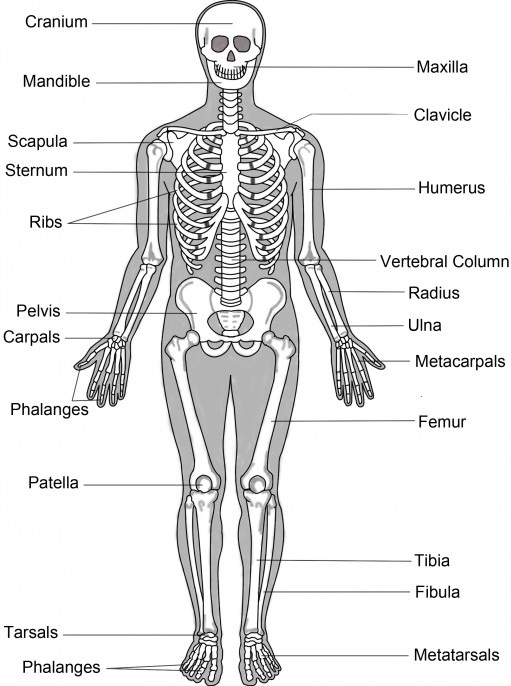
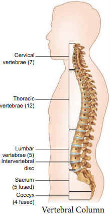
The bones of the upper and lower limbs along with their girdles constitute the appendicular skeleton. The appendicular skeleton is composed of 126 bones.

(a) The Pectoral Girdle

The upper limbs are attached to the pectoral girdles. These are very light and allow the upper limbs a degree of mobility not seen anywhere else in the body. The girdle is formed of two halves. Each half of the pectoral girdle (Figure 9.8) consists of a clavicle or collar bone and a scapula.

The scapula is a large, thin, triangular bone situated in the dorsal surface of the ribcage between the second and seventh ribs. It has a slightly elevated ridge called the spine which projects as a flat, expanded process called the acromion.
The Appendicular Skeleton img 1

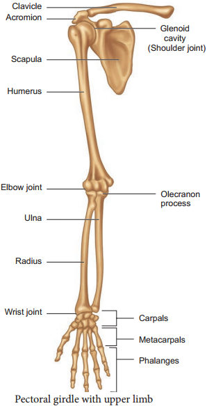
The clavicle articulates with this process. Below the acromion is a depression called the glenoid cavity which articulates with the head of the humerus to form the shoulder joint. Each clavicle is a long slender bone with two curvatures which lies horizontally and connects axial skeleton with appendicular skeleton.

The Upper Limb

The upper limb consists of 30 separate bones and is specialized for mobility. The skeleton of the arm, the region between the shoulder and elbow is the humerus. The head of humerus articulates with the glenoid cavity of the scapula and forms the shoulder joint.

The distal end of humerus articulates with the two forearm bones the radius and ulna. The forearm is the region between the elbow and the wrist. Olecranon process is situated at the upper end of the ulna which forms the pointed portion of the elbow. The hand consists of carpals, metacarpals and phalanges.

Carpals,the wrist bones, 8 in number are arranged in two rows of four each. The anterior surface of the wrist has tunnellike appearance, due to the arrangement of carpals with the ligaments. This tunnel is termed as carpal tunnel. Metacarpals,the palm bones are 5 in number and phalanges the digits bones are 14 in number.

(b) Pelvic Girdle

The pelvic girdle is a heavy structure specialized for weight bearing. It is composed of two hip bones called coxal bones that secure the lower limbs to the axial skeleton (Figure 9.9). Together, with the sacrum and coccyx, the hip bones form the basin-like bony pelvis.

Each coxal bone consists of three fused bones, ilium, ischium and pubis. At the point of fusion of ilium, ischium, and pubis a deep hemispherical socket called the acetabulum is present on the lateral surface of the pelvis. It receives the head of the femur or thigh bone at the hip joint and helps in the articulation of the femur. Ventrally the two halves of the pelvic girdle meet and form the pubic symphysis containing firous
cartilage.
The Appendicular Skeleton img 2

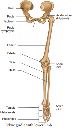
The ilium is the superior floring portion of the hip bone. Each ilium forms a secure joint with the sacrum posteriorly. The ischium is a curved bar of bone. The V-shaped pubic bones articulate anteriorly at the pubic symphysis. The pelvis of male is deep and narrow with larger heavier bones and the female is shallow, wide and flexible in nature, and this helps during pregnancy which is influenced by female hormones.

The Lower Limb

The lower limb consists of 30 bones which carries the entire weight of the erect body and is subjected to exceptional forces when we jump or run. The bones of the lower limbs are thicker and stronger than the upper limbs.

The three segments of each lower limb are the thigh, the leg or the shank and the foot. The femur is the single bone of the thigh. It is the largest, longest and strongest bone in the body. The head of femur articulates with the acetabulum of the pelvis to form the hip joint.

Two parallel bones, the tibia and fibula, form the skeleton of the shank. A thick, triangular patella forms the knee cap, which protects the knee joint anteriorly and improves the leverage of thigh muscles acting across the knee.

The foot includes the bones of ankle, the tarsus, the metatarsus and the phalanges or toe bones. The foot supports our body weight and acts as a lever to propel the body forward, while walking and running. The tarsus is made up of seven bones called tarsals. The metatarsus consists of five bones called metatarsals. The arrangement of the metatarsals is parallel to each other. There are 14 phalanges in the toes which are smaller than those of the fingers.

Structure of a Typical Long Bone

A typical long bone has a diaphysis, epiphyses (singular-epiphysis) and membranes (Figure 9.10). A tubular diaphysis or shaft forms the long axis of the bone. It is constructed of a thick collar of compact bone that surrounds a central medullary cavity or marrow cavity.

The epiphyses are the bone ends. Compact bone forms the exterior of epiphyses and their interior contains spongy bone with red marrow. The region where the diaphysis and epiphyses meet is called the metaphysis.
The Appendicular Skeleton img 3

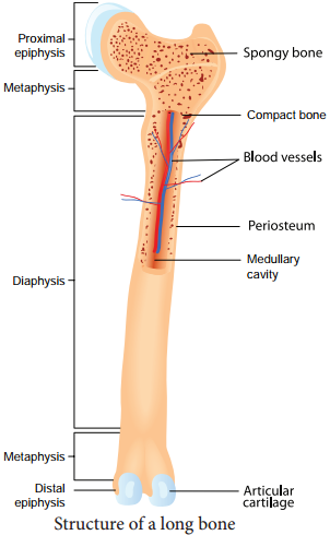
The external surface of the entire bone except the joint surface is covered by a doublelayered membrane called the periosteum. The outer firous layer is dense irregular connective tissue. The inner osteogenic layer consists of osteoblasts (bone- forming cells) which secrete bone matrix elements and osteoclasts (bone-destroying cells).

In addition, there are primitive stem cells, osteogenic cells, that give rise to the osteoblasts. The periosteum is richly supplied with nerve fires, lymphatic vessels and blood vessels. Internal bone surfaces are covered with a delicate connective tissue membrane called the endosteum. The endosteum covers the trabeculae of spongy bone and lines the canals that pass through the compact bone. It also contains both osteoblasts and osteoclasts. Between the epiphysis and diaphysis epiphyseal plate or growth plate is present.

The Skeletal System – The Axial Skeleton

Learninsta presents the core concepts of Biology with high-quality research papers and topical review articles.

The Skeletal System – The Axial Skeleton

Axial skeleton forms the main axis of the body. It consists of the skull, hyoid bone, vertebral column and thoracic cage.

(a) The Skull

The skull is composed of two sets of bones – cranial and facial bones. It consists of 22 bones of which 8 are cranial bones and 14 are facial bones (Figure 9.5). The cranial bones form the hard protective outer covering of the brain and called the brain box.

The capacity of the cranium is 1500 cm3. These bones are joined by sutures which are immovable. They are a paired parietal, paired temporal and individual bones such as the frontal, sphenoid, occipital and ethmoid.

The large hole in the temporal bone is the external auditory meatus. In the facial bones maxilla, zygomatic, palatine, lacrimal, nasal are paired bones whereas mandible or lower jaw and vomer are unpaired bones. They form the front part of the skull. A single U-shaped hyoid bone is present at the base of the buccal cavity.

It is the only one bone without any joint. Each middle ear contains three tiny bonesmalleus, incus and stapes collectively are called ear ossicles. The upper jaw is formed of the maxilla and the lower jaw is formed of the mandible. The upper jaw is fused with the cranium and is immovable.

The lower jaw is connected to the cranium by muscles and is movable. The most prominent openings in the skull are the orbits and the nasal cavity. Foramen magnum is a large opening found at the posterior base of the skull. Though this opening the medulla oblongata of the brain descends down as the spinal cord.
The Skeletal System - The Axial Skeleton img 1

(b) The Vertebral Column

Vertebral column is also called the back bone. It consists of 33 serially arranged vertebrae which are inter connected by cartilage known as intervertebral disc (Figure 9.6). The vertebral column extends from the base of the skull to the pelvis and forms the main frame work of the trunk.

The vertebral column has five major regions. They are, the Cervical, Thoracic, Lumbar, Sacrum (5 sacral vertebrae found in the infant which are fused to form one bone in the adult) and Coccyx (4 coccygeal vertebrae found in the infant which are fused to form one bone in the adult).
The Skeletal System - The Axial Skeleton img 2

Each vertebra has a central hollow portion, the neural canal, through which the spinal cord passes. The first vertebra is called as the atlas and the second vertebra is called as the axis. Atlas is articulated with the occipital condyles. The vertebral column protects the spinal cord, supports the head and serves as the point of attachment for the ribs and musculature of the back.

(c) The Sternum (Chest bone)

Sternum is a flat bone on the mid ventral line of the thorax. It provides space for the attachment of the thoracic ribs and abdominal muscles.

(d) The Rib Cage

There are 12 pairs of ribs (Figure 9.7). Each rib is a thin flat bone connected dorsally to the vertebral column and ventrally to the sternum. It has two articulation surfaces on its dorsal end, hence called bicephalic. The first seven pairs of ribs are called ‘true ribs’ or vertebro-sternal ribs.

Dorsally they are attached to the thoracic vertebrae and ventrally connected to the sternum with the help of hyaline cartilages. The 8th, 9th and 10th pairs of ribs do not articulate directly with the sternum but joined with the cartilaginous (hyaline cartilage) part of the seventh rib.

These are called ‘false ribs’ or vertebro-chondral ribs. The last 11th and 12th pairs of ribs are not connected ventrally. Therefore, they are called as ‘flating ribs’ or vertebral ribs. Thoracic vertebrae, ribs and sternum together form the ribcage. Rib cage protects the lungs, heart, liver and also plays a role in breathing.
The Skeletal System - The Axial Skeleton img 3