Reflex Action and Reflex Arc

Learninsta presents the core concepts of Biology with high-quality research papers and topical review articles.

Reflex Action and Reflex Arc

When dust falls in our eyes, the eyelids close immediately not waiting for our willingness; on touching a hot pan, the hand is withdrawn rapidly. Do you know how this happens?

The spinal cord remains as a connecting functional nervous structure in between the brain and effector organs. But sometimes when a very quick response is needed, the spinal cord can effect motor initiation as the brain and brings about an effect. This rapid action by spinal cord is called reflex action. It is a fast, involuntary, unplanned sequence of actions that occurs in response to a particular stimulus.

The nervous elements involved in carrying out the reflex action constitute a reflex arc or in other words the pathway followed by a nerve impulse to produce a reflex action is called a reflex arc (Figure 10.12).
Reflex Action and Reflex Arc img 1

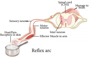
Functional Components of a Reflex Arc

Sensory Receptor:
It is a sensory structure that responds to a specific stimulus.

Sensory Neuron:
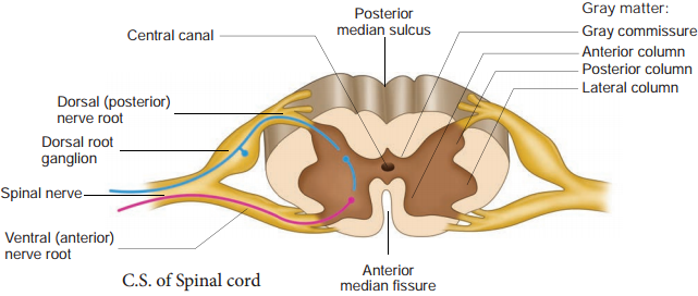
This neuron takes the sensory impulse to the grey (afferent) matter of the spinal cord through the dorsal root of the spinal cord.

Interneurons:
One or two interneurons may serve to transmit the impulses from the sensory neuron to the motor neuron.

Motor Neuron:
It transmits impulse from CNS to the effector organ.

Effector Organs:
It may be a muscle or gland which responds to the impulse received. There are two types of reflexes. They are:-

(1) Unconditional Reflex:

Is an inborn reflex for an unconditioned stimulus. It does not need any past experience, knowledge or training to occur; Ex: blinking of an eye when a dust particle about to fall into it, sneezing and coughing due to foreign particle entering the nose or larynx.

(2) Conditioned Reflex:

Is a respone to a stimulus that has been acquired by learning. This does not naturally exists in animals. Only an experience makes it a part of the behaviour. Example: excitement of salivary gland on seeing and smelling a food. The conditioned reflex was first demonstrated by the Russian physiologist Pavlov in his classical conditioning experiment in a dog. The cerebral cortex controls the conditioned reflex.

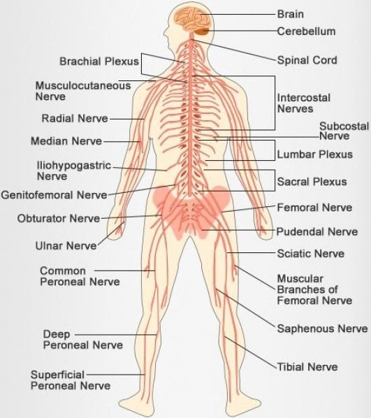
Peripheral Neural System (PNS)

PNS consists of all nervous tissue outside the CNS. Components of PNS include nerves, ganglia, enteric plexuses and sensory receptors. A nerve is a chord like structure that encloses several neurons inside. Ganglia (singular-ganglion) are small masses of nervous tissue, consisting primarily of neuron cell bodies and are located outside the brain and spinal cord.

Enteric plexuses are extensive networks of neurons located in the walls of organs of the gastrointestinal tract. The neurons of these plexuses help in regulating the digestive system. The specialized structure that helps to respond to changes in the environment i.e. stimuli are called sensory receptor which triggers nerve impulses along the afferent fires to CNS. PNS comprises of cranial nerves arising from the brain and spinal nerves arising from the spinal cord.

(A) Cranial Nerves:

There are 12 pairs of cranial nerves, of which the first two pairs arise from the fore brain and the remaining 10 pairs from the mid brain. Other than the Vagus nerve, which extends into the abdomen, all cranial nerves serve the head and face.

(B) Spinal Nerves:

31 pairs of spinal nerves emerge out from the spinal cord through spaces called the intervertebral foramina found between the adjacent vertebrae. The spinal nerves are named according to the region of vertebral column from which they originate

  • Cervical nerves (8 pairs)
  • Thoracic nerves (12 pairs)
  • Lumbar nerves (5 pairs)
  • Sacral nerves (5 pairs)
  • Coccygeal nerves (1 pair)

Each spinal nerve is a mixed nerve containing both afferent (sensory) and efferent (motor) fibres. It originates as two roots:

  • A posterior dorsal root with a ganglion outside the spinal cord and
  • An anterior ventral root with no external ganglion.

Somatic Neural System (SNS)

The somatic neural system (SNS or voluntary neural system) is the part of the peripheral neural system associated with the voluntary control of body movements via skeletal muscles. The sensory and motor nerves that innervate striated muscles form the somatic neural system. Major functions of the somatic neural system include voluntary movement of the muscles and organs, and reflex movements.

Autonomic Neural System

The autonomic neural system is auto functioning and self governed. It is a part of peripheral neural system that innervates smooth muscles, glands and cardiac muscle. This system controls and coordinates the involuntary activities of various organs. ANS controlling centre is in the hypothalamus. Autonomic neural system comprises the following components:

Preganglionic Neuron

Whose cell body is in the brain or spinal cord; its myelinated axon exits the CNS as part of cranial or spinal nerve and ends in an autonomic ganglion.

Autonomic Ganglion

Consists of axon of preganglionic neuron and cell bodies of postganglionic neuron.

Postganglionic Neuron

Conveys nerve impulses from autonomic ganglia to visceral effector organs. The autonomic neural system consists of Sympathetic neural system and Parasympathetic neural system.

Central Neural System (CNS) | Brain | Spinal Cord

Learninsta presents the core concepts of Biology with high-quality research papers and topical review articles.

Central Neural System (CNS) | Brain | Spinal Cord

The CNS includes the brain and the spinal cord, which are protected by the bones of the skull and vertebral column. During its embryonic development, CNS develops from the ectoderm.

Brain

The brain acts as the command and control system. It is the site of information processing. It is located in the cranial cavity and is covered by three cranial meninges. The outer thick layer is Duramater which lines the inner surface of the cranial cavity; the median thin layer is Arachnoid mater which is separated from the duramater by a narrow subdural space.

The innermost layer is Piamater which is closely adhered to the brain but separated from the arachnoid mater by the subarachnoid space. The brain is divided into three major regions: Forebrain, Midbrain and Hindbrain.

Fore Brain

It Comprises the Following Regions:

Cerebrum and Diencephalon. Cerebrum is the ‘seat of intelligence’ and forms the major part of the brain. The cerebrum consists of an outer cortex, inner medulla and basal nuclei. The superficial region of the cerebrum is called cerebral cortex, which looks grey due to the presence of unmyelinated nerve cells. Cerebral cortex consists of neuronal cell body, dendrites, associated glial and blood vessels. The surface of the cerebrum shows many convolutions (folds) and grooves.

The folds are called gyri (singular gyrus); the shallow grooves between the gyri are called sulci (singular sulcus) and deep grooves are called fissures. These sulci and gyri increase the surface area of the cerebral cortex. Several sulci divide the cerebrum into eight lobes: a pair of frontals, parietals, temporals and occipital lobes (Figure 10.7 & Table 10.2).
Central Neural System (CNS) - Brain - Spinal Cord img 1
Central Neural System (CNS) - Brain - Spinal Cord img 2

A median longitudinal fissure divides the cerebrum longitudinally into two cerebral hemispheres (Figure 10.7). A transverse fissure separates the cerebral hemispheres from the cerebellum. The hemispheres are connected by a tract of nerve fires called corpus callosum. Cerebral cortex has three functional areas namely sensory areas occur in the parietal, temporal and occipital lobes of the cortex.
Central Neural System (CNS) - Brain - Spinal Cord img 1

They receive and interpret the sensory impulses. Motor area of the cortex which controls voluntary muscular movements lies in the posterior part of the frontal lobes. The areas other than sensory and motor areas are called Association areas that deal with integrative functions such as memory, communications, learning and reasoning. Inner to the cortex is medulla which is white in colour and acts as a nerve tract between the cortex and the diencephalon.

Diencephalon consists largely of following three paired structures. Epithalamus forms the roof of the diencephalon and it is a non-nervous tissue. The anterior part of epithalamus is vascular and folded to form the anterior choroid plexus. Just behind the choroid plexus, the epithalamus forms a short stalk which ends in a rounded body called pineal body which secretes the hormone, melatonin which regulates sleep and wake cycle.

Thalamus is composed of grey mater which serves as a relay centre for impulses between the spinal cord, brain stem and cerebrum. Within the thalamus, information is sorted and edited and plays a key role inlearning and memory. It is a major coordinating centre for sensory and motor signalling.
Central Neural System (CNS) - Brain - Spinal Cord img 3

Hypothalamus forms the floor of the diencephalon. The downward extension of the hypothalamus, the infundibulum connects the hypothalamus with the pituitary gland. The hypothalamus contains a pair of small rounded body called mammillary bodies that are involved in olfactory reflxes and emotional responses to odour.

Hypothalamus maintains homeostasis and has many centres which control the body temperature, urge for eating and drinking. It also contains a group of neurosecretory cells which secrete the hypothalamic hormones. Hypothalamus also acts as the satiety centre.

Limbic System

The inner part of the cerebral hemisphere constitutes the limbic system. The main components of limbic system are oldfactory bulbs, cingulate gyrus, mammillary body, amygdala, hippocampus and hypothalamus. The limbic system is called ‘emotional brain’ because it plays a primary role in the regulation of pleasure, pain, anger, fear, sexual feeling and affection. The hippocampus and amygdala also play a role in memory (Figure 10.9).
Central Neural System (CNS) - Brain - Spinal Cord img 4

Brain stem is the part of the brain between the spinal cord and the diencephalon. It consists of mid brain, pons varolii and medulla oblongata (Figure 10.10).
Central Neural System (CNS) - Brain - Spinal Cord img 5

Mid Brain

The mid brain is located between the diencephalon and the pons. The lower portion of the midbrain consists of a pair of longitudinal bands of nervous tissue called cerebral peduncles which relay impulses back and forth between cerebrum, cerebellum, pons and medulla. The dorsal portion of the midbrain consists of four rounded bodies called corpora quadrigemina which acts as a reflex centre for vision and hearing.

Hind Brain

Rhombencephalon forms the hind brain. It comprises of cerebellum, pons varolii and medulla oblongata. Cerebellum is the second largest part of the brain. It consists of two cerebellar hemispheres and central worm shaped part, the vermis. The cerebellum controls and coordinates muscular movements and body equilibrium. Any damage to cerebellum often results in uncoordinated voluntary muscle movements.

Pons varoli lies infront of the cerebellum between the midbrain and the medulla oblongata. The nerve fires in the pons varolii form a bridge between the two cerebellar hemispheres and connect the medulla oblongata with the other region of the brain. The respiratory nuclei found in the pons cooperate with the medulla to control respiration.

Medulla oblongata forms the posterior most part of the brain. It connects the spinal cord with various parts of the brain. It receives and integrates signals from spinal cord and sends it to the cerebellum and thalamus. Medulla contains vital centres that control cardio vascular reflexes, respiration and gastric secretions.

Ventricles of the Brain

The brain has four hollow, fluid filled spaces. The C – shaped space found inside each cerebral hemisphere forms the lateral ventricles I and II which are separated from each other by a thin membrane called theseptum pellucidum.

Each lateral ventricle communicates with the narrow III ventricle in the diencephalon through an opening called interventricular foramen (foramen of Monro). The ventricle III is continuous with the ventricle IV in the hind brain through a canal called aqueduct of Sylvius (cerebral aqueduct).

Choroid plexus is a network of blood capillaries found in the roof of the ventricles and forms cerebro spinal fluid (CSF) from the blood. CSF provides buoyancy to the CNS structures; CSF acts as a shock absorber for the brain and spinal cord; it nourishes the brain cells by transporting constant supply of food and oxygen; it carries harmful metabolic wastes from the brain to the blood; and maintains a constant pressure inside the cranial vessels.

Spinal Cord

The spinal cord is a long, slender, cylindrical nervous tissue. It is protected by the vertebral column and surrounded by the three membranes as in the brain. The spinal cord that extends from the brain stem into the vertebral canal of the vertebral column up to the level of 1st or 22nd lumbar vertebra.

So the nerve roots of the remaining nerves are greatly elongated to exit the vertebral column at their appropriate space. The thick bundle of elongated nerve roots within the lower vertebral canal is called the cauda equina (horse’s tail) because of its appearance.

In the cross section of spinal cord (Figure 10.11), there are two indentations: the posterior median sulcus and the anterior median fissure. Although there might be slight variations, the cross section of spinal cord is generally the same throughout its length. In contrast to the brain, the grey matter in the spinal cord forms an inner butterfly shaped region surrounded by the outer white matter.
Central Neural System (CNS) - Brain - Spinal Cord img 6

The grey matter consists of neuronal cell bodies and their dendrites, interneurons and glial cells. White matter consists of bundles of nerve fibres. In the center of the grey matter there is a central canal which is filled with CSF. Each half of the grey matter is divided into a dorsal horn, a ventral horn and a lateral horn.

The dorsal horn contains cell bodies of interneurons on which afferent neurons terminate. The ventral horn contains cell bodies of the efferent motor neurons supplying the skeletal muscle. Autonomic nerve fires, supplying cardiac and smooth muscles and exocrine glands, originate from the cell bodies found in the lateral horn.

In the white matter, the bundles of nerve fires form two types of tracts namely ascending tracts which carry sensory impulses to the brain and descending tracts which carry motor impulses from the brain to the spinal nerves at various levels of the spinal cord. The spinal cord shows two enlargements, one in the cervical region and another one in the lumbosacral region. The cervical enlargement serves the upper limb and lumbar enlargement serves the lower limbs.

Neuron as a Structural and Functional Unit of Neural System

Learninsta presents the core concepts of Biology with high-quality research papers and topical review articles.

Neuron as a Structural and Functional Unit of Neural System

A neuron is a microscopic structure composed of three major parts namely cell body (soma), dendrites and axon. The cell body is the spherical part of the neuron that contains all the cellular organelles as a typical cell (except centriole). The plasma membrane covering the neuron is called neurilemma and the axon is axolemma.

The repeatedly branched short fibres coming out of the cell body are called dendrites, which transmit impulses towards the cell body. The cell body and the dendrites contain cytoplasm and granulated endoplasmic reticulum called Nissl’s granules.

An axon is a long fibre that arises from a cone shaped area of the cell body called the Axon hillock and ends at the branched distal end. Axon hillock is the place where the nerve impulse is generated in the motor neurons. The axon of one-neuron branches and forms connections with many other neurons. An axon contains the same organelles found in the dendrites and cell body but lacks Nissl’s granules and Golgi apparatus.

The axon, particularly of peripheral nerves is surrounded by Schwann cells (a type of glial cell) to form myelin sheath, which act as an insulator. Myelin sheath is associated only with the axon; dendrites are always non-myelinated.

Schwann cells are not continuous along the axon; so there are gaps in the myelin sheath between adjacent Schwann cells. These gaps are called Nodes of Ranvier. Large myelinated nerve fibres conduct impulses rapidly, whereas nonmyelinated fibres conduct impulses quite slowly (Figure 10.1).
Neuron as a Structural and Functional Unit of Neural System img 1

Each branch at the distal end of the axon terminates into a bulb like structure called synaptic knob which possesses synaptic vesicles filled with neurotransmitters. The axon transmits nerve impulses away from the cell body to an inter neural space or to a neuro-muscular junction. The neurons are divided into three types based on number of axon and dendrites they possess (Figure 10.2).
Neuron as a Structural and Functional Unit of Neural System img 2

1. Multipolar Neurons

Have many processes with one axon and two or more dendrites. They are mostly interneurons.

2. Bipolar Neurons

Have two processes with one axon and one dendrite. These are found in the retina of the eye, inner ear and the oldfactory area of the brain.

3. Unipolar Neurons

Have a single short process and one axon. Unipolar neurons are located in the ganglia of cranial and spinal nerves.

Generation and Conduction of Nerve Impulses

This section deals with how the nerve impulses are produced and conducted in our body. Sensation felt in the sensory organs are carried by the nerve fibres in the form of electrical impulses. A nerve impulse is a series of electrical impulses, which travel along the nerve fibre.

Inner to the axolemma, the cytoplasm contains the intracellular fluid (ICF) with large amounts of potassium and magnesium phosphate along with negatively charged proteins and other organic molecules.

The extra cellular fluid (ECF) found outside the axolemma contains large amounts of sodium chloride, bicarbonates, nutrients and oxygen for the cell; and carbon dioxide and metabolic wastes released by the neuronal cells. The ECF and ICF (cytosol) contains negatively charged particles (anions) and positively charged particles (cations). These charged particles are involved in the conduction of impulses.

The neurons maintain an uneven distribution of various inorganic ions across their axolemma for transmission of impulses. This unequal distribution of ions establishes the membrane potential across the axolemma. The axolemma contains a variety of membrane proteins that act as ionic channels and regulates the movement of ions across the axolemma. (Shown in Table 10.1).
Neuron as a Structural and Functional Unit of Neural System img 3

Transmission of Impulses

The transmission of impulse involves two main phases; Resting membrane potential and Action membrane potential.

Resting Membrane Potential:

The electrical potential difference across the plasma membrane of a resting neuron is called the resting potential during which the interior of the cell is negative due to greater efflux of K+ outside the cell than Na+ influx into the cell.

When the axon is not conducting any impulses i.e. in resting condition, the axon membrane is more permeable to K+ and less permeable to Na+ ions, whereas it remains impermeable to negatively charged protein ions.
Neuron as a Structural and Functional Unit of Neural System img 4

The axoplasm contains high concentration of K+ and negatively charged proteins and low concentration of Na+ ions. In contrast, fluid outside the axon (ECF) contains low concentration of K+ and high concentration of Na+, and this forms a concentration gradient. This ionic gradient across the resting membrane is maintained by ATP driven Sodium Potassium pump, which exchanges 3Na+ outwards for 2K+ into the cells.

In this state, the cell membrane is said to be polarized. In neuron, the resting membrane potential ranges from -40mV to -90mV, and its normal value is -70mV. The minus sign indicates that the inside of the cell is negative with respect to the Figure 10.3 Ionic channels outside (Figure 10.4).
Neuron as a Structural and Functional Unit of Neural System img 5

Action Membrane Potential

An action potential occurs when a neuron sends information down an axon, away from the cell body. It includes following phases, depolarization, repolarisation and hypo polarization.

Depolarization – Reversal of Polarity

When a nerve fire is stimulated, sodium voltage-gate opens and makes the axolemma permeable to Na+ ions; meanwhile the potassium voltage gate closes. As a result, the rate of flow of Na+ ions into the axoplasm exceeds the rate of flow of K+ ions to the outside fluid [ECF].

Therefore, the axolemma becomes positively charged inside and negatively charged outside. This reversal of electrical charge is called Depolarization. During depolarization, when enough Na+ ions enter the cell, the action potential reaches a certain level, called threshold potential [-55 mV]. The particular stimulus which is able to bring the membrane potential to threshold is called threshold stimulus.

The action potential occurs in response to a threshold stimulus but does not occur at subthreshold stimuli. This is called all or none principle. Due to the rapid influx of Na+ ions, the membrane potential shoots rapidly up to +45mV which is called the Spike potential.

Repolarisation [Falling Phase]

When the membrane reaches the spike potential, the sodium voltage-gate closes and potassium voltage-gate opens. It checks influx of Na+ ions and initiates the efflux of K+ ions which lowers the number of positive ions within the cell. This, the potential falls back towards the resting potential. The reversal of membrane potential inside the axolemma to negative occurs due to the efflux of K+ ions. This is called Repolarisation.

Hyperpolarization

If repolarization becomes more negative than the resting potential -70 mV to about -90 mV, it is called Hyperpolarization. During this, K+ ion gates are more permeable to K+ even after reaching the threshold level as it closes slowly; hence called Lazy gates. The membrane potential returns to its original resting state when K+ ion channels close completely. During hyperpolarization the Na+ voltage gate remains closed (Figure 10.5).
Neuron as a Structural and Functional Unit of Neural System img 6

Conduction Speed of a Nerve Impulse

The conduction speed of a nerve impulse depends on the diameter of axon. The greater the axon’s diameter, the faster is the conduction. The myelinated axon conducts the impulse faster than the non-myelinated axon. The voltage-gated Na+ and K+ channels are concentrated at the nodes of Ranvier.

As a result, the impulse jumps node to node, rather than travelling the entire length of the nerve fire. This mechanism of conduction is called Saltatory Conduction. Nerve impulses travel at the speed of 1-300 m/s.

Synaptic Transmission

The junction between two neurons is called a Synapse through which a nerve impulse is transmitted. The first neuron involved in the synapse forms the presynaptic neuron and the second neuron is the post synaptic neuron. A small gap between the pre and postsynaptic membranes is called Synaptic Cleft that forms a structural gap and a functional bridge between neurons.

The axon terminals contain synaptic vesicles filled with neurotransmitters. When an impulse [action potential] arrives at the axon terminals, it depolarizes the presynaptic membrane, opening the voltage gated calcium channels. Influx of calcium ions stimulates the synaptic vesicles towards the pre-synaptic membrane and fuses with it.

In the neurilemma, the vesicles release their neurotransmitters into the synaptic cleft by exocytosis. The released neurotransmitters bind to their specific receptors on the post-synaptic membrane, responding to chemical signals.

The entry of the ions can generate a new potential in the post-synaptic neuron, which may be either excitatory or inhibitory. Excitatory post-synaptic potential causes depolarization whereas inhibitory post synaptic potential causes hyperpolarization of post-synaptic membrane (Figure 10.6).
Neuron as a Structural and Functional Unit of Neural System img 7

Human Neural System Definition and its Function

Learninsta presents the core concepts of Biology with high-quality research papers and topical review articles.

Human Neural System Definition and its Function

The human neural system is divided into two, the central neural system (CNS) and the peripheral neural system (PNS). The structural and functional units of the neural system are neurons that transmit nerve impulses. The non-nervous special cells called neuroglia form the supporting cells of the nervous tissue.

There are three functional classes of neurons. They are the afferent neurons that take sensory impulses to the Central Neural system (CNS) from the sensory organs; the efferent neurons that carry motor impulses from the CNS to the effector organs; and interneurons that lie entirely within the CNS between the afferent and efferent neurons.

The central neural system lacks connective tissue, so the interneuron space is filled by neuroglia. They perform several functions such as providing nourishment to the surrounding neurons; involving the memory process; repairing the injured tissues due to their dividing and regenerating capacity; and acting as phagocyte cells to engulf the foreign particles at the time of any injury to the brain.

The human nervous system consists of two main parts: the central nervous system (CNS) and the peripheral nervous system (PNS). The CNS contains the brain and spinal cord. The PNS consists mainly of nerves, which are long fibers that connect the CNS to every other part of the body.

The central nervous system is made up of the brain and spinal cord, and the peripheral nervous system is made up of the Somatic and the Autonomic nervous systems.

The nervous system of vertebrates (including humans) is divided into the central nervous system (CNS) and the peripheral nervous system (PNS). The (CNS) is the major division, and consists of the brain and the spinal cord. The spinal canal contains the spinal cord, while the cranial cavity contains the brain.

The nervous system is the major controlling, regulatory, and communicating system in the body. It is the center of all mental activity including thought, learning, and memory. Together with the endocrine system, the nervous system is responsible for regulating and maintaining homeostasis.

The Four Main Functions of the Nervous System are:

Control of body’s internal environment to maintain ‘homeostasis’ An example of this is the regulation of body temperature. Programming of spinal cord reflexes. An example of this is the stretch reflex. Memory and learning. Voluntary control of movement.

The Nervous System has two main Parts:

The central nervous system is made up of the brain and spinal cord. The peripheral nervous system is made up of nerves that branch off from the spinal cord and extend to all parts of the body.

The nervous system includes the brain, nerves and spinal cord. It is the communication center for the body, sending and receiving messages, regulating body functions and serving as the control center for the five senses and for emotions, speech, coordination, balance, and learning.

The 11 organ systems include the integumentary system, skeletal system, muscular system, lymphatic system, respiratory system, digestive system, nervous system, endocrine system, cardiovascular system, urinary system, and reproductive systems.
Human Neural System img 1

Neural System Definition, Function, Structure and its Types

Learninsta presents the core concepts of Biology with high-quality research papers and topical review articles.

Neural System Definition, Function, Structure and its Types

The neural system comprises of highly specialized cells called neurons, which can detect, receive, process and transmit different kinds of stimuli. Simple form of neural system as nerve net is seen in lower invertebrates. The neural system of higher animals are well developed and performs the following basic functions:

Sensory Functions:
It receives sensory input from internal and external environment.

Motor Functions:
It transmits motor commands from the brain to the skeletal and muscular system.

Autonomic Functions:
Reflex actions.

The nervous system is the part of an animal’s body that coordinates its behavior and transmits signals between different body areas. In vertebrates it consists of two main parts, called the central nervous system (CNS) and the peripheral nervous system (PNS). The CNS contains the brain and spinal cord.

The nervous system takes in information through our senses, processes the information and triggers reactions, such as making your muscles move or causing you to feel pain. For example, if you touch a hot plate, you reflexively pull back your hand and your nerves simultaneously send pain signals to your brain.

The central nervous system is made up of the brain and spinal cord, and the peripheral nervous system is made up of the Somatic and the Autonomic nervous systems.

The nervous system consists of the brain, spinal cord, sensory organs, and all of the nerves that connect these organs with the rest of the body.

The nervous system in a human is made of the brain, spinal cord, sensory organs and all the neurons that serve as communication channels between the various organs of the body.

The peripheral nervous system carries messages to and from the central nervous system. It sends information to the brain and carries out orders from the brain. Messages travel through the cranial nerves, those which branch out from the brain and go to many places in the head such as the ears, eyes and face.

The nervous system has three broad functions: sensory input, information processing, and motor output. In the PNS, sensory receptor neurons respond to physical stimuli in our environment, like touch or temperature, and send signals that inform the CNS of the state of the body and the external environment.

The neural or nervous system is a complex network of nerve cells or neurons. The nervous system is specialized to carry messages while the endocrine system provides chemical integration through hormones. To better understand the nervous system, one must realize the difference between a neuron and a nerve.

The Structure of a Neuron:

The above image shows the basic structural components of an average neuron, including the dendrite, cell body, nucleus, Node of Ranvier, myelin sheath, Schwann cell, and axon terminal.

The gap between two neurons called synapse, helps in quick transmission of impulses from one neuron to another. Always one-way communication i.e. unidirectional, transmitting from pre-synaptic to post-synaptic neurons. Can be used to calsculate timing of sensory inputs. Greater plasticity.

Neurons have specialized projections called dendrites and axons. The synapse contains a small gap separating neurons. The synapse consists of: a presynaptic ending that contains neurotransmitters, mitochondria and other cell organelles.
Neural Tissue img 1