Mechanism of Hormone Action

Learninsta presents the core concepts of Biology with high-quality research papers and topical review articles.

Mechanism of Hormone Action

Hormones circulate in the blood but their concentration can increase or decrease based on the requirement of the body. This is controlled by feedback mechanisms. These mechanisms control the secretion of endocrine glands by stimulating the hypothalamus, pituitary or both, which inturn governs the secretion of a particular hormone.

In positive feedback, the secretion of the hormone increases where as in negative feedback further secretion of hormone slows down. Feedback mechanisms are the key factors for maintaining homeostasis in our body.

Hormones are classified into three major groups as peptide hormones, steroid hormones and amino acid derived hormones based on their chemical structure.

Peptide hormones cannot cross the phospolipid cell membrane and bind to the receptors on the exterior cell surface. They are are transported to the golgi, which is the site of modification. It acts as a first messenger in the cell. Hormones on binding to their receptors do not enter the target cell but generate the production of second messengers such as cyclic AMP (c AMP), which in turn regulates cellular metabolism.

This is catalyzed by the enzyme adenylate cyclase. The interaction between the hormone at the surface and the effect brought out by cAMP within the cell is known as signaling cascade. At each step there is a possibility of amplification. (Figure 11.17)
Mechanism of Hormone Action img 1

  • One hormone molecule may bind to multiple receptor molecules before it is degraded.
  • Each receptor may activate several adenylate cyclases each of which make much cAMP.
  • Thus there is more signal after each step.

The actions of cAMP are terminated by phosphodiesterases. The effect of peptide hormones like insulin, glucagon, somatotropin are usually short lived because they work through second messenger system.

Steroid hormones can easily cross the cell membrane, and bind to their receptors, which are intracellular or intranuclear. Upon binding to the receptors, they pair up with another receptor – hormone complex (dimerize). This dimer can then bind to DNA and alter its transcription. (Figure 11.18)
Mechanism of Hormone Action img 2

The effect of steroid hormones such as aldosterone, oestrogen, FSH are long lived, as they alter the amount of mRNA and protein in a cell. Amino acid derived hormones are derived from one or two aminoacid with a few additional modifications. Thyroid hormone is synthesised from tyrosine and includes the addition of several iodine atoms.

Epinephrine an amino acid derivative may function through second messenger system like peptide hormones or they may actually enter the cell and function like steroid hormones.

Hormones activate target cells by diffusing through the plasma membrane of the target cells (lipid-soluble hormones) to bind a receptor protein within the cytoplasm of the cell, or by binding a specific receptor protein in the cell membrane of the target cell (water-soluble proteins).

There are two modes of hormonal action. A: Activation of cell-surface receptors and coupled second-messenger systems, with a variety of intracellular consequences.

Hormone levels are primarily controlled through negative feedback, in which rising levels of a hormone inhibit its further release. The three mechanisms of hormonal release are humoral stimuli, hormonal stimuli, and neural stimuli.

This type of mechanism is shown by lipid soluble hormones such as fatty acids and steroids that can easily pass through the plasma membrane. They possess intracellular receptors. The hormones bind to the target receptor that activates the enzymatic activity of the cell to bring about biochemical changes.

Mechanism of hormone action is not the same in all categories of hormones is a proteinaceous hormone, has large molecular weight and is insoluble in lipids, therefore, it cannot enter the target cell. Thus, it binds with the membrane bound receptor present on ovarian cell membrane.

The action involves secretion of by the thyroid gland into the circulation, uptake of target tissues, activation or inactivation of by deiodinase enzymes, binding of to nuclear receptors that act as ligand-regulated transcription factors, and regulation of expression of target genes.

What is the most common mechanism of hormone control? With negative feedback, the most common mechanism of hormone control, some feature of hormone action directly or indirectly inhibits further hormone secretion so that the hormone level returns to an ideal level or set point.

Hormones bind to specific proteins (hormone receptors) in the target tissues and produce effect on them. There are two types of receptors: membrane bound receptors (hormone receptors present on the cell membrane of the target cell) and intracellular receptors (receptors present inside the target cell).

The mechanism by which peptide hormones act upon specific target tissues to evoke characteristic functional responses is believed to be initiated by interaction with a highly special- ized portion of the plasma membrane, the so called hormone receptor site.

Hypo and Hyper Activity of Endocrine Glands and Related Disorders

Learninsta presents the core concepts of Biology with high-quality research papers and topical review articles.

Hypo and Hyper Activity of Endocrine Glands and Related Disorders

The hyper secretion and hypo secretion of hormones leads to several disorders. Dwarfism is due to hyposecretion of growth hormone (GH) in children, skeletal growth and sexual maturity is arrested. They attain a maximum height of 4 feet only (Figure 11.8).
Hypo and Hyper Activity of Endocrine Glands and Related Disorders img 1

Gigantism is due to hypersecretion of growth hormone (GH) in children. Overgrowth of skeletal structure occurs (up to 8 feet) and the visceral growth is not appropriate with that of limbs. Figure 11.9.
Hypo and Hyper Activity of Endocrine Glands and Related Disorders img 2

Acromegaly is due to excessive secretion of growth hormone in adults. Over growth of hand bones, feet bones, jaw bones, malfunctioning of gonads, enlargement of viscera, tongue, lungs, heart, liver, spleen and endocrine gland like thyroid, adrenal etc., are the symptoms of acromegaly. (Figure 11.10)
Hypo and Hyper Activity of Endocrine Glands and Related Disorders img 3

In infants, hypothyroidism causes cretinism. A cretin shows retarded skeletal growth, absence of sexual maturity, retarded mental ability, thick wrinkled skin, protruded enlarged tongue, bloated face, thick and short limbs occurs. The other symptoms are low BMR, slow pulse rate, subnormal body temperature and elevated blood cholesterol levels. (Figure 11.11)
Hypo and Hyper Activity of Endocrine Glands and Related Disorders img 4

Hyposecretion of thyroid in adults causes myxodema. It is otherwise called Gull’s disease. This disease is characterised by decreased mental activity, memory loss, slowness of movement, speech, and general weakness of body, dry coarse skin, scarce hair, puffy appearance, disturbed sexual function, low BMR, poor appetite, and subnormal body temperature. (Figure 11.12)
Hypo and Hyper Activity of Endocrine Glands and Related Disorders img 5

Grave’s disease also called as thyrotoxicosis or exophthalmic goitre. This disease is caused due to hyper secretion of thyroid. It is characterised by enlargement of thyroid gland, increased BMR (50% – 100%), elevated respiratory and excretory rates, increased heart beat, high BP, increased body temperature, protrusion of eyeball and weakness of eye muscles and weight loss. (Figure 11.13)
Hypo and Hyper Activity of Endocrine Glands and Related Disorders img 6

Simple goitre is also known asEndemic goitre. It is caused due to hyposecretion of thyroxine. The symptoms includes enlargement of thyroid gland, fall in serum thyroxine level, increased TSH secretion. (Figure 11.14)
Hypo and Hyper Activity of Endocrine Glands and Related Disorders img 7

Tetany is caused due to the hyposecretion of parathyroid hormone (PTH). Due to hyposecretion of PTH serum calcium level decreases (Hypocalcemia), as a result serum phosphate level increases. Calcium and phosphate excretion level decreases. Generalized convulsion, locking of jaws increased heart beat rate, increased body temperature, muscular spasm are the major symptoms of tetany.

Hyperparathyroidism is caused due to excess PTH in blood. Demineralisation of bone, cyst formation, softening of bone, loss of muscle tone, general weakness, renal disorders are the symptoms of hyperparathyroidism.

Addison’s disease is caused due to hyposecretion of glucocorticoids and mineralocorticoids from the adrenal cortex. Muscular weakness, low BP., loss of appetite, vomiting, hyper pigmentation of skin, low metabolic rate, subnormal temperature, reduced blood volume, weight loss are the symptoms that occur in Addison’s disease (Figure 11.15). Reduced aldosterone secretion increases urinary excretion of NaCl and water and decreases potassium excretion leading to dehydration.
Hypo and Hyper Activity of Endocrine Glands and Related Disorders img 8

Cushing’s syndrome is caused due to excess secretion of cortisol. Obesity of the face and trunk, redness of face, hand, feet, thin skin, excessive hair growth, loss of minerals from bone (osteoporosis) systolic hypertension are features of Cushing’s syndrome. Suppression of sexual function like atrophy of gonads are the other symptoms of Cushing’s syndrome. (Figure 11.16)
Hypo and Hyper Activity of Endocrine Glands and Related Disorders img 9

Hypoglycaemia is due to increased secretion of insulin thereby blood glucose level decreases. In this disorder blood glucose level lowers than normal fasting index. Increased heartbeat, weakness, nervousness, headache, confusion, lack of co-ordination, slurred speech, serious brain defects like epilepsy and coma occurs.

Hyperglycaemia is otherwise known as Diabetes mellitus. It is caused due to reduced secretion of insulin. As the result, blood glucose level is elevated. Diabetes mellitus is of two types, Type I Diabetes and Type II Diabetes. Type I diabetes is also known Insulin dependent diabetes, caused by the lack of insulin secretion due to illness or viral infections.

Type II diabetes is also known as Non – Insulin dependent diabetes, caused due to reduced sensitivity to insulin, often called as insulin resistance. Symptoms of diabetes includes, polyurea (excessive urination), polyphagia (excessive intake of food), polydipsia (excessive consumption liquids due to thirst), ketosis (breakdown of fat into glucose results in accumulation of ketone bodies) in blood. Gluconeogenesis (Conversion of non – carbohydrate form like amino acids and fat into glucose) also occur in diabetes.

Diabates insipidus is caused due to hyposecretion of vasopressin (ADH) from neurohypophysis. The symptom includes frequent urination (polyurea) and excessive consumption of liquids due to thirst (polydipsia).

Human Endocrine System Various Types and its Function

Learninsta presents the core concepts of Biology with high-quality research papers and topical review articles.

Human Endocrine System Various Types and its Function

There are two glandular systems such as the exocrine glands and the endocrine glands. The exocrine glands secrete enzymes, saliva and sweat and have ducts that carry their substances to the membrane surfaces. Example: salivary gland and gastric gland. The endocrine glands, called ductless glands produce hormones and lack ducts; they release their hormone to the surrounding tissue fluid.

The hormones circulate around the body and eventually reach the target organs. Endocrine glands (Figure: 11. 1) include the pituitary, thyroid, parathyroid, pineal, adrenal, thymus and are also known as exclusive endocrine glands.

The hypothalamus along with its neural function also produces hormones and is considered as a neuro endocrine gland. In addition several organs such as pancreas, gastro intestinal tract epithelium, kidney, heart, gonads and placenta are also have endocrine tissues and are known as partial endocrine glands.
Human Endocrine System img 1
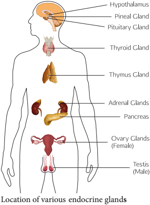
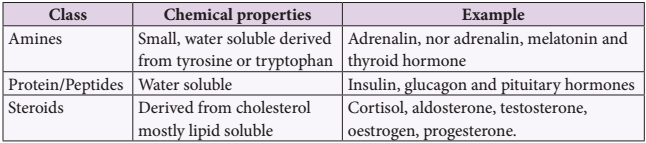
Table 11.1 Chemical Nature of Hormones
Human Endocrine System img 2

Hypothalamus

Hypothalamus is a small cone shaped structure that projects downward from the brain ending into the pituitary stalk. It interlinks both the nervous system and endocrine system. Though pituitary gland is known as master endocrine glands that controls the other endocrine glands, but it is, in turn controlled by the hypothalamus. Hypothalamus contains groups of neurosecretory cells. It produces neurotransmitters which regulate the secretions of the pituitary (Figure 11. 2). The hormones produced by the hypothalamus act either as a releasing hormone or as an inhibitory hormone.
Human Endocrine System img 3
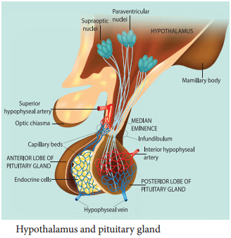
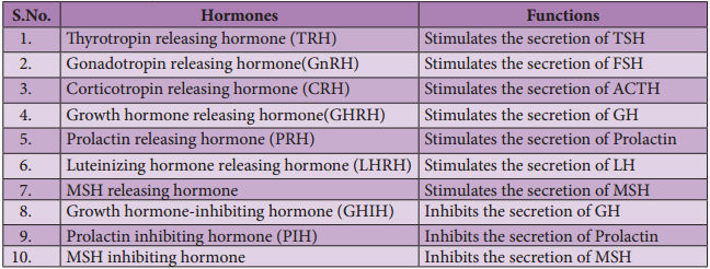
Table 11.2 The major hypothalamic hormones and their functions
Human Endocrine System img 4

In the basal region of the brain, the hypothalamic hypophyseal portal blood vessel connects hypothalamus and anterior pituitary. It allows hypothalamic hormones to control anterior pituitary secretion. The posterior pituitary is connected with hypothalamus by a nerve bundle called hypothalamic hypophyseal axis. It produces nerve signal that control the posterior pituitary secretion. Hypothalamus maintains homeostasis, blood pressure, body temperature, cardio and fluid electrolyte balance of the body. As the part of limbic system it influences various emotional responses.

Pituitary Gland or Hypophysis

The pituitary gland (means to grow under) is ovoid in shape and is located in the sella turcica, a bony cavity of the sphenoid bone at the base of brain and connected to the hypothalamic region of the brain by a stalk called infundibulum. It is about one centimetre in diameter and 0.5 gm in weight. The pituitary consists of two lobes, anterior glandular adenohypophysis and posterior neural neurohypophysis.

The anterior lobe originates from the embryonic invagination of pharyngeal epithelium called Rathke’s pouch and the posterior lobe is originates from the base of the brain as an outgrowth of hypothalamus. Anatomically the adenohypophysis has three lobes or zones namely pars intermedia, pars distalis and pars tuberalis. The neurohypophysis is otherwise known as pars nervosa.

The anterior lobe of pituitary secretes six tropic hormones such as growth hormone (GH), thyroid stimulating hormone (TSH), adreno corticotropic hormone (ACTH), follicle stimulating hormone (FSH), luteinizing hormone (LH), luteotropic hormone (LTH) and melanocyte stimulating hormone (MSH) (in lower animals only). The posterior lobe of pituitary secretes the hormones namely vasopressin and oxytocin.

Hormones of Adenohypophysis

(i) Growth Hormone (GH):

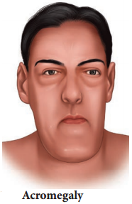
It is also known as somatotropic hormone (STH) or Somatotropin. It is a peptide hormone. Growth hormone promotes growth of all the tissues and metabolic process of the body. It influences the metabolism of carbohydrates, proteins and lipids and increases the rate of protein biosynthesis in the cells.

It stimulates chondrogenesis (cartilage formation), osteogenesis (bone formation) and helps in the retention of minerals like nitrogen, potassium, phosphorus, sodium etc., in the body. GH increases the release of fatty acid from adipose tissue and decreases the rate of glucose utilization for energy by the cells. Thus it conserves glucose for glucose dependent tissues, such as the brain.

(ii) Throid Stimulating Hormone (TSH) or Thyrotropin:

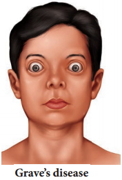
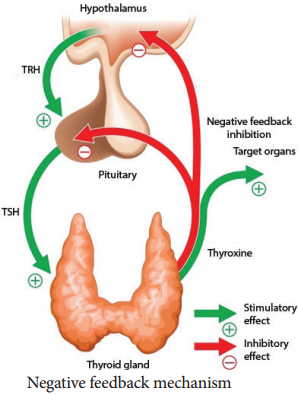
TSH is a glycoprotein hormone, which stimulates the thyroid gland to secrete Tri-iodothyronine (T3) and thyroxine (T4). TSH secretion is regulated by negative feedback mechanism. It’s release from the anterior pituitary is induced by the thyrotropin releasing hormone (TRH). When thyroxine level in the blood increases, TRH acts on both the pituitary and hypothalamus to inhibit TSH secretion.

(iii) Adreno Cortico Tropic Hormone

(ACTH):

ACTH is a peptide hormone that stimulates the adrenal cortex to secrete glucocorticoids and mineralocorticoids. It stimulates melanin synthesis in melanocytes, induces the release of fatty acids from adipose tissues and stimulates insulin secretion. ACTH secretion is regulated by negative feedback mechanism.

(iv) Follicle Stimulating Hormone (FSH):

FSH is a glycoprotein hormone which regulates the functions of the gonads (ovary and testis). In males, FSH along with androgens acts on the germinal epithelium of seminiferous tubules and stimulates the production and release of sperms (spermatogenesis). In females, FSH acts on the ovaries and brings about the development and maturation of graffian follicles.

(v) Luteinizing Hormone (LH):

LH is a glycoprotein hormone which is also known as interstitial cell stimulating hormone (ICSH). In males, ICSH acts on the interstitial cells of testis to produce the male sex hormone, testosterone. In females, LH along with FSH matures the ovarian follicles.

LH independently induces ovulation, maintains the corpus luteum and promotes synthesis and release of ovarian hormones. FSH and LH are collectively referred as gonadotropins. FSH and LH are not produced during childhood. The secretion of FSH and LH starts only during pre pubertal period.

(vi) Luteotropic Hormone (LTH):

LTH is also called luteotropin or lactogenic hormone or prolactin or mammotropin. It is a protein hormone which stimulates milk secretion after the child birth in females. High prolactin secretion during lactation suppresses LH secretion and ovulation since it induces the corpus luteum hence named as luteo tropic hormone.

Hormones of Neurohypophysis

(i) Vasopressin or Antidiuretic Hormone (ADH) :

ADH is a peptide hormone which promotes reabsorption of water and electrolytes by distal tubules of nephron and thereby reduces loss of water through urine. Hence it is called as anti diuretic hormone. It also causes constriction of blood vessels when released in large amount and increases blood pressure. ADH deficiency causes Diabetes insipidus which induces the production of large amount of urine.

(ii) Oxytocin (Means Quick Birth):

It is a peptide hormone which stimulates vigorous contraction of the smooth muscles of uterus during child birth and ejection of milk from the mammary glands.

Pineal Gland

In human, the pineal gland or epiphysis cerebri or conarium is located behind the third ventricle of brain and is formed of parenchymal cells and interstitial cells. It secretes the hormone, melatonin, which plays a central role in the regulation of circadian rhythm of our body and maintains the normal sleep wake cycle. It also regulates the timing of sexual maturation of gonads. In addition melatonin also influences metabolism, pigmentation, menstrual cycle and defence mechanism of our body.

Thyroid Gland

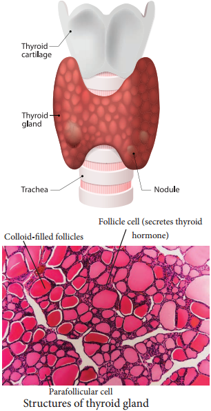
The butterfly shaped thyroid gland is a bilobed gland located below the larynx on each side of upper trachea. It is the largest endocrine gland in the body. Its two lateral lobes are connected by a median tissue mass called isthmus.

Each lobe is made up of many lobules. The lobules consist of follicles called acini (acinus in singular). Each acinus is lined with glandular, cuboidal or squamous epithelial cells. The lumen of acinus is filled with colloid, a thick glycoprotein mixture consisting of thyroglobulin molecules.

Hormones of the thyroid gland are often called the major metabolic hormones. The follicular cells of thyroid gland secrete two hormones namely tri-iodothyronine (T3) and thyroxine or tetra – iodothyronine (T4). The parafollicular cells or ‘C’ cells of thyroid gland secrete a hormone called thyrocalcitonin. Iodine is essential for the normal synthesis of thyroid hormones.

Thyroid releasing hormone from the hypothalamus stimulates the adenohypophysis to secrete TSH, which inturn stimulates the thyroid gland to secrete the thyroid hormones. Thyroid hormones show a negative feedback effect on the hypothalamus and pituitary (Figure 11. 3).
Human Endocrine System img 5

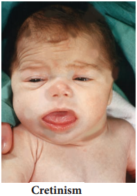
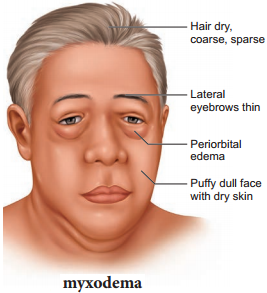
Functions of Thyroxine or Tetraiodothyronine (T4):

Thyroxine regulates the basal metabolic rate (BMR) and body heat production. It stimulates protein synthesis and promotes growth. It is essential for the development of skeletal and nervous system. Thyroxine plays an important role in maintaining blood pressure. It reduces serum cholesterol levels, Optimum levels of thyroxine in blood is necessary for gonadial functions.

TCT is a polypeptide hormone, which regulates the blood calcium and phosphate levels. It reduces the blood calcium level and opposes the effects of parathyroid hormone.
Human Endocrine System img 6

Parathyroid Gland

In human, four tiny parathyroid glands are found in the posterior wall of the thyroid glands. This gland is composed of two types of cells, the chief cells and oxyphil cells. The chief cells secrete parathyroid hormone (PTH) and the functions of oxyphil cells are not known.

Parathyroid Hormone or Parathormone (PTH)

PTH is a hypercalcemic hormone. It is a peptide hormone involved in controlling the calcium and phosphate homeostasis. The secretion of PTH is controlled by calcium level in the blood. It increases the blood calcium level by stimulating osteoclasts to dissolve the bone matrix.

As a result calcium and phosphate are released into the blood. PTH enhances the reabsorption of calcium and excretion of phosphates by the renal tubules and promotes activation of vitamin D to increase calcium absorption by intestinal mucosal cells.

Thymus Gland

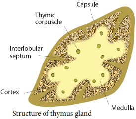
Thymus gland is partially an endocrine and partially a lymphoid organ. It is a bilobed structure located just above the heart and aorta, behind the sternum. It is covered by firous capsule and anatomically it is divisible into an outer cortex and an inner medulla.

It secretes four hormones such as thymulin, thymosin, thymopoietin and thymic humoral factor (THF). The primary function of thymus is the production of immuno competent ‘T’ lymphocytes which provides cell mediated immunity.
Human Endocrine System img 7

Adrenal Gland

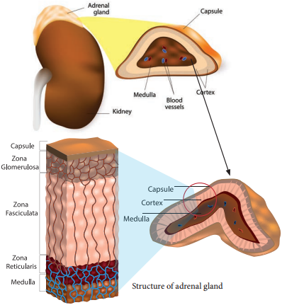
A pair of adrenal glands are located at the anterior end of the kidneys, hence also called suprarenal glands. Anatomically the outer region is the cortex and the inner region is the medulla. Histologically the adrenal cortex has three distinct zones, zona glomerulosa, zona fasciculata and zona reticularis.

Zona glomerulosa an outer thin layer constitutes about 15% of adrenal cortex, and secretes mineralocorticoids. Zona fasciculata, the middle widest layer constitutes about 75% of adrenal cortex and secretes glucocorticoids such as cortisol, corticosterone and trace amounts of adrenal androgen and oestrogen. Zona reticularis, an inner zone of adrenal cortex constitute about 10% of adrenal cortex and secretes the adrenal androgen, trace amount of oestrogen and glucocorticoids.

Adrenal Medulla:

It is the central part of adrenal gland and is composed of ovoid and columnar cells, which are found around the network of blood capillaries. Adrenalin (epinephrine) and nor adrenalin (nor epinephrine) are the two hormones secreted by the adrenal medulla. Both adrenalin and nor adrenalin are catecholamines.

Function of Adrenal Hormones:

Glucocorticoids stimulate gluconeogensis, lipolysis and proteolysis (the life saving activity). Cortisol is a glucocorticoid involved in maintaining cardio vascular and kidney functions. It produces anti inflammatory reactions and suppresses the immune response.

It stimulates the RBC production. It is also known as stress combat hormone. Mineralocorticoids regulates water and electrolyte balance of our body. Aldosterone stimulates the reabsorption of sodium and water and eliminates potassium and phosphate ions through excretion, thus it helps in maintaining electrolytes, osmotic pressure and blood pressure. Adrenal androgen plays a role in hair growth in the axial region, pubis and face during puberty.

The adrenal medulla secretes the hormones adrenalin and noradrenalin and are referred as “3F hormone” (fight, flight and fright hormone). Adrenalin increases liver glycogen breakdown into glucose and increases the release of fatty acids from fat cells.

During emergency it increases heart beat rate and blood pressure. It stimulates the smooth muscles of cutaneous and visceral arteries to decrease blood flow. It increases blood flow to the skeletal muscles thereby increases the metabolic rate of skeletal muscles, cardiac muscles and nervous tissue.
Human Endocrine System img 8
Human Endocrine System img 9

Pancreas

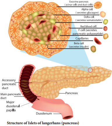
Pancreas is a composite gland which performs both exocrine and endocrine functions. It is located just below the stomach as a leaf like structure. The pancreas is composed of two major tissues such the acini and islets of langerhans.

Acini secretes digestive enzymes and the islets of langerhans secretes hormones like insulin and glucagon. Human pancreas has one to two million islets of langerhans. In each islet about 60% cells are beta cells, 30% cells are alpha cells and 10% cells are delta cells. The alpha cells secrete glucagon, the beta cells secrete insulin and delta cells secrete somatostatin.

Insulin:

Insulin is a peptide hormone and plays an important role in glucose homeostasis. It’s main effect is to lower blood glucose levels by increasing the uptake of glucose into the body cells, especially muscle and fat cells. Insulin also inhibits the breakdown of glycogen to glucose, the conversion of amino acids or fats to glucose, so insulin is rightly called a hypoglycemic hormone.

Glucagon:

Glucagon is a polypeptide hormone. It is a potent hyperglycemic hormone that acts on the liver and promotes the breakdown of glycogen to glucose (Glygogenolysis), synthesis of glucose from lactic acid and from non-carbohydrate molecules (Gluconeogenesis).

Glucagon releases glucose from the liver cells, increasing the blood glucose levels. Since glucagon reduces the cellular uptake and utilisation of glucose it is called a hyperglycemic hormone. Prolonged hyperglycemia leads to the disorder called diabetes mellitus.

Gonads

Testis:

A pair of testis is present in the scrotal sac of males. The testis functions as a sex organ and also as an endocrine gland. The testis is composed of seminiferous tubules and interstitial cells or Leydig cells. The Leydig cells secrete several male sex hormones, collectively called androgens, mainly testosterone.

Functions of Testosterone:

Under the influence of FSH and LH, testosterone initiates maturation of male reproductive organs, and the appearance of secondary sexual characters, muscular growth, growth of facial and axillary hair, masculine voice and male sexual behaviour. It enhances the total bone matrix and plays a stimulating role in the process of spermatogenesis.

Ovary:

Females have a pair of ovaries located in the pelvic region of the abdomen. The ovary is composed of ovarian follicles and stromal tissues. It produces the eggs or ova. The ovaries secrete the steroid hormones oestrogen and progesterone. Oestrogen is responsible for the maturation of reproductive organs and the development of secondary sexual characters at puberty.

Along with progesterone, oestrogens promotes breast development and initiate the cyclic changes during menstrual cycle. Progesterone prepares the uterus for implantation of the fertilized ovum. It decreases the uterine contraction during pregnancy and stimulates the development of mammary glands and milk secretion. It is responsible for premenstrual changes in the uterus and is essential for the formation of placenta.

Hormones of Heart, Kidney and Gastro Intestinal Tract

Some tissues of the heart, kidney and gastro intestinal tract acts as partial endocrine glands. In the heart, cardiocytes on the atrial wall’s secretes an important peptide hormone called atrial natriuretic factor (ANF). When blood pressure is increased, ANF is secreted and causes dilation of the blood vessels to reduce the blood pressure.

In kidneys, hormones such as renin, erythropoietin and calcitriol are secreted. Renin is secreted by juxta glomerular cells (JGA), which increases blood pressure when angiotensin is formed in blood. Erythropoietin is also secreted by the JGA cells of the kidney and stimulates erythropoiesis (formation of RBC) in bone marrow. Calcitriol is sercreted by proximal tubules of nephron. It is an active form of vitamin D3 which promotes calcium and phosphorus absorption from intestine and accelerates bone formation.

Gastro Intestinal Tract Hormones

Group of specialized endocrine cells present in gastro-intestinal tract secretes hormones such as gastrin, cholecystokinin (CCK), secretin and gastric inhibitory peptides (GIP). Gastrin acts on the gastric glands and stimulates the secretion of HCl and pepsinogen.

Cholecystokinin (CCK) is secreted by duodenum in response to the presence of fat and acid in the diet. It acts on the gall bladder to release bile into duodenum and stimulates the secretion of pancreatic enzymes and its discharge. Secretin acts on acini cells of pancreas to secrete bicarbonate ions and water to neutralize the acidity. Gastric inhibitory peptide (GIP) inhibits gastric secretion and motility.

Endocrine Glands and Hormones

Learninsta presents the core concepts of Biology with high-quality research papers and topical review articles.

Endocrine Glands and Hormones

Physiological functions of our human body is regulated and coordinated by both neural and endocrine systems. The endocrine system influences the metabolic activities by means of hormones (hormone means to excite) which are chemical messengers released into the blood and circulated as chemical signals and acts specifically on certain organs or tissues called target organs or target tissues.

Hormones may speed up or slow down or alter the activity of the target organs. The hormones secreted do not remain permanently in the blood but are converted by the liver into inactive compounds and excreted by the kidneys.

Hormones are chemical messengers because they act as organic catalysts and coenzymes to perform specific functions in the target organs. The target organs contain receptor molecules either on the surface or within the cell.

Although different hormones come in contact with cells, only the cells that contain receptor molecules specific for the hormone are physiologically activated. A single hormone may have multiple effects on a single target tissue or on different target tissues.

Many hormones exhibit long term changes like growth, puberty and pregnancy. Hormones often influence many organs and organ systems at the same time. Serious deficiency or excess secretion of hormones leads to disorders. Hormones coordinate different physical, physiological, mental activities and maintain homeostasis. Hormones are composed of water soluble proteins or peptides or amines or fat soluble steroids.

While many parts of the body make hormones, the major glands that make up the endocrine system are the:

  • Hypothalamus
  • Pituitary
  • Thyroid
  • Parathyroids
  • Adrenals
  • Pineal body
  • The ovaries
  • The testes

Endocrine glands are ductless glands and release the substances that they make (hormones) directly into the bloodstream. These glands form part of the endocrine system and information on them is included in this website. There is another type of gland called an exocrine gland (e.g. sweat glands, lymph nodes).

The following are integral parts of the endocrine system:

Hypothalamus. The hypothalamus is located at the base of the brain, near the optic chiasm where the optic nerves behind each eye cross and meet.

  • Pineal body
  • Pituitary
  • Thyroid and parathyroid
  • Thymus
  • Adrenal gland
  • Pancreas
  • Ovary

The symptoms of an endocrine disorder vary widely and depend on the specific gland involved. However, most people with endocrine disease complain of fatigue and weakness. Blood and urine tests to check your hormone levels can help your doctors determine if you have an endocrine disorder.

When you are facing a diagnosis of a hormonal condition, like diabetes or thyroid disease, your doctor may suggest you see an endocrinologist. You may be wondering why you need to see a specialist instead of simply sticking with your primary doctor.

Endocrinologists are qualified to diagnose and treat conditions like diabetes, thyroid diseases, infertility, growth issues, metabolic disorders, osteoporosis, some cancers, and disorders in the hormone-producing adrenal glands and pituitary glands.

Beginning the examination with the hands establishes a personal connection with the patient and reinforces the trust between physician and patient that was initiated during the history. It opens the possibility for an equal exchange between doctor and patient as both observe and talk about the hands.
Endocrine Glands and Hormones img 1

Sensory Reception and Processing

Learninsta presents the core concepts of Biology with high-quality research papers and topical review articles.

Sensory Reception and Processing

Our senses make us aware of changes that occur in our surroundings and also within our body. Sensation [awareness of the stimulus] and perception [interpretation of the meaning of the stimulus] occur in the brain.

Receptors are Classified Based on Their Location:

1. Exteroceptors are located at or near the surface of the body. These are sensitive to external stimuli and receive sensory inputs for hearing, vision, touch, taste and smell.

2. Interoceptors are located in the visceral organs and blood vessels. They are sensitive to internal stimuli. Proprioceptors are also a kind of interoceptors. They provide information about position and movements of the body.

These are located in the skeletal muscles, tendons, joints, ligaments and in connective tissue coverings of bones and muscles. Receptors based on the type of stimulus are shown in Table 10.3.
Sensory Reception and Processing img 1

Photoreceptor – Eye

Eye is the organ of vision; located in the orbit of the skull and held in its position with the help of six extrinsic muscles. They are superior, inferior, lateral, median rectus muscles, superior oblique and inferior oblique muscles. These muscles aid in the movement of the eyes and they receive their nerve innervation from III, IV and VI cranial nerves.

Eyelids, eye lashes and eye brows are the accessory structures useful in protecting the eyes. The eye lids protect the eyes from excessive light and foreign objects and spread lubricating secretions over the eyeballs.

Eyelashes and the eyebrows help to protect the eyeballs from foreign objects, perspiration and also from the direct rays of sunlight. Sebaceous glands at the base of the eyelashes are called ciliary glands which secrete a lubricating fluid into the hair follicles.

Lacrymal glands, located in the upper lateral region of each orbit, secrete tears. Tears are secreted at the rate of 1mL/day and it contains salts, mucus and lysozyme enzyme to destroy bacteria. The conjunctiva is a thin, protective mucous membrane found lining the outer surface of the eyeball (Figure 10.13).
Sensory Reception and Processing img 2

The eye has two compartments, the anterior and posterior compartments. The anterior compartment has two chambers, first one lies between the cornea and iris and the second one lies between the iris and lens. These two chambers are filled with watery fluid called aqueous humor.

The posterior compartment lies between the lens and retina and it is filled with a jelly like fluid called vitreous humor that helps to retain the spherical nature of the eye. Eye lens is transparent and biconvex, made up of long columnar epithelial cells called lens fires. These cells are accumulated with the proteins called crystalline.

The Eye Ball

The eye ball is spherical in nature. The anterior one – sixth of the eyeball is exposed; the remaining region is fitted well into the orbit. The wall of the eye ball consists of three layers: firous Sclera, vascular Choroid and sensory Retina (Figure 10.14).
Sensory Reception and Processing img 3

The outer coat is composed of dense non-vascular connective tissue. It has two regions: the anterior cornea and the posterior sclera. Cornea is a non-vascular transparent coat formed of stratified squamous epithelium which helps the cornea to renew continuously as it is very vulnerable to damage from dust. Sclera forms the white of the eye and protects the eyeball.

Posteriorly the sclera is innervated by the optic nerve. At the junction of the sclera and the cornea, is a channel called ‘canal of schlemm’ which continuously drains out the excess of aqueous humor.

Choroid

Is highly vascularized pigmented layer that nourishes all the eye layers and its pigments absorb light to prevent internal reflection. Anteriorly the choroid thickens to form the ciliary body and iris. Iris is the coloured portion of the eye lying between the cornea and lens. The aperture at the centre of the iris is the pupil through which the light enters the inner chamber.

Iris is made of two types of muscles the dilator papillae (the radial muscle) and the sphincter papillae (the circular muscle). In the bright light, the circular muscle in the iris contract; so that the size of pupil decreases and less light enters the eye.

In dim light, the radial muscle in the iris contract; so that the pupil size increases and more light enters the eye. Smooth muscle present in the ciliary body is called the ciliary muscle which alters the convexity of the lens for near and far vision.

The ability of the eyes to focus objects at varying distances is called accommodation which is achieved by suspensory ligament, ciliary muscle and ciliary body. The suspensory ligament extends from the ciliary body and helps to hold the lens in its upright position. The ciliary body is provided with blood capillaries that secrete a watery fluid called aqueous humor that fills the anterior chamber.

Retina Forms the Inner Most Layer of the Eye and it Contains Two Regions:

A sheet of pigmented epithelium (non visual part) and neural visual regions. The neural retina layer contains three types of cells: photoreceptor cells – cones and rods (Figure 10.15 and Table 10.4), bipolar cells and ganglion cells.

The yellow flat spot at the centre of the posterior region of the retina is called macula lutea which is responsible for sharp detailed vision. A small depression present in the centre of the yellow spot is called fovea centralis which contains only cones.

The optic nerves and the retinal blood vessels enter the eye slightly below the posterior pole, which is devoid of photo receptors; hence this region is called blind spot.
Sensory Reception and Processing img 4
Differences between rod and cone cells
Sensory Reception and Processing img 5

Mechanism of Vision

When light enters the eyes, it gets refracted by the cornea, aqueous humor and lens and it is focused on the retina and excites the rod and cone cells. The photo pigment consists of Opsin, the protein part and Retinal, a derivative of vitamin A.

Light induces dissociation of retinal from opsin and causes the structural changes in opsin. This generates an action potential in the photoreceptor cells and is transmitted by the optic nerves to the visual cortex of the brain, via bipolar cells, ganglia and optic nerves, for the perception of vision.

Refractive Errors of Eye

Myopia (near sightedness):

The affected person can see the nearby objects but not the distant objects. This condition may result due to an elongated eyeball or thickened lens; so that the image of distant object is formed in front of the yellow spot. This error can be corrected using concave lens that diverge the entering light rays and focuses it on the retina.

Hypermetropia (Long Sightedness):

The affected person can see only the distant objects clearly but not the objects nearby. This condition results due to a shortened eyeball and thin lens; so the image of closest object is converged behind the retina. This defect can be overcome by using convex lens that converge the entering light rays on the retina.

Presbyopia:

Due to aging, the lens loses elasticity and the power of accommodation. Convex lenses are used to correct this defect.

Astigmatism

Is due to the rough (irregular) curvature of cornea or lens. Cylindrical glasses are used to correct this error (Figure 10.16).
Sensory Reception and Processing img 6

Cataract:

Due to the changes in nature of protein, the lens becomes opaque. It can be corrected by surgical procedures.

Phonoreceptor

The ear is the site of reception of two senses namely hearing and equilibrium. Anatomically, the ear is divided into three regions: the external ear, the middle ear and internal ear.

The external ear consists of pinna, external auditory meatus and ear drum. The pinna is flap of elastic cartilage covered by skin. It collects the sound waves. The external auditory meatus is a curved tube that extends up to the tympanic membrane [the ear drum]. The tympanic membrane is composed of connective tissues covered with skin outside and with mucus membrane inside.

There are very fine hairs and wax producing sebaceous glands called ceruminous glands in the external auditory meatus. The combination of hair and the ear wax [cerumen] helps in preventing dust and foreign particles from entering the ear.

The middle ear is a small air-filled cavity in the temporal bone. It is separated from the external ear by the eardrum and from the internal ear by a thin bony partition; the bony partition contains two small membrane covered openings called the oval window and the round window.

The Middle Ear Contains Three Ossicles:

Malleus [hammer bone], incus [anvil bone] and stapes [stirrup bone] which are attached to one another. The malleus is attached to the tympanic membrane and its head articulates with the incus which is the intermediate bone lying between the malleus and stapes.

The stapes is attached to the oval window in the inner ear. The ear ossicles transmit sound waves to the inner ear. A tube called Eustachian tube connects the middle ear cavity with the pharynx. This tube helps in equalizing the pressure of air on either sides of the ear drum.

Inner ear is the fluid filled cavity consisting of two parts, the bony labyrinth and the membranous labyrinths. The bony labyrinth consists of three areas: cochlea, vestibule and semicircular canals. The cochlea is a coiled portion consisting of 3 chambers namely: scala vestibuli and scala tympani – these two are filled with perilymph; and the scala media is filled with endolymph.

At the base of the cochlea, the scala vestibule ends at the ‘oval window’ whereas the scala tympani ends at the ‘round window’ of the middle ear. The chambers scala vestibuli and scala media are separated by a membrane called Reisner’s membrane whereas the scala media and scala tympani are separated by a membrane called Basilar membrane (Figure 10.17)
Sensory Reception and Processing img 7

Organ of Corti

The organ of Corti (Figure.10.18) is a sensory ridge located on the top of the Basilar membrane and it contains numerous hair cells that are arranged in four rows along the length of the basilar membrane. Protruding from the apical part of each hair cell is hair like structures known as stereocilia. During the conduction of sound wave, stereocilia makes a contact with the stiff gel membrane called tectorial membrane, a roof like structure overhanging the organ of corti throughout its length.

Mechanism of Hearing

Sound waves entering the external auditory meatus fall on the tympanic membrane. This causes the ear drum to vibrate, and these vibrations are transmitted to the oval window through the three auditory ossicles. Since the tympanic membrane is 17-20 times larger than the oval window, the pressure exerted on the oval window is about 20 times more than that on the tympanic membrane.

This increased pressure generates pressure waves in the fluid of perilymph. This pressure causes the round window to alternately bulge outward and inward meanwhile the basilar membrane along with the organ of Corti move up and down.

These movements of the hair alternately open and close the mechanically gated ion channels in the base of hair cells and the action potential is propagated to the brain as sound sensation through cochlear nerve.
Sensory Reception and Processing img 8

Defects of Ear

Deafness may be temporary or permanent. It can be further classified into conductive deafness and sensory-neural deafness. Possible causes for conductive deafness may be due to

  • The blockage of ear canal with earwax
  • Rupture of eardrum
  • Middle ear infection with fluid accumulation
  • Restriction of ossicular movement. In sensory-neural deafness, the defect may be in the organ of Corti or the auditory nerve or in the ascending auditory pathways or auditory cortex.

Organ of Equilibrium

Balance is part of a sense called proprioception, which is the ability to sense the position, orientation and movement of the body. The organ of balance is known as the vestibular system which is located in the inner ear next to the cochlea. The vestibular system is composed of a series of fluid filled sacs and tubules.

These sacs and tubules contain endolymph and are kept in the surrounding perilymph (Figure 10.19). These two fluids, perilymph and endolymph, respond to the mechanical forces, during changes occurring in body position and acceleration.
Sensory Reception and Processing img 8

The utricle and saccule are two membranous sacs, found nearest the cochlea and contain equilibrium receptor regions called maculae that are involved in detecting the linear movement of the head. The maculae contain the hair cells that act as mechanorecptors.

These hair cells are embeded in a gelatinous otolithic membrane that contains small calcareous particles called otoliths. This membrane adds weight to the top of the hair cells and increase the inertia.

The canals that lie posterior and lateral to the vestibule are semicircular canals; they are anterior, posterior and lateral canals oriented at right angles to each other. At one end of each semicircular canal, at its lower end has a swollen area called ampulla. Each ampulla has a sensory area known as crista ampullaris which is formed of sensory hair cells and supporting cells. The function of these canals is to detect rotational movement of the head.

Oldfactory Receptors

The receptors for taste and smell are the chemoreceptors. The smell receptors are excited by air borne chemicals that dissolve in fluids. The yellow coloured patches of oldfactory epithelium form the oldfactory organs that are located on the roof of the nasal cavity.

The oldfactory epithelium is covered by a thin coat of mucus layer below and oldfactory glands bounded connective tissues, above. It contains three types of cells: supporting cells, Basal cells and millions of pin shaped oldfactory receptor cells (which are unusual bipolar cells).

The oldfactory glands and the supporting cells secrete the mucus. The unmyelinated axons of the oldfactory receptor cells are gathered to form the filaments of oldfactory nerve [cranial nerve I] which synapse with cells of oldfactory bulb. The impulse, through the oldfactory nerves, is transmitted to the frontal lobe of the brain for identification of smell and the limbic system for the emotional responses to odour.

Gustatory Receptor:

The sense of taste is considered to be the most pleasurable of all senses. The tongue is provided with many small projections called papillae which give the tongue an abrasive feel. Taste buds are located mainly on the papillae which are scattered over the entire tongue surface.

Most taste buds are seen on the tongue (Figure 10.20) few are scattered on the sof palate, inner surface of the cheeks, pharynx and epiglottis of the larynx. Taste buds are flask-shaped and consist of 50 – 100 epithelial cells of two major types.
Sensory Reception and Processing img 10

Gustatory epithelial cells (taste cells) and Basal epithelial cells (Repairing cells) Long microvilli called gustatory hairs project from the tip of the gustatory cells and extends through a taste pore to the surface of the epithelium where they are bathed by saliva.

Gustatory hairs are the sensitive portion of the gustatory cells and they have sensory dendrites which send the signal to the brain. The basal cells that act as stem cells, divide and differentiate into new gustatory cells (Figure 10.20).
Sensory Reception and Processing img 11

Skin-Sense of Touch

Skin is the sensory organ of touch and is also the largest sense organ. This sensation comes from millions of microscopic sensory receptors located all over the skin and associated with the general sensations of contact, pressure, heat, cold and pain.

Some parts of the body, such as the finger tips have a large number of these receptors, making them more sensitive. Some of the sensory receptors present in the skin (Figure 10.21) are:

Tactile Merkel Disc

Is light touch receptor lying in the deeper layer of epidermis.

Hair Follicle Receptors

Are light touch receptors lying around the hair follicles.

Meissner’s Corpuscles

Are small light pressure receptors found just beneath the epidermis in the dermal papillae. They are numerous in hairless skin areas such as finger tips and soles of the feet.

Pacinian Corpuscles

Are the large egg shaped receptors found scattered deep in the dermis and monitoring vibration due to pressure. It allows to detect different textures, temperature, hardness and pain.

Ruffi Endings

Which lie in the dermis responds to continuous pressure.

Krause End Bulbs

Are thermoreceptors that sense temperature.