Bryophytes Definition and its Economic Importance

Learninsta presents the core concepts of Biology with high-quality research papers and topical review articles.

Bryophytes Definition and its Economic Importance

Amphibians of Plant Kingdom

In the previous chapter, we noticed a wide range of thallus organization in Algae. Majority of them are aquatic. The development of heterotrichous habit, development of parenchyma tissue and dichotomous branching in some algae supports the view that colonization of plants in land occurred in the past.

Bryophytes are simplest and most primitive plant groups descended from alga – like ancestors. They are simple embryophytes. Let us learn about the structure and reproduction of these primitive land plants called Bryophytes in detail.
Bryophytes img 1

Bryophytes are simplest land inhabiting cryptogams and are restricted to moist, shady habitats. They lack vascular tissue and hence called ‘Non- vascular cryptogams’. They are also called as ‘amphibians of plant kingdom’ because they need water for completing their life cycle.

General Characteristic Features

  • The plant body of bryophyte is gametophyte and is not differentiated into root, stem and leaf like structure.
  • Most of them are primitive land dwellers. Some of them are aquatic (Riella, Ricciocarpus).
  • The gametophyte is conspicuous, long lived phase of the life cycle. Thalloid forms are present in liverworts and Hornworts.

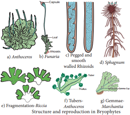
In Mosses leaf like, stem like structures are present. In Liverworts thallus grows prostrate on the ground and is attached to the substratum by means of rhizoids. Two types of rhizoids are present namely smooth walled and pegged or tuberculate.

Multicellular scales are also present. In Moss the plant body is erect with central axis bearing leaf like expansions. Multicellular rhizoids are present. The structure and reproduction in Bryophytes is given in Figure 2.5.
Bryophytes img 2

Vascular tissue like xylem and phloem are completely absent, hence called ‘Non vascular cryptogams’. Vegetative reproduction takes place by the formation of adventitious buds (Riccia fluitans) tubers develop in Anthoceros.

In some forms small detachable branches or brood bodies are formed, they help in vegetative reproduction as in Bryopteris fruticulosa. In Marchantia propagative organs called gemmae are formed and help in reproduction. Sexual reproduction is oogamous. Antheridia and Archegonia are produced in a protective covering and are multicellular.

The antheridia produces biflagellate antherozoids which swims in thin film of water and reach the archegonium and fuse with the egg to form diploid zygote.

Water is Essential for Fertilization

The zygote is the first cell of the sporophyte generation. It undergoes mitotic division to form multicellular undifferentiated embryo. The embryogeny is exoscopic (the first division of the zygote is transverse and the apex of the embryo develops from the outer cell). The embryo divides and give rise to sporophyte.

The sporophyte is dependent on gametophyte. It is differentiated into three recognizable parts namely foot, seta and capsule.

Foot is the basal portion and is embedded in the gametophyte through which water and nutrients are supplied for the sporophyte. The diploid spore mother cells found in the capsule region undergoes meiotic division and give rise to haploid spores. Bryophytes are homosporous. In some sporophytes elaters are present and help in dispersal of spores (Example: Marchantia). The spores germinate to produce gametophyte.

The zygote, embryo and the sporogonium constitute sporophytic phase. The green long living haploid phase is called gametophytic phase. The haploid gametophytic phase alternates with diploid sporophyte and shows heterologous alternation of generation. Proskauer in the year 1957 classified Bryophytes into 3 Classes namely:-

(i) Hepaticopsida:-
(Riccia, Marchantia, Porella and Riella)

(ii) Anthocerotopsida:-
(Anthoceros and Dendroceros)

(iii) Bryopsida:-
(Funaria, Polytrichum and Sphagnum).

Economic Importance

Dead thalli of Sphagnum gets accumulated and compressed, hardened to form peat. In northern Europe (Netherlands) peat is used as fuel in commercial scale. Apart from this nitrates, brown dye and tanning materials are derived from peat.

Sphagnum and peat are also used in horticulture as packing material because of their water holding capacity. Marchantia polymorpha is used to cure pulmonary tuberculosis. Sphagnum, Bryum and Polytrichum are used as food. Bryophytes play a major role in soil formation through succession and help in soil conservation.

Algae Classification and its Importance

Learninsta presents the core concepts of Biology with high-quality research papers and topical review articles.

Algae Classification and its Importance

Rain brings joy and life to various organisms on earth. Have you noticed some changes in and around you after the rain? Could you identify the reason for the slippery nature of the terrace and green patches on the wall of our home, green colour of puddles and ponds? Why should we clean our water tanks very often? The reason is algae.

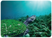
Algae are simple plants that lack true roots, true stems and true leaves. Two-third of our earth’s surface is covered by oceans and seas. The photosynthetic plants called algae are present here. More than half of the total primary productivity of the world depends on this plant group. Further, other aquatic organisms also depend upon them for their existence.
Algae Classification and its Importance img 1

Algae are autotrophs, and grow in a wide range of habitats. Majority of them are aquatic, marine (Gracilaria, and Sargassum) and freshwater (Oedogonium, and Ulothrix) and also found in soils (Fritschiella, and Vaucheria). Chlorella lead an endozoic life in hydra and sponges whereas Cladophora crispata grow on the shells of molluscs. Algae are adapted to thrive in harsh environment too.

Dunaliella salina grows in salt pans (Halophytic alga). Algae growing in snow are called Cryophytic algae. Chlamydomonas nivalis grow in snow covered mountains and impart red colour to the snow (Red snow). A few algae grow on the surface of aquatic plants and are called epiphytic algae (Coleochaete, and Rhodymenia). The study of algae is called algology or phycology. Some of the eminent algologists include F.E. Fritsch, F.E. Round, R.E. Lee, M.O. Parthasarathy Iyengar, M.S. Randhawa, Y. Bharadwaja, V.S. Sundaralingam and T.V. Desikachary.

General Characteristic Features

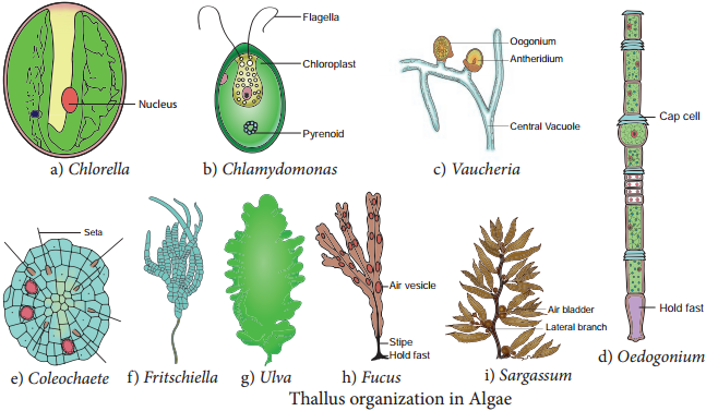
The algae show a great diversity in size, shape and structure. A wide range of thallus organisation is found in algae. Unicellular motile (Chlamydomonas), unicellular non-motile (Chlorella), Colonial motile (Volvox), Colonial non motile (Hydrodictyon), siphonous (Vaucheria), unbranched filamentous (Spirogyra), branched filamentous (Cladophora), discoid (Coleochaete) heterotrichous (Fritschiella), Foliaceous (Ulva) to giant kelps (Laminaria and Macrocystis). The thallus organization in algae is given in Figure 2.3.
Algae Classification and its Importance img 2

Algae are eukaryotes except blue green algae. The plant body does not show differentiation into tissue systems. The cell wall of algae is made up of cellulose and hemicellulose. Siliceous walls are present in diatoms. In Chara the thallus is encrusted with calcium carbonate.

Some algae possess algin, polysulphate esters of polysaccharides which are the sources for the alginate, agar agar and carrageenan. The cell has a membrane bound nucleus and cell organelles like chloroplast, mitochondria, endoplasmic reticulum, golgi bodies etc., Pyrenoids are present. They are proteinaceous bodies found in chromatophores and assist in the synthesis and storage of starch.

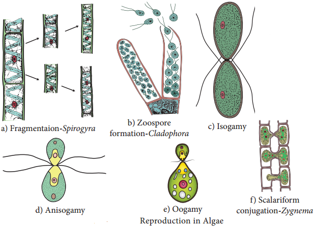
The pigmentation, reserve food material and flagellation differ among the algal groups. Algae reproduces by vegetative, asexual and sexual methods (Figure 2.4).

Vegetative reproduction includes fission (In unicellular forms the cell divides mitotically to produce two daughter cells Example: Chlamydomonas); Fragmentation (fragments of parent thallus grow into new individual Example: Ulothrix) budding (A lateral bud is formed in some members like Protosiphon and helps in reproduction) bulbils, (a wedge shaped modified branch develop in Sphacelaria) akinetes (Thick walled spores meant for perennation and germinates with the advent of favourable condition Example: Pithophora) and Tubers (Structures found on the rhizoids and the lower nodes of Chara which store food materials).
Algae Classification and its Importance img 3

Asexual reproduction takes place by the production of zoospores motile spores (Ulothrix, Oedogonium) aplanospore (thin walled non motile spores Example: Vaucheria); autospores (spores which look similar to parent cell Example: Chlorella); hypnospore (thick walled aplanospore – Example: Chlamydomonas nivalis) and Tetraspores (Diploid thallus of Polysiphonia produce haploid spores after meiosis).

Sexual Reproduction in Algae is of Three Types

  • Isogamy (Fusion of morphologically and Physiologically similar gametes Example: Ulothrix)
  • Anisogamy (Fusion of either morphologically or physiologically dissimilar gametes Example: Pandorina)
  • Oogamy (Fusion of both morphologically and physiologically dissimilar gametes. Example: Sargassum).

The life cycle shows distinct alternation of generation.

Classification

F.E. Fritsch proposed a classification for algae based on pigmentation, types of flagella, reserve food materials, thallus structure and reproduction. He published his classification in the book “The structure and reproduction of the Algae”(1935). He classified algae into 11 classes namely Chlorophyceae, Xanthophyceae, Chrysophyceae, Bacillariophyceae, Cryptophyceae, Dinophyceae, Chloromonadineae, Euglenophyceae, Phaeophyceae, Rhodophyceae, Cyanophyceae. The salient features of Chlorophyceae, Phaeophyceae and Rhodophyceae are given below.

Chlorophyceae

The members are commonly called ‘Green algae’. Most of the species are aquatic (Fresh water-Spirogyra, Marine – Ulva). A few are terrestrial (Trentipohlia). Variation among the shape of the chloroplast is found in members of algae. It is cup shaped (Chlamydomonas), discoid (Chara), girdle shaped, (Ulothrix), reticulate (Oedogonium), spiral (Spirogyra), stellate (Zygnema) and plate like (Mougeoutia).

Chlorophyll ‘a’ and Chlorophyll ‘b’ are the major photosynthetic pigments. Storage bodies called pyrenoids are present in the chloroplast and store starch. They also contain proteins. The cell wall is made up of inner layer of cellulose and outer layer of pectin.

Vegetative reproduction takes place by means of fragmentation and asexual reproduction is by the production of zoospores, aplanospores and akinetes. Sexual reproduction is present and may be isogamous, anisogamous or oogamous. Examples for this group of algae includes Chlorella, Chlamydomonas, Volvox, Spirogyra, Ulothrix, Chara and Ulva.

Phaeophyceae

The members of this class are called ‘Brown algae’. Majority of the forms are found in marine habitats. Pleurocladia is a fresh water form. The thallus is filamentous (Ectocarpus) frond like (Dictyota) or may be giant kelps (Laminaria and Macrocystis). The thallus is differentiated into leaf like photosynthetic part called fronds, a stalk like structure called stipe and a holdfast which attach thallus to the substratum.

The Pigments include Chlorophyll a, c, Carotenoids and Xanthophylls. A golden brown pigment called fucoxanthin is present and it gives shades of colour from olive green to brown to the algal members of this group. Mannitol and Laminarin are the reserve food materials.

Motile reproductive structures are present. Two laterally inserted unequal flagella are present. Among these one is whiplash and another is tinsel. Although sexual reproduction ranges from isogamy to oogamy, Most of the forms show oogamous type. Alternation of generation is present (isomorphic, heteromorphic or diplontic). Examples for this group include Sargassum, Laminaria, Fucus and Dictyota.

Rhodophyceae

Members of this group include ‘Red algae’ and are mostly marine. The thallus is multicellular, macroscopic and diverse in form. Porphyridium is the unicellular form. Filamentous (Goniotrichum) ribbon like (Porphyra) are also present.

Corallina and Lithothamnion are heavily impregnated with lime and form coral reefs. Apart from chlorophyll a, r-phycoerythrin and r-phycocyanin are the photosynthetic pigments. Asexual reproduction takes place by means of monospores, neutral spores and tetraspores.

The storage product is floridean starch. Sexual reproduction is oogamous. Male sex organ is spermatangium which produces spermatium. Female sex organ is called carpogonium. The spermatium is carried by the water currents and fuse with egg nucleus to form zygote.

The zygote develops into carpospores. Meiosis occurs during carpospore formation. Alternation of generation is present. Examples for this group of algae include Ceramium, Polysiphonia, Gelidium, Cryptonemia and Gigartina.

Economic Importance

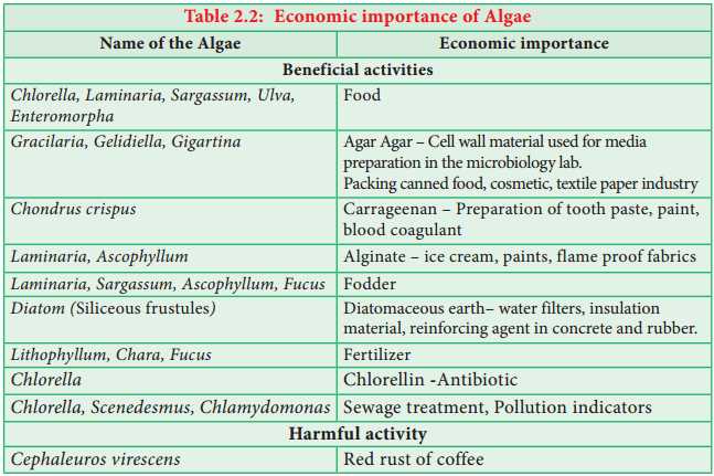
The Economic importance of Algae is given in Table 2.2
Algae Classification and its Importance img 4

Life Cycle Patterns in Plants

Learninsta presents the core concepts of Biology with high-quality research papers and topical review articles.

Life Cycle Patterns in Plants

Alternation of Generation

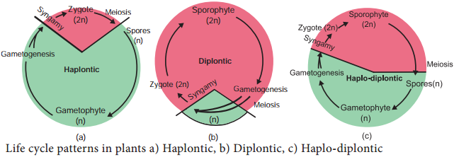
Alternation of generation is common in all plants. Alternation of the haploid gametophytic phase (n) with diploid sporophytic phase (2n) during the life cycle is called alternation of generation. Following type of life cycles are found in plants (Figure 2.2).
Life Cycle Patterns in Plants img 1

Haplontic Life Cycle

Gametophytic phase is dominant, photosynthetic and independent, whereas sporophytic phase is represented by the zygote. Zygote undergoes meiosis to restore haploid condition. Example: Volvox, Spirogyra.

Diplontic Life Cycle

Sporophytic phase (2n) is dominant, photosynthetic and independent. The gametophytic phase is represented by the single to few celled gametophyte. The gametes fuse to form zygote which develops into sporophyte. Example: Fucus, gymnosperms and angiosperms.

Haplodiplontic Life Cycle

This type of life cycle is found in bryophytes and pteridophytes which is intermediate between haplontic and diplontic type. Both the phases are multicellular but they differ in their dominant phase.

In bryophytes dominant independent phase is gametophyte and it alternates with short-lived multicellular sporophyte totally or partially dependent on the gametophyte. In pteridophytes sporophyte is the independent phase. It alternates with multicellular saprophytic or autotrophic, independent, short lived gametophyte (n).

There are three different plant life cycles: haploid (1n), diploid (2n), and the more common haploid-diploid (1n-2n). A haploid organism consists of a multicellular structure of cells that contain only one set of chromosomes, whereas, a diploid organism’s multicellular stage contains two sets of chromosomes.

Plants have two distinct stages in their lifecycle: the gametophyte stage and the sporophyte stage. The haploid gametophyte produces the male and female gametes by mitosis in distinct multicellular structures. Fusion of the male and females gametes forms the diploid zygote, which develops into the sporophyte.

Alternation of generations is a type of life cycle found in terrestrial plants and some algae in which subsequent generations of individuals alternate between haploid and diploid organisms. This can be contrasted to sexual reproduction in animals, in which both haploid and diploid cells are found in every generation.

Plants alternate between the diploid sporophyte and haploid gametophyte, and between asexual and sexual reproduction. Therefore, the life cycle of plants is known as alternation of generations. In tracheophytes, the dominant generation is diploid and the sporophyte comprises the main plant.

Flowering plants all go through the same stages of a life cycle, but the length of time they take varies a lot between species. Some plants go though their complete cycle in a few weeks – others take many years. Annuals are plants that grow from a seed, then flower and make new seeds, then die, all in less than a year.

Some animals lay eggs with shells as the first stage of their life cycle. Birds and reptiles lay eggs that are covered by protective shells. The eggs hatch when the baby animal breaks through the protective shell. The young animal that emerges has many of the same features as the adult.

Bacteria – Types, Characteristics and its Silent Features

Learninsta presents the core concepts of Biology with high-quality research papers and topical review articles.

Bacteria – Types, Characteristics and its Silent Features

Bacteria Friends or Foes?

Have you noticed the preparation of curd in our home? A little drop of curd turns the milk into curd after some time. What is responsible for this change? Why it Sours? The change is brought by Lactobacillus lactis, a bacterium present in the curd. The sourness is due to the formation of Lactic acid. Have you been a victim of Typhoid? It is a bacterial disease caused by Salmonella typhi, a bacterium. So we can consider this prokaryotic organism as friend and foe, due to their beneficial and harmful activities.

Milestones in Bacteriology

1829 – C.G. Ehrenberg coined the term Bacterium
1884 – Christian Gram introduced Gram staining method
1923 – David H. Bergy published First edition of Bergey’s Manual
1928 – Fredrick Griffith discovered Bacterial transformation
1952 – Joshua Lederberg discovered of Plasmid

Bacteria are prokaryotic, unicellular, ubiquitous, microscopic organisms. The study of Bacteria is called Bacteriology. Bacteria were first discovered by a Dutch scientist, Anton van Leeuwenhoek in 1676 and were called “animalcules”.

General Characteristic Features of Bacteria

  • They are Prokaryotic organisms and lack nuclear membrane and membrane bound organelles
  • The Genetic material is called nucleoid or genophore or incipient nucleus
  • The cell wall is made up of Polysaccharides and proteins
  • Most of them lack chlorophyll, hence they are heterotrophic (Vibrio cholerae) but some are autotrophic and possess Bacteriochlorophyll (Chromatium)
  • They reproduce vegetatively by Binary fission and endospore formation.
  • They exhibit variations which are due to genetic recombination and is achieved through conjugation, transformation and transduction.

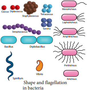
The shape and flagellation of the bacteria varies and is given in Figure 1.8.
Bacteria img 1

Ultrastructure of a Bacterial Cell

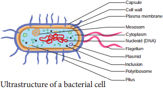
The bacterial cell reveals three layers

  • Capsule/Glycocalyx
  • Cell wall and
  • Cytoplasm (Figure 1.9).

Capsule/Glycocalyx

Some bacteria are surrounded by a gelatinous substance which is composed of polysaccharides or polypeptide or both. A thick layer of glycocalyx bound tightly to the cell wall is called capsule. It protects cell from desiccation and antibiotics. The sticky nature helps them to attach to substrates like plant root surfaces, Human teeth and tissues. It helps to retain the nutrients in bacterial cell.
Bacteria img 2

Cell Wall

The bacterial cell wall is granular and is rigid. It provides protection and gives shape to the cell. The chemical composition of cell wall is rather complex and is made up of peptidoglycan or mucopeptide (N-acetyl glucosamine, N-acetyl muramic acid and peptide chain of 4 or 5 aminoacids). One of the most abundant polypeptide called porin is present and it helps in the diffusion of solutes.

Plasma Membrane

The plasma membrane is made up of lipoprotein. It controls the entry and exit of small molecules and ions. The enzymes involved in the oxidation of metabolites (i.e., the respiratory chain) as well as the photosystems used in photosynthesis are present in the plasma membrane.

Cytoplasm

Cytoplasm is thick and semitransparent. It contains ribosomes and other cell inclusions. Cytoplasmic inclusions like glycogen, poly-β-hydroxybutyrate granules, sulphur granules and gas vesicles are present.

Bacterial Chromosome

The bacterial chromosome is a single circular DNA molecule, tightly coiled and is not enclosed in a membrane as in Eukaryotes. This genetic material is called Nucleoid or Genophore. It is amazing to note that the DNA of E.coli which measures about 1mm long when uncoiled, contains all the genetic information of the organism. The DNA is not bound to histone proteins.

The single chromosome or the DNA molecule is circular and at one point it is attached to the plasma membrane and it is believed that this attachment may help in the separation of two chromosomes after DNA replication.

Plasmid

Plasmids are extra chromosomal double stranded, circular, self-replicating, autonomous elements. The size of a plasmid varies from 1 to 500 kb usually plasmids contribute to about 0.5 to 5.0% of the total DNA of bacteria.

They contain genes for fertility, antibiotic resistant and heavy metals. It also help in the production of bacteriocins and toxins which are not found in bacterial chromosome. The number of plasmids per cell varies. Plasmids are classified into different types based on the function. Some of them are F (Fertility) factor, R (Resistance) plasmids, Col (Colicin) plasmids, Ri (Root inducing) plasmids and Ti (Tumour inducing) plasmids.

Mesosomes

These are localized infoldings of plasma membrane produced into the cell in the form of vesicles, tubules and lamellae. They are clumped and folded together to maximize their surface area and helps in respiration and in binary fission.

Polysomes / Polyribosomes

The ribosomes are the site of protein synthesis. The number of ribosome per cell varies from 10,000 to 15,000. The ribosomes are 70S type and consists of two subunits (50S and 30S). The ribosomes are held together by mRNA and form polyribosomes or polysomes.

Flagella

Certain motile bacteria have numerous thin hair like projections of variable length emerge from the cell wall called flagella. It is 20-30 μm in diameter and 15 μm in length.

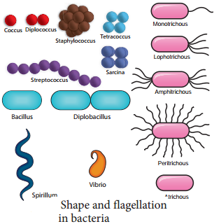
The flagella of Eukaryotic cells contain 9+2 microtubles but each flagellum in bacteria is made up of a single fibril. Flagella are used for locomotion. Based on the number and position of flagella there are different types of bacteria (Figure 1.8)
Bacteria img 3

Fimbriae or Pili

Pili or fimbriae are hair like appendages found on surface of cell wall of gram-negative bacteria (Example: Enterobacterium). The pili are 0.2 to 20 µm long with a diameter of about 0.025µm. In addition to normal pili there are special type of pili which help in conjugation called sex pili are also found.

Gram Staining Procedure

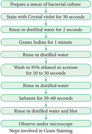
The Gram staining method to differentiate bacteria was developed by Danish Physician Christian Gram in the year 1884. It is a differential staining procedure and it classifies bacteria into two classes – Gram positive and Gram negative.

The steps involved in Gram staining procedure is given in Figure 1.10. The Gram positive bacteria retain crystal violet and appear dark violet whereas Gram negative type loose the crystal violet and when counterstained by safranin appear red under a microscope.
Bacteria img 4

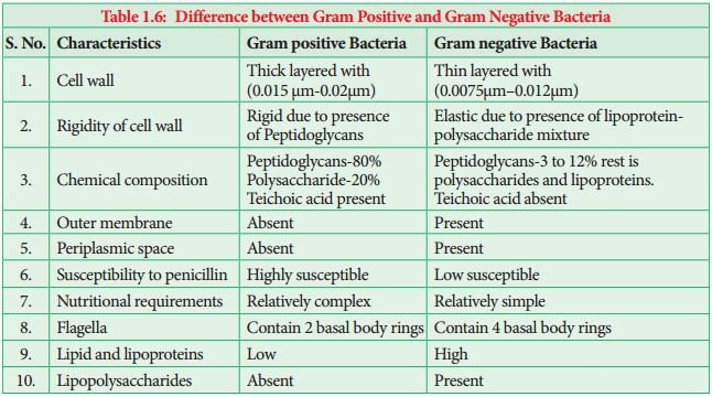
Most of the gram positive cell wall contain considerable amount of teichoic acid and teichuronic acid. In addition, they may contain polysaccharide molecules. The gram negative cell wall contains three components that lie outside the peptidoglycan layer.

  • Lipoprotein
  • Outer membrane
  • Lipopolysaccharide.

Thus the different results in the gram stain are due to differences in the structure and composition of the cell wall. The difference between Gram Positive and Gram negative bacteria is given in Table 1.6.
Bacteria img 5

Life Processes in Bacteria

Respiration

Two types of respiration are found in Bacteria. They are:-

  1. Aerobic Respiration
  2. Anaerobic Respiration.

1. Aerobic Respiration

These bacteria require oxygen as terminal acceptor and will not grow under anaerobic conditions. (i.e. in the absence of (O2) Example: Streptococcus.

Obligate Aerobes:
Some Micrococcus species are obligate aerobes (i.e. they must have oxygen to survive).

2. Anaerobic Respiration

These bacteria do not use oxygen for growth and metabolism but obtain their energy from fermentation reactions. Example: Clostridium.

Facultative Anaerobes

There are bacteria that can grow either using oxygen as a terminal electron acceptor or anaerobically using fermentation reaction to obtain energy. When a facultative anaerobe such as E. coli is present at a site of infection like an abdominal abscess, it can rapidly consume all available O2 and change to anaerobic metabolism producing an anaerobic environment and thus allow the anaerobic bacteria that are present to grow and cause disease. Example: Escherichia coli and Salmonella.

Capnophilic Bacteria

Bacteria which require CO2 for their growth are called as capnophilic bacteria. Example: Campylobacter

Nutrition

On the basis of their mode of nutrition bacteria are classified into two types namely autotrophs and heterotrophs.

I. Autotrophic Bacteria

Bacteria which can synthesise their own food are called autotrophic bacteria. They may be further subdivided as:-

A. Photoautotrophic Bacteria

Bacteria use sunlight as their source of energy to synthesize food. They may be

1. Photolithotrophs

In photolithotrophs the hydrogen donor is an inorganic substance.

a. Green Sulphur Bacteria:

In this type of bacteria the hydrogen donor is H2S and possess pigment called Bacterioviridin. Example: Chlorobium.

b. Purple Sulphur Bacteria:

For bacteria belong to this group the hydrogen donor is thiosulphate, Bacteriochlorophyll is present. Chlorophyll containing chlorosomes are present Example: Chromatium.

2. Photoorganotrophs

They utilize organic acid or alcohol as hydrogen donor. Example: Purple non sulphur bacteria – Rhodospirillum.

B. Chemoautotrophic Bacteria

They do not have photosynthetic pigment hence they cannot use sunlight energy. This type of bacteria obtain energy from organic or inorganic substance.

1. Chemolithotrophs

This type of bacteria oxidize inorganic compound to release energy.

Examples:

  • Sulphur bacteria – Thiobacillus thiooxidans
  • Iron bacteria – Ferrobacillus ferrooxidans
  • Hydrogen bacteria – Hydrogenomonas
  • Nitrifying bacteria – Nitrosomonas and Nitrobacter

2. Chemoorganotrophs

This type of bacteria oxidize organic compounds to release energy.

Examples:

  • Methane bacteria – Methanococcus
  • Acetic acid bacteria – Acetobacter
  • Lactic acid bacteria – Lactobacillus

II. Heterotrophic Bacteria

They are Parasites (Mycobacterium) Saprophytes (Bacillus mycoides) or Symbiotic (Rhizobium in root nodules of leguminous crops).

Reproduction in Bacteria

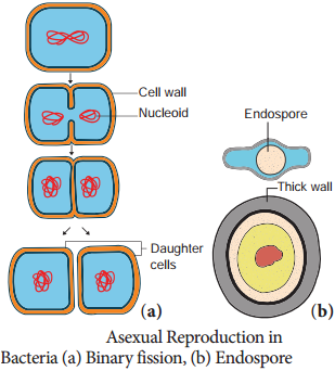
Bacteria reproduces asexually by binary fission, conidia and endospore formation (Figure 1.11). Among these, binary fission is the most common one.

Binary Fission

Under favourable conditions the cell divides into two daughter cells. The nuclear material divides first and it is followed by the formation of a simple median constriction which finally results in the separation of two cells.
Bacteria img 6

Endospores

During unfavourable condition bacteria produce endospores. Endospores are produced in Bacillus megaterium, Bacillus sphaericus and Clostridium tetani. Endospores are thick walled resting spores. During favourable condition, they germinate and form bacteria.

Sexual Reproduction

Typical sexual reproduction involving the formation and fusion of gametes is absent in bacteria. However gene recombination can occur in bacteria by three different methods they are:-

  • Conjugation
  • Transformation
  • Transduction

1. Conjugation

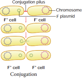
J. Lederberg and Edward L. Tatum demonstrated conjugation in E. coli. in the year 1946. In this method of gene transfer the donor cell gets attached to the recipient cell with the help of pili. The pilus grows in size and forms the conjugation tube.

The plasmid of donor cell which has the F+ (fertility factor) undergoes replication. Only one strand of DNA is transferred to the recipient cell through conjugation tube. The recipient completes the structure of double stranded DNA by synthesizing the strand that complements the strand acquired from the donor (Figure 1.12).
Bacteria img 7

2. Transformation

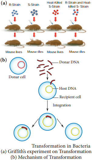
Transfer of DNA from one bacterium to another is called transformation (Figure 1.13). In 1928 the bacteriologist Frederick Griffith demonstrated transformation in Mice using Diplococcus pneumoniae. Two strains of this bacterium are present. One strain produces smooth colonies and are virulent in nature (S-type).

In addition another strain produce rough colonies and are avirulent (R-type). When S-type of cells were injected into the mouse, the mouse died. When R-type of cells were injected, the mouse survived. He injected heat killed S-type cells into the mouse.

The mouse did not die. When the mixture of heat killed S-type cells and R-type cells were injected into the mouse, the mouse died. The avirulent rough strain of Diplococcus had been transformed into S-type cells. The hereditary material of heat killed S-type cells had transformed R-type cell into virulent smooth strains. Thus the phenomenon of changing the character of one strain by transferring the DNA of another strain into the former is called Transformation.
Bacteria img 8

3. Transduction

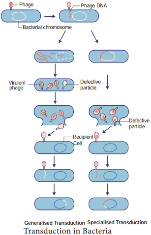
Zinder and Lederberg (1952) discovered Transduction in Salmonella typhimurum. Phage mediated DNA transfer is called Transduction (Figure 1.14).
Bacteria img 9

Transduction is of Two Types

  1. Generalized transduction
  2. Specialized or Restricted transduction

(i) Generalized Transduction

The ability of a bacteriophage to carry genetic material of any region of bacterial DNA is called generalised transduction.

(ii) Specialized or Restricted

Transduction

The ability of the bacteriophage to carry only a specific region of the bacterial DNA is called specialized or restricted transduction.

Economic Importance of Bacteria
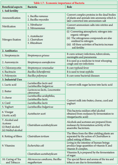
Bacteria are both beneficial and harmful. The beneficial activities of bacteria are given in table 1.7.
Bacteria img 10

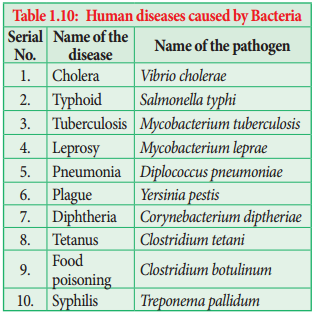
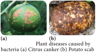
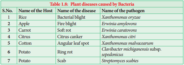
Bacteria are known to cause disease in plants, animals and Human beings. The List is given in Table 1.8, 1.9, 1.10 and Figure 1.15.
Bacteria img 11
Bacteria img 12
Bacteria img 26
Bacteria img 14

Activity 1.3

Collect some root nodules of leguminous crops. Draw diagram. Wash it in tap water and prepare a smear by squeezing the content into a clean slide. Follow Gram staining method and identify the bacteria.

Archaebacteria

Archaebacteria are primitive prokaryotes and are adapted to thrive in extreme environments like hot springs, high salinity, low pH and so on. They are mostly chemoautotrophs.

The unique feature of this group is the presence of lipids like glycerol & isopropyl ethers in their cell membrane. Due to the unique chemical composition the cell membrane show resistance against cell wall antibiotics and lytic agents. Example: Methanobacterium, Halobacterium, Thermoplasma.

Cyanobacteria (Blue Green Algae)

How old are Cyanobacteria? Stromatolites reveals the truth.

Stromatolites are deposits formed when colonies of cyanobacteria bind with calcium carbonate. They have a geological age of 2.7 billion years. Their abundance in the fossil record indicates that cyanobacteria helped in raising the level of free oxygen in the atmosphere.

Cyanobacteria are popularly called as ‘Blue green algae’ or ‘Cyanophyceae’. They are photosynthetic, prokaryotic organisms. According to evolutionary record Cyanobacteria are primitive forms and are found in different habitats. Most of them are fresh water and few are marine (Trichodesmium and Dermacarpa) Trichodesmium erythraeum a cyanobacterium imparts red colour to Red sea.

Species of Nostoc, Anabaena lead an endophytic life in the coralloid root of Cycas, leaves of aquatic fern Azolla by establishing a symbiotic association and fix atmospheric nitrogen. Members like Gloeocapsa, Nostoc, Scytonema are found as phycobionts in lichen thalli.

Salient Features

  1. The members of this group are prokaryotes and lack motile reproductive structures.
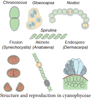
  2. The thallus is unicellular in Chroococcus, Colonial in Gloeocapsa and filamentous trichome in Nostoc.
  3. Gliding movement is noticed in some species (Oscillatoria).
  4. The protoplasm is differentiated into central region called centroplasm and peripheral region bearing chromatophore called chromoplasm.
  5. The photosynthetic pigments include c-phyocyanin and c-phycoerythrin along with myxoxanthin and myxoxanthophyll.
  6. The reserve food material is Cyanophycean starch.
  7. In some forms a large colourless cell is found in the terminal or intercalary position called Heterocysts. They are involved in nitrogen fixation.
  8. They reproduce only through vegetative methods and produce Akinetes (thick wall dormant cell formed from vegetative cell), Hormogonia (a portion of filament get detached and reproduce by cell division), fission and endospores.
  9. The presence of mucilage around the thallus is characteristic feature of this group. Therefore, this group is also called Myxophyceae.
  10. Sexual reproduction is absent.
  11. Microcystis aeruginosa, Anabaena flos-aquae cause water blooms and release toxins and affect the aquatic organism.

Most of them fix atmospheric nitrogen and are used as biofertilizers (Example: Nostoc, Anabaena). Spirulina is rich in protein hence it is used as single cell protein. The thallus organisation and methods of reproduction is given in Figure 1.16.
Bacteria img 15

Mycoplasma or Mollicutes

The Mycoplasma are very small (0.1-0.5µm), pleomorphic gram negative microorganisms. They are first isolated by Nocard and coworkers in the year 1898 from pleural fluid of cattle affected with bovine pleuropneumonia.

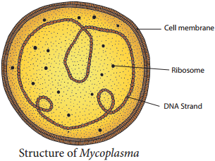
They lack cell wall and appear like “Fried Egg” in culture. The DNA contains low Guanine and Cytosine content than true bacteria. They cause disease in animals and plants. Little leaf of brinjal, witches broom of legumes phyllody of cloves, sandal spike are some plant diseases caused by mycoplasma. Pleuropneumonia is caused by Mycoplasma mycoides. The structure of Mycoplasma is given in Figure 1.17.
Bacteria img 16

Actinomycetes (Actinobacteria)

Actinomycetes are also called ‘Ray fungi’ due to their mycelia like growth. They are anaerobic or facultative anaerobic microorganisms and are Gram positive. They do not produce an aerial mycelium. Their DNA contains high guanine and cytosine content (Example: Streptomyces).

Frankia is a symbiotic actinobacterium which produces root nodules and fixes nitrogen in non – leguminous plants such as Alnus and Casuarina. They produce multicellular sporangium. Actinomyces bovis grows in oral cavities and cause lumpy jaw.

Streptomyces is a mycelial forming Actinobacteria which lives in soil, they impart “earthy odour” to soil after rain which is due to the presence of Geosmin (volatile organic compound). Some important antibiotics namely, Streptomycin, Chloramphenicol, and Tetracycline are produced from this genus.
Classification of Living World

Learninsta presents the core concepts of Biology with high-quality research papers and topical review articles.

Classification of Living World

From the previous chapter we know that the planet earth is endowed with living and non-living things. In our daily life we see several things in and around us. Imagine, you are on a trip to Hill station. You are enjoying the beauty of mountains, dazzling colour of the flowers, and melodious sound of the birds. You may be capturing most of the things you come across in the form of photography.

Now, from this experience can you mention the objects you have come across? Can you record your observations and tabulate them? How will you organize the things? Will you place mountain and flowers together or tall trees and trailing herbs in one category or place it in different category?

If you place it in different category, what made you to place them in different category? So classification is essential and could be done only by understanding and comparing the things based on some characters. In this chapter we shall learn about classification of living world.

Many attempts have made in the past to classify the organisms on earth. Theophrastus, “Father of Botany” used the morphological characters to classify plants into trees, shrubs and herbs. Aristotle classified animals into two groups. i.e., Enaima (with red blood) and Anaima (without red blood).

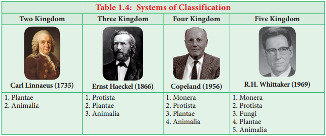
Carl Linnaeus classified living world into two groups namely Plants and Animals based on morphological characters. His classification faced major setback because Prokaryotes and Eukaryotes were grouped together. Similarly fungi, heterotrophic organisms were placed along with the photosynthetic plants.

In course of time, the development of tools compelled taxonomists to look for different areas like cytology, anatomy, embryology, molecular biology, phylogeny etc., for classifying organisms on earth. Thus, new dimensions to classifications were put forth from time to time.

Need of Classification

Classification is essential to achieve following needs:

  • To relate things based on common characteristic features.
  • To define organisms based on the salient features.
  • Helps in knowing the relationship amongst different groups of organisms.
  • It helps in understanding the evolutionary relationship between organisms.

Classification of Living World

A comparison of classification proposed for classification of living world is given in Table 1.4.
Classification of Living World img 1

Five Kingdom Classification

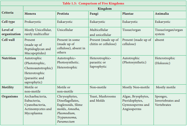
R.H.Whittaker, an American taxonomist proposed five Kingdom classification in the year 1969. The Kingdoms include Monera, Protista, Fungi, Plantae and Animalia (Figure 1.7). The criteria adopted for the classification include cell structure, thallus organization, mode of nutrition, reproduction and phylogenetic relationship. A comparative account of the salient features of each Kingdom is given in Table 1.5
Classification of Living World img 2

Merits

  • The classification is based on the complexity of cell structure and organization of thallus.
  • It is based on the mode of nutrition
  • Separation of fungi from plants
  • It shows the phylogeny of the organisms

Demerits

  • The Kingdom Monera and protista accommodate both autotrophic and heterotrophic organisms, cell wall lacking and cell wall bearing organisms thus making these two groups more heterogeneous.
  • Viruses were not included in the system.

Carl Woese and co-workers in the year 1990 introduced three domains of life viz., Bacteria, Archaea and Eukarya based on the difference in rRNA nucleotide sequence, lipid structure of the cell membrane. A revised six Kingdom classification for living world was proposed by Thomas Cavalier-Smith in the year 1998 and the Kingdom Monera is divided in to Archaebacteria and Eubacteria.

Recently Ruggierio et al., 2015 published a seven Kingdom classification which is a practical extension of Thomas Cavalier’s six Kingdom scheme. According to this classification there are two Super Kingdoms. (Prokaryota and Eukaryota) Prokaryota includes two Kingdoms namely Archaebacteria and Eubacteria. Eukaryota includes the Protozoa, Chromista, Fungi, Plantae and Animalia.

A new Kingdom, the Chromista was erected and it included all algae whose chloroplasts contain chlorophyll a and c, as well as various colourless forms that are closely related to them. Diatoms, Brown algae, Cryptomonads and Oomycetes were placed under this Kingdom.

Activity 1.2

Visit to a pond and record the names of the biotic components of it with the help of your teacher. Tabulate the data and segregate them according to Five Kingdom Classification.