Plant and Animal Cell Structure and its Types

Learninsta presents the core concepts of Biology with high-quality research papers and topical review articles.

Plant and Animal Cell Structure and its Types

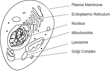
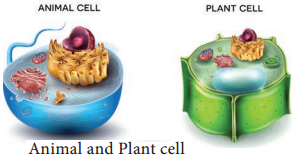
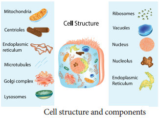
An eukaryotic cell is highly distinct in its organisation. It shows several variations in different organisms. For instance, eukaryotic cells in plants and animals vary greatly (Figure 6.7)
Plant and Animal Cell img 1

Animal Cell

Animal cells are surrounded by cell membrane or plasma membrane. Inside this membrane a gelatinous matrix called protoplasm is seen to contain nucleus and other organelles which include the endoplasmic reticulum, mitochondria, golgi bodies, centrioles, lysosomes, ribosomes and cytoskeleton.
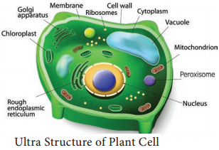
Plant Cell

A typical plant cell has prominent cell wall, a large central vacuole and plastids in addition to other organelles present in animal cell (Figure 6.8).
Plant and Animal Cell img 2

Protoplasm

Protoplasm is the living content of cell that is surrounded by plasma membrane. It is a colourless material that exists throughout the cell together with cytoplasm, nucleus and other organelles. Protoplasm is composed of a mixture of small particles, such as ions, amino acids, monosaccharides, water, macromolecules like nucleic acids, proteins, lipids and polysaccharides.

It appears colourless, jelly like gelatinous, viscous elastic and granular. It appears foamy due to the presence of large number of vacuoles. It responds to the stimuli like heat, electric shock, chemicals and so on.
Plant and Animal Cell img 3

Difference Between Plant and Animal Cells

Plant Cell

Animal Cell

1. Usually they are larger than animal cells1. Usually smaller than plant cells
2. Cell way present in addition to plasma membrane and consists of middle lamellae, primary and secondary walls2. Cell wall absent
3. Plasmodesmata present3. Plasmodesmata absent
4. Chloroplast present4. Chloroplast absent
5. Vacuole large and permanent5. Vacuole small and temporary
6. Tonoplast present around vacuole6. Tonoplast absent
7. Centrioles absent except motile cells of lower plants7. Centrioles present
8. Nucleus present along the periphery of the cell8. Nucleus at the centre of the cell
9. Lysosomes are rare9. Lysosomes present
10. Storage material is starch grains10. Storage material is a glycogen granules

Cell Wall

Cell wall is the outermost protective cover of the cell. It is present in bacteria, fungi and plants whereas it is absent in animal cell. It was first observed by Robert Hooke. It is an actively growing portion. It is made up of different complex material in various organism.

In bacteria it is composed of peptidoglycan, in fungi chitin and fungal cellulose, in algae cellulose, galactans and mannans. In plants it is made up of cellulose, hemicellulose, pectin, lignin, cutin, suberin and silica.

In plant, cell wall shows three distinct regions

  1. Primary Wall
  2. Secondary Wall
  3. Middle Lamellae (Figure 6.10).
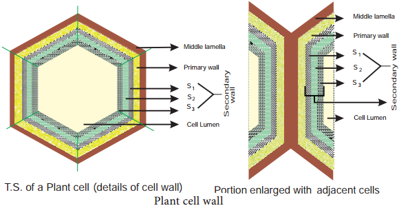
Plant and Animal Cell img 4

1. Primary Wall

It is the first layer inner to middle lamella, primarily consisting of loose network of cellulose microfibrils in a gel matrix. It is thin, elastic and extensible.In most plants the microfibrils are made up of cellulose oriented differently based on shape and thickness of the wall. The matrix of the primary wall is composed of hemicellulose, pectin, glycoprotein and water. Hemicellulose binds the microfibrils with matrix and glycoproteins control the orientation of microfibrils while pectin serves as filling material of the matrix. Cells such as parenchyma and meristems have only primary wall.

b. Secondary Wall

Secondary wall is laid during maturation of the cell. It plays a key role in determining the shape of a cell. It is thick,inelastic and is made up of cellulose and lignin. The secondary wall is divided into three sublayers termed as S1, S2 and S3 where the cellulose microfibrils are compactly arranged with different orientation forming a laminated structure and the cell wall strength is increased.

c. Middle Lamellae

It is the outermost layer made up of calcium and magnesium pectate, deposited at the time of cytokinesis. It is a thin amorphous layer which cements two adjacent cells. It is optically inactive (isotropic).

Plasmodesmata and Pits

Plasmodesmata act as a channel between the protoplasm of adjacent cells through which many substances pass through. Moreover, at few regions, the secondary wall layer is laid unevenly whereas the primary wall and middle lamellae are laid continuously such regions are called pits. The Pits of adjacent cells are opposite to each other. Each pit has a pit chamber and a pit membrane. The pit membrane has many minute pores and thus they are permeable. The pits are of two types namely simple and bordered pit.

Functions of Cell Wall

The cell wall plays a vital role in holding several important functions given below

  1. Offers definite shape and rigidity to the cell.
  2. Serves as barrier for several molecules to enter the cells.
  3. Provides protection to the internal protoplasm against mechanical injury.
  4. Prevents the bursting of cells by maintaining the osmotic pressure.
  5. Plays a major role by acting as a mechanism of defense for the cells.
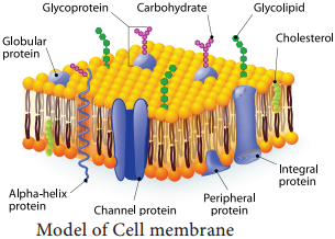
Cell Membrane

The cell membrane is also called cell surface (or) plasma membrane. It is a thin structure which holds the cytoplasmic content called ‘cytosol’. It is extremely thin (less than 10nm).

Fluid Mosaic Model

Jonathan Singer and Garth Nicolson (1972) proposed fluid mosaic model. It is made up of lipids and proteins together with a little amount of carbohydrate. The lipid membrane is made up of phospholipid. The phospholipid molecule has a hydrophobic tail and hydrophilic head.

The hydrophobic tail repels water and hydrophilic head attracts water. The proteins of the membrane are globular proteins which are found intermingled between the lipid bilayer most of which are projecting beyond the lipid bilayer.

These proteins are called as integral proteins. Few are superficially attached on either surface of the lipid bilayer which are called as peripheral proteins. The proteins are involved in transport of molecules across the membranes and also act as enzymes, receptors (or) antigens.

Carbohydrate molecules of cell membrane are short chain polysaccharides. These are either bound with ‘glycoproteins’ or ‘glycolipids’ and form a ‘glyocalyx’ (Figure 6.11). The movement of membrane lipids from one side of the membrane to the other side by vertical movement is called flip flopping or flip flop movement.

This movement takes place more slowly than lateral diffusion of lipid molecule. The Phospholipids can have flip flop movement because they have smaller polar regions, whereas the proteins cannot flip flop because the polar region is extensive.
Plant and Animal Cell img 5

Function of Cell Membrane

The functions of the cell membrane is enormous which includes cell signalling, transporting nutrients and water, preventing unwanted substances entering into the cell, and so on.

Cytoplasm

Cytoplasm is the main arena of various activities of a cell. It is the semifluid gelatinous substance that fills the cell. It is made up of eighty percent water and is usually clear and colourless. The cytoplasm is sometimes described as non nuclear content of protoplasm.

The cytoplasm serves as a molecular soup where all the cellular organelles are suspended and bound together by a lipid bilayer plasma membrane. It constitutes dissolved nutrients, numerous salts and acids to dissolve waste products.

It is a very good conductor of electricity. It gives support and protection to the cell organelles. It helps movement of the cellular materials around the cell through a process called cytoplasmic streaming. Further, most cellular activities such as many metabolic pathways including glycolysis and cell division occur in cytoplasm.

Types of Cells and its Importance

Learninsta presents the core concepts of Biology with high-quality research papers and topical review articles.

Types of Cells and its Importance

On the basis of the cellular organization and the nuclear characteristics, the cell can be classified into:-

  • Prokaryotes
  • Mesokaryotes and
  • Eukaryotes

Prokaryotes

Those organisms with primitive nucleus are called as prokaryotes (pro – primitive; karyon – nucleus). The DNA lies in the ‘nucleoid’ which is not bound by the nuclear membrane and therefore it is not a true nucleus and is also a primitive type of nuclear material. The DNA is without histone proteins. Example: Bacteria, blue green algae, Mycoplasma, Rickettsiae and Spirochaetae.

Mesokaryotes

In the year 1966, scientist Dodge and his coworkers proposed another kind of organisms called mesokaryotes. These organisms which shares some of the characters of both prokaryotes and eukaryotes. In other words these are organisms intermediate between pro and eukaryotes.

These contains well organized nucleus with nuclear membrane and the DNA is organized into chromosomes but without histone protein components divides through amitosis similar with prokaryotes. Certain Protozoa like Noctiluca, some phytoplanktons like Gymnodinium, Peridinium and Dinoflagellates are representatives of mesokaryotes.

Eukaryotes

Those organisms which have true nucleus are called Eukaryotes (Eu – True; karyon – nucleus). The DNA is associated with histones forming the chromosomes. Membrane bound organelles are present. Few organelles may have risen by endosymbiosis which is a cell living inside another cell. The Organelles like mitochondria and chloroplast well support this theory.

Origin of Eukaryotic cell:

Endosymbiont Theory:
Two eukaryotic organelles believed to be the descendants of the endosymbiotic prokaryotes. The ancestors of the eukaryotic cell engulfed a bacterium and the bacteria continued to function inside the host cell.

Comparison Between Types of Cellular Organisation
Types of Cells img 1

Cell Theory Various Types and its Shapes

Learninsta presents the core concepts of Biology with high-quality research papers and topical review articles.

Cell Theory Various Types and its Shapes

In 1833, German botanist Matthias Schleiden and German zoologist Theodor Schwann proposed that all plants and animals are composed of cells and that cells were the basic building blocks of life.

These observations led to the formulation of modern cell theory.

  • All organisms are made up of cells.
  • New cells are formed by the division of pre-existing cells.
  • Cells contains genetic material, which is passed on from parents to daughter cells.
  • All metabolic reactions take place inside the cells.

Exception to Cell Theory

Viruses are puzzle in biology. Viruses, viroids and prions are the exception to cell theory. They lack protoplasm, the essential part of the cell and exists as obligate parasites which are sub-cellular in nature.

Protoplasm Theory

Corti first observed protoplasm. Felix Dujardin (1835) observed a living juice in animal cell and called it “Sarcode”. Purkinje (1839) coined the term protoplasm for sap inside a plant cell. Hugo Van Mohl (1846) indicated importance of protoplasm.

Max Schultze (1861) established similarity between Protoplasm and Sarcode and proposed a theory which later on called “Protoplasm Theory” by O. Hertwig (1892). Huxley (1868) proposed Protoplasm as a “physical basis of life”.

Protoplasm as a Colloidal System

Protoplasm is a complex colloidal system which was suggested by Fisher in 1894 and Hardy in 1899. It is primarily made of water and various other solutes of biological importance such as glucose, fatty acids, amino acids, minerals, vitamins, hormones and enzymes. These solutes may be homogeneous (soluble in water) or heterogeneous mass (insoluble in water) which forms the basis for its colloidal nature.

Physical Properties of Protoplasm

The protoplasm exists either in semisolid (jelly-like) state called ‘gel᾿ due to suspended particles and various chemical bonds or may be liquid state called ‘sol᾿. The colloidal protoplasm which is in gel form can change into sol form by solation and the sol can change into gel by gelation. These gel-sol conditions of colloidal system are prime basis for mechanical behaviour of cytoplasm.

1. Protoplasm is translucent, odourless and polyphasic fluid.

2. It is a crystal colloid solution which is a mixture of chemical substances forming crystalloid i.e. true solution (sugars, salts, acids, bases) and others forming colloidal solution (Proteins and lipids).

3. It is the most important property of the protoplasm by which it exhibits three main phenomena namely Brownian movement, amoeboid movement and cytoplasmic streaming or cyclosis. Viscosity of protoplasm is 2-20 centipoises. The Refractive index of the protoplasm is 1.4.

4. The pH of the protoplasm is around 6.8, contain 90% water (10% in dormant seeds)

5. Approximately 34 elements are present in protoplasm but only 13 elements are main or universal elements i.e. C, H, O, N, Cl, Ca, P, Na, K, S, Mg, I and Fe. Carbon, Hydrogen, Oxygen and Nitrogen form the 96% of protoplasm.

6. Protoplasm is neither a good nor a bad conductor of electricity. It forms a delimiting membrane in contact with water and solidifies when heated.

7. Cohesiveness:
Particles or molecules of protoplasm are adhered with each other by forces, such as Vander Waal’s bonds, that hold long chains of molecules together. This property varies with the strength of these forces.

8. Contractility:
The contractility of protoplasm is important for the absorption and removal of water especially for stomatal operations.

9. Surface tension:
The proteins and lipids of the protoplasm have less surface tension, hence they are found at the surface forming the membrane. On the other hand the chemical substances (NaCl) have high surface tension, so they occur in deeper parts of the protoplasm.

Cell Sizes and Shapes

Cell greatly vary in size, shape and also in function. Group of cells with similar structures are called tissue they integrate together to perform similar function, group of tissue join together to perform similar function called organ, group of organs with related function called organ system, organ system coordinating together to form an organism.

Shape

The shape of cell vary greatly from organism to organism and within the organism itself. In bacteria, cell shape vary from round (cocci) to rectangular (rod). In virus, shape of the envelope varies from round to hexagonal or ‘T’ shaped. In fungi, globular to elongated cylindrical cells and the spores of fungi vary greatly in shape. In plants and animals cells vary in shape according to cell types such as parenchyma, mesophyll, palisade, tracheid, fiber, epithelium and others (Figure 6.6).
Cell Theory img 1

Microscopy – Bright Field Microscope and Electron Microscope

Learninsta presents the core concepts of Biology with high-quality research papers and topical review articles.

Microscopy – Bright Field Microscope and Electron Microscope

Microscope is an inevitable instrument in studying the cell and subcellular structures. It offers scope in studying microscopic organisms therefore it is named as microscope (mikros – small; skipein – to see) in Greek terminology. Compound microscope was invented by Z. Jansen.

Microscope basically works on the lens system and its properties of light and lens such as reflection, magnification and numerical aperture. The common light microscope which has many lenses are called as compound microscope. The microscope transmits visible light from sources to eye or camera through sample.

Bright Field Microscope

Bright field microscope is the routinely used microscope in studying various aspects of cells. It allows light to pass directly through specimen and shows a well distinguished image from different portions of the specimen. The contrast can be increased by staining the specimen with reagent that reacts with cells and tissue components of the object.

The light rays are focused by condenser on to the specimen on a microslide placed upon the adjustable platform called stage. Light comes from the Compact Flourescent Lamp (CFL) or Light Emitting Diode (LED). Then it passes through two lens systems namely objective lens (closer to the object) and the eye piece (closer to eye).

There are four objective lenses (5X, 10X, 45X and 100X) which can be rotated and fixed at certain point to get required magnification. It works on the principle of numerical aperture value and its own resolving power.

The first magnification of the microscope is done by the objective lens which is called primary magnification and it is real, inverted image. The second magnification of the microscope is obtained through eye piece lens called as secondary magnification and it is virtual and inverted image (Figure 6.2 a, b and c).
Microscopy img 1

Electron Microscope

Electron Microscope was first introduced by Ernest Ruska (1931) and developed by G Binning and H Roher (1981). It is used to analyse the fine details of cell and organelles called ultrastructure. It uses beam of accelerated electrons as source of illumination and therefore the resolving power is 1,00,000 times greater than that of light microscope.

The specimen to be viewed under electron microscope is dehydrated and impregnated with electron opaque chemicals like gold or palladium. This is essential for withstanding electrons and also for contrast of the image.

There are two kinds of electron microscopes namely:

  1. Transmission Electron Microscope (TEM)
  2. Scanning Electron Microscope (SEM)

1. Transmission Electron Microscope:

This is the most commonly used electron microscope which provides two dimensional image. The components of the microscope are as follows:

  • Electron generating system
  • Electron condensor
  • Specimen objective
  • Tube lens
  • Projector

A beam of electron passes through the specimen to form an image on fluorescent screen. The magnification is 1-3 lakhs times and resolving power is 2-10 Å. It is used for studying detailed structrue of viruses, mycoplasma, cellular organelles, etc (Figure 6.3 a and b).
Microscopy img 2

2. Scanning Electron Microscope:

This is used to obtain three dimensional image and has a lower resolving power than TEM. In this, electrons are focused by means of lenses into a very fine point.

The interaction of electrons with the specimen results in the release of different forms of radiation (such as auger electrons, secondary electrons, back scattered electrons) from the surface of the specimen. These radiations are then captured by an appropriate detector, amplified and then imaged on fluorescent screen. The magnification is 2,00,000 times and resolution is 5-20 nm (Figure 6.4 a and b).
Microscopy img 3

Discovery of a Cell Definition and its Structure

Learninsta presents the core concepts of Biology with high-quality research papers and topical review articles.

Discovery of a Cell Definition and its Structure

Aristotle (384 – 322BC), was the one who first recognised that animals and plants consists of organised structural units but unable to explain what it was. In 1660’s Robert Hooke observed something which looks like ‘honeycomb with a great numbers of little boxes’ which was later called as ‘cell’ from the cork tissue. In 1665, He compiled his work as Micrographia.

Later, Anton Van Leeuwenhoek observed unicellular particles which he named as ‘animalcules’. Robert Brown (1831 – 39) described the spherical body in plant cell as nucleus. H. J. Dutrochet (1824), a French scientist, was the first to give an idea on cell theory. Later, Matthias Schleiden (German Botanist) and Theodor Schwann (German Zoologist) (1833) outlined the basic features of the cell theory.

Rudolf Virchow (1858) explained the cell theory by adding a feature stating that all living cells arise from pre-existing living cells by ‘cell division’. Cells were first discovered by Robert Hooke in 1665. He observed the cells in a cork slice with the help of a primitive microscope. The cell theory, that all the plants and animals are composed of cells and that the cell is the basic unit of life, was presented by two biologists, Schleiden (1838) and Schwann (1839).

A cell is the smallest and most basic form of life. Robert Hooke, one of the first scientists to use a light microscope, discovered the cell in 1665. In all life forms, including bacteria, plants, animals, and humans, the cell was defined as the most basic structural and functional unit.

The cell (from Latin cella, meaning “small room”) is the basic structural, functional, and biological unit of all known organisms. Cells are the smallest units of life, and hence are often referred to as the “building blocks of life”. The study of cells is called cell biology, cellular biology, or cytology.

The levels, from smallest to largest, are: molecule, cell, tissue, organ, organ system, organism, population, community, ecosystem, biosphere.

A cell consists of a nucleus and cytoplasm and is contained within the cell membrane, which regulates what passes in and out. The nucleus contains chromosomes, which are the cell’s genetic material, and a nucleolus, which produces ribosomes.

The cell is the smallest structural and functional unit of living organisms, which can exist on its own. Therefore, it is sometimes called the building block of life.

The cell is the structural and functional unit of all known living organisms. So, the entire functioning of the living organisms begins from the basic unit called cell. Hence, cell is called the fundamental unit of life.

They provide structure for the body, take in nutrients from food, convert those nutrients into energy, and carry out specialized functions. Cells also contain the body’s hereditary material and can make copies of themselves.

The largest cells is an egg cell of ostrich. The longest cell is the nerve cell. The largest cell in the human body is female ovum. Smallest cell in the human body is male gametes, that is, sperm.
Discovery of a Cell img 1