Cell Cycle Definition and its Types

Learninsta presents the core concepts of Biology with high-quality research papers and topical review articles.

Cell Cycle Definition and its Types

Definition:
A series of events leading to the formation of new cell is known as cell cycle. The series of events include several phases.

History of a Cell

Year

Scientist  

Events

1665Robert HookeCoined word “Cell”
1670-74Anthony van LaeeuwenhoekFirst living cells observed in microscope – Structure of bacteria
1831-33Robert BrownPresence of nucleus in cells of orchid roots
1839Jan Evangelista Purkyne (J.E. Purkinje)Coined “Protoplasm”
1838-39Schleiden & SchwannCell theory
1858Rudolph Ludwig Carl VirchowCell theory ‘omnis cellula e cellula’
1873Anton SchneiderDescribed chromosomes (Nuclear filaments) for the first time
1882Walther FlemmingCoined the word mitosis; chromosome behaviour
1883Edouard Van BenedenCell division in round worm
1888Theodor BoveriCentrosome; Chromosome Theory

Duration of Cell Cycle

Different kinds of cells have varied duration for cell cycle phases. Eukaryotic cell divides every 24 hours. The cell cycle is divided into mitosis and interphase. In a cell cycle 95% is spent for interphase whereas the mitosis and cytokinesis last only for an hour.

Cell Cycle of a Proliferating Human Cell

Interphase
The different phases of cell cycle are as follows (Figure 7.1).

Phase

Time Duration (in hrs)

G111
S8
G24
M1

Longest part of the cell cycle, but it is of extremely variable length. At first glance the nucleus appears to be resting but this is not the case at all. The chromosomes previously visible as thread like structure, have dispersed. Now they are actively involved in protein synthesis, at least for most of the interphase.

G1 Phase

The first gap phase – 2C amount of DNA in cells of G1. Cells become metabolically active and grows by producing proteins, lipids, carbohydrates and cell organelles including mitochondria and endoplasmic reticulum. Many checkpoints control the cell cycle.

The check point are also called as the restriction point. First check point at the end of G1, determines a cells fate whether it will continue in the cell cycle and divide or enter a stage called G0 a quiescent stage, probably as specified cell or die. Cells are arrested in G1 due to:

  • Nutrient deprivation
  • Lack of growth factors or density dependant inhibition
  • Undergo metabolic changes and enter into G0 state.

Biochemicals inside cell activates the cell division. The proteins called kinases and cyclins activate genes and their proteins to perform cell division. Cyclins act as major checkpoint which operates in G1 to determine whether or not a cell divides.

G0 Phase

Some cells exit G1 and enters a quiescent stage called G0, where the cell remains metabolically active without proliferation. Cells can exist for long periods in G0 phase. In G0 cells cease growth with reduced rate of RNA and protein synthesis.

The G0 phase is not permanent. Mature neuron and skeletal muscle cell remain permanently in G0. Many
cells in animals remains in G0 unless called on to proliferate by appropriate growth factors or other extracellular signals. G0 cells are not dormant. S phase – Synthesis phase – cells with intermediate amounts of DNA.

Growth of the cell continues as replication of DNA occur, protein molecules called histones are synthesised and attach to the DNA. The centrioles duplicate in the cytoplasm. DNA content increases from 2C to 4C. G2 – The second Gap phase – 4C amount of DNA in cells of G2 and mitosis

Cell growth continues by protein and cell organelle synthesis, mitochondria and chloroplasts divide. DNA content remains as 4C. Tubulin is synthesised and microtubules are formed. Microtubles organise to form spindle fire. The spindle begins to form and nuclear division follows.

One of the proteins synthesized only in the G2 period is known as Maturation Promoting Factor (MPF). It brings about condensation of interphase chromosomes into the mitotic form. DNA damage checkpoints operates in G1S and G2 phases of the cell cycle.

Nuclear Divisions – Definition and its Difference

Learninsta presents the core concepts of Biology with high-quality research papers and topical review articles.

Nuclear Divisions – Definition and its Difference

There are two types of nuclear division, as mitosis and meiosis. In mitosis, the daughter cells formed will have the same number of chromosomes as the parent cell, typically diploid (2n) state. Mitosis is the nuclear division that occurs when cells grow or when cells need to be replaced and when organism reproduces asexually.

In meiosis, the daughter cells contain half the number of chromosomes of the parent cell and is known as haploid state (n). Whichever division takes place, it is normally followed by division of the cytoplasm to form separate cells, called as cytokinesis.

The process by which a nucleus divides, resulting in the segregation of the genome to opposite poles of a dividing cell. Supplement, Nuclear divisions occur in both mitosis and meiosis. In mitosis, the result is the division of duplicated copies of genome into two.

There are two kinds of nuclear division-mitosis and meiosis. Mitosis divides the nucleus so that both daughter cells are genetically identical. In contrast, meiosis is a reduction division, producing daughter cells that contain half the genetic information of the parent cell.

Mitosis is a process of nuclear division in eukaryotic cells that occurs when a parent cell divides to produce two identical daughter cells. Mitosis is conventionally divided into five stages known as prophase, prometaphase, metaphase, anaphase, and telophase.

Mitosis is a single nuclear division that results in two nuclei, usually partitioned into two new cells. The nuclei resulting from a mitotic division are genetically identical to the original. They have the same number of sets of chromosomes: one in the case of haploid cells, and two in the case of diploid cells. Mitosis is a single nuclear division that results in two nuclei that are usually partitioned into two new daughter cells.

The process by which a nucleus divides, resulting in the segregation of the genome to opposite poles of a dividing cell. Nuclear divisions occur in both mitosis and meiosis. In mitosis, the result is the division of duplicated copies of genome into two.

Cytokinesis is the physical process of cell division, which divides the cytoplasm of a parental cell into two daughter cells. It occurs concurrently with two types of nuclear division called mitosis and meiosis, which occur in animal cells.

Meiosis I, the first meiotic division, begins with prophase I. During prophase I, the complex of DNA and protein known as chromatin condenses to form chromosomes. The pairs of replicated chromosomes are known as sister chromatids, and they remain joined at a central point called the centromere.

Under the microscope, you will now see the chromosomes lined up in the middle of the cell. You will probably also see thin-stranded structures that appear to radiate outward from the chromosomes to the outer poles of the cell.

Nuclear division occures twice during meiosis as four haploid gametes are produced; each of which are genetically different from each other. In both processes the nuclear envelope is fragmented and completley broken down into small vesicles during prophase, to allow the chromosomes to segregate. Cell division occurs during phase, which consists of nuclear division (mitosis) followed by cytoplasmic division (cytokinesis).

They are also genetically identical to the parental cell. Mitosis has five different stages: interphase, prophase, metaphase, anaphase and telophase. The process of cell division is only complete after cytokinesis, which takes place during anaphase and telophase.
Nuclear Divisions img 1

Flagella – Definition Structure and its Types

Learninsta presents the core concepts of Biology with high-quality research papers and topical review articles.

Flagella – Definition Structure and its Types

Prokaryotic Flagellum

Bacterial flagella are helical appendages helps in motility. They are much thinner than flagella or cilia of eukaryotes. The filament contains a protein called flagellin. The structure consists of a basal body associated with cytoplasmic membrane and cell wall with short hook and helical filament. Bacteria rotates their helical flagella and propels rings present in the basal body which are involved in the rotary motor that spins the flagellum.

Structure of Flagella in Bacteria

The gram positive bacteria contain only two basal rings. S-ring is attached to the inside of peptidoglycan and M-ring is attached to the cell membrane. In Gram negative bacteria two pairs of rings proximal and distal ring are connected by a central rod.

They are L-Lipopolysaccharide ring, P-Peptidoglycan ring, S-Super membrane ring and M-membrane ring. The outer pair L and P rings is attached to cell wall and the inner pair S and M rings attached to cell membrane (Figure 6.27).
Flagella img 1

Mechanism of Flagellar Movement – Proton Motive Force

In flagellar rotation only proton movements are involved and not ATP. Protons flowing back into the cell through the basal body rings of each flagellum drives it to rotate. These rings constitute the rotary motor.The proton motive force (The force derived from the electrical potential and the hydrogen ion gradient across the cytoplasmic membrane) drives the flagellar motor.

For the rotation of flagellum the energy is derived from proton gradient across the plasma membrane generated by oxidative phosphorylation. In bacteria flagellar motor is located in the plasma membrane where the oxidative phosphorylation takes place. Therefore, plasma membrane is a site of generation of proton motive force.

Eukaryotic Flagellum – Cell Motility Structure

Eukaryotic Flagella are enclosed by unit membrane and it arises from a basal body. Flagella is composed of outer nine pairs of microtubules with two microtubules in its centre (9+2 arrangement). Flagella are microtubule projection of the plasma membrane. Flagellum is longer than cilium (as long as 200µm). The structure of flagellum has an axoneme made up microtubules and protein tubulin (Figure 6.28)
Flagella img 2

Movement

Outer microtubule doublet is associated with axonemal dynein which generates force for movement. The movement is ATP driven. The interaction between tubulin and dynein is the mechanism for the contraction of cilia and flagella. Dynein molecules uses energy from ATP to shift the adjacent microtubules. This movement bends the cilium or flagellum.

Cilia

Cilia (plural) are short cellular, numerous microtubule bound projections of plasma membrane. Cilium (singular) is membrane bound structure made up of basal body, rootlets, basal plate and shaft.

The shaft or axoneme consists of nine pairs of microtubule doublets, arranged in a circle along the periphery with a two central tubules, (9+2) arrangement of microtubules is present. Microtubules are made up of tubulin. The motor protein dynein connects the outer microtubule pair and links them to the central pair. Nexin links the peripheral doublets of microtubules (Figure 6.29).
Flagella img 3

Nucleus Definition and Various Types of Functions

Learninsta presents the core concepts of Biology with high-quality research papers and topical review articles.

Nucleus Definition and Various Types of Functions

Nucleus is an important unit of cell which controls all activities of the cell. Nucleus holds the hereditary information. It is the largest among all cell organelles. It may be spherical, cuboidal, ellipsoidal or discoidal. It is surrounded by a double membrane structure called nuclear envelope, which has the inner and outer membrane.

The inner membrane is smooth without ribosomes and the outer membrane is rough by the presence of ribosomes and it continues with irregular and infrequent intervals with the endoplasmic reticulum.

The membrane is perforated by pores known as nuclear pores which allows materials such as mRNA, ribosomal units, proteins and other macromolecules to pass in and out of the nucleus. The pores enclosed by circular structures called annuli. The pore and annuli form the pore complex. The space between two membranes is called perinuclear space.

Nuclear space is filled with nucleoplasm, a gelatinous matrix has uncondensed chromatin network and a conspicuous nucleolius. The Chromatin network is an uncoiled, indistinct and remain thread like during the interphase. It has little amount of RNA and DNA bound to histone proteins in eukaryotic cells (Figure 6.22).
Nucleus img 1

During cell division chromatin is condensed into an organized form called chromosome. The portion an eukaryotic chromosome which is transcribed into mRNA contains active genes that are nottightly condensed during interphase is called Euchromatin.

The portion of an eukaryotic chromosome that is not transcribed into mRNA which remains condensed during interphase and stains intensely is called Heterochromatin. Nucleolus is a small, dense, spherical structure either present singly or in multiples inside the nucleus and it’s not membrane bound. Nucleoli possess genes for rRNA and tRNA.

Functions of the Nucleus

  • Controlling all cellular activities
  • Storing the genetic or hereditary information.
  • Coding the information from DNA for the production of enzymes and proteins.
  • DNA duplication and transcription takes place in the nucleus.
  • In nucleolus ribosomal biogenesis takes place.

Chromosomes

Strasburger (1875) first reported its present in eukaryotic cell and the term ‘chromosome’ was introduced byWaldeyerin 1888. Bridges (1916) first proved that chromosomes are the physical carriers of genes. It is made up of DNA and associated proteins.

Structure of Chromosome

The chromosomes are composed of thread like strands called chromatin which is made up of DNA, protein and RNA. Each chromosome consists of two symmetrical structures called chromatids. During cell division the chromatids forms a well organized chromosomes with definite size and shape.

They are identical and are called sister chromatids. A typical chromosome has narrow zones called constrictions. There are two types of constrictions, namely primary constriction and secondary constriction. The primary constriction is made up of centromere and kinetochore.

Both the chromatids are united at centromere, whose number varies. The monocentric chromosome has one centromere and the polycentric chromosome has many centromeres. Centromere contains a complex system of protein fibres called kinetochore. Kinetochore is the region of chromosome which is attached to the spindle fibre during mitosis.

Besides primary there are few secondary constrictions, are present. Nucleoli develop from these secondary constrictions are called nucleolar organizers. Secondary constrictions contain the genes for ribosomal RNA which induce the formation of nucleoli and are called nucleolar organizer regions (Figure 6.23).
Nucleus img 2

A satellite or SAT Chromosome is a short chromosomal segment or rounded body separated from main chromosome by a relatively elongated secondary constriction. It is a morphological entity in certain chromosomes.

Telomere is the terminal part of chromosome. It offers stability to the chromosome. DNA of the telomere has specific sequence of nucleotides. Telomere in all eukaryotes are composed of many repeats of short DNA sequences (5’TTAGGG3’ sequence in Neurospora crassa and human beings).

Maintenance of telomeres appears to be an important factor in determining the life span and reproductive capacity of cells, so studies of telomeres and telomerase have the promise of providing new insights into conditions such as ageing and cancer. Telomeres prevent the fusion of chromosomal ends with one another.

Types of Chromosomes

Based on the position of centromere, chromosomes are called telocentric (terminal centromere), acrocentric (terminal centromere capped by telomere), sub metacentric (centromere subterminal) and metacentric (centromere median). The eukaryotic chromosome may be rod shaped (telocentric and acrocentric), L-shaped (sub-metacentric) and V-shaped (metacentric) (Figure 6.24).
Nucleus img 3

Based on the functions of chromosome it can be divided into autosomes and sex chromosomes. Autosomes are present in all cells controlling somatic characteristics of an organism. In human diploid cell, 44 chromosomes are autosomes whereas two are sex chromosomes. Sex chromosomes are involved in the determination of sex.

Special Types of Chromosomes

These chromosomes are larger in size and are called giant chromosomes in certain plants and they are found in the suspensors of the embryo. The polytene chromosome and lamp brush chromosome occur in animals and are also called as giant chromosomes.

Polytene chromosomes observed in the salivary glands of Drosophila (fruit fly) by E.G. Balbiani in 1881. In larvae of many flies, midges (Dipthera) and some insects the interphase chromosomes duplicates and reduplicates without nuclear division.

A single chromosome which is present in multiple copies form a structure called polytene chromosome which can be seen in light microscope. They are genetically active. There is a distinct alternating dark bands and light inter-bands. About 95% of DNA are present in bands and 5% in inter-bands.

The polytene chromosome has extremely large puff called Balbiani rings which is seen in Chironomous larvae. It is also known as chromosomal puff. Puffing of bands are the sites of intense RNA synthesis. As this chromosome occurs in the salivary gland it is known as salivary gland chromosomes. Gene expression, transcription of genes and RNA synthesis occurs in the bands along the polytene chromosomes.
Nucleus img 4

Lampbrush chromosomes occur at the diplotene stage of first meiotic prophase in oocytes of an animal Salamandar and in giant nucleus of the unicellular alga Acetabularia. It was first observed by Flemming in 1882. The highly condensed chromosome forms the chromosomal axis, from which lateral loops of DNA extend as a result of intense RNA synthesis.
Nucleus img 5

Cell Organelles Definition, Functions and Various Types of Organelles

Learninsta presents the core concepts of Biology with high-quality research papers and topical review articles.

Cell Organelles Definition, Functions and Various Types of Organelles

Endomembrane System

System of membranes in a eukaryotic cell, comprises the plasma membrane, nuclear membrane, endoplasmic reticulum, golgi apparatus, lysosomes and vacuolar membranes (tonoplast). Endomembranes are made up of phospholipids with embedded proteins that are similar to cell membrane which occur within the cytoplasm. The endomembrane system is evolved from the inward growth of cell membrane in the ancestors of the first eukaryotes (Figure 6.12).
Cell Organelles img 1

Endoplasmic Reticulum

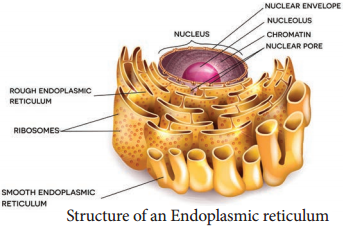
The largest of the internal membranes is called the endoplasmic reticulum (ER). The name endoplasmic reticulum was given by K.R. Porter (1948). It consists of double membrane. Morphologically the structure of endoplasmic reticulum consists of the following:

  1. Cisternae are long, broad, flat, sac like structures arranged in parallel bundles or stacks to form lamella. The space between membranes of cisternae is filled with fluid.
  2. Vesicles are oval membrane bound vacuolar structure.
  3. Tubules are irregular in shape, branched, smooth walled, enclose a space.

Endoplasmic reticulum is associated with nuclear membrane and cell surface membrane. It forms a network in cytoplasm and gives mechanical support to the cell. Its chemical environment enables protein folding and undergo modification necessary for their function. Misfolded proteins are pulled out and are degraded in endoplasmic reticulum.

When ribosomes are present in the outer surface of the membrane it is called as rough endoplasmic reticulum(RER), when the ribosomes are absent in the endoplasmic reticulum it is called as smooth Endoplasmic reticulum(SER).

Rough endoplasmic reticulum is involved in protein synthesis and smooth endoplasmic reticulum are the sites of lipid synthesis. The smooth endoplasmic reticulum contains enzymes that detoxify lipid soluble drugs, certain chemicals and other harmful compounds.

Golgi Body (Dictyosomes)

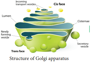
In 1898, Camillo Golgi visualized a netlike reticulum of fibrils near the nucleus, were named as Golgi bodies. In plant cells they are found as smaller vesicles termed as dictyosomes. Golgi apparatus is a stack of flat membrane enclosed sacs.

It consist of cisternae, tubules, vesicles and golgi vacuoles. In plants, the cisternae are 10-20 in number placed in piles separated from each other by a thin layer of inter cisternal cytoplasm often flat or curved.

Peripheral edge of cisternae forms a network of tubules and vesicles. Tubules interconnect cisternae and are 30 – 50nm in dimension. Vesicles are large round or concave sac. They are pinched off from the tubules. They are smooth/secretary or coated type.

Golgi vacuoles are large spherical structures filled with granular or amorphous substance, some function like lysosomes. Golgi apparatus compartmentalises a series of steps leading to the production of functional protein.

Small pieces of rough endoplasmic reticulum are pinched off at the ends to form small vesicles. A number of these vesicles then join up and fuse together to make a Golgi body. Golgi complex plays a major role in post translational modification of proteins and glycosylation of lipids (Figure 6.13 and 6.14).
Cell Organelles img 2
Cell Organelles img 3

Functions:

  • Production of glycoproteins and glycolipids
  • Transporting and storing of lipids.
  • Formation of lysosomes.
  • Production of digestive enzymes.
  • Cell plate and cell wall formation
  • Secretion of carbohydrates for the formation of plant cell walls and insect cuticles.
  • Zymogen granules (proenzyme/precursor of all enzyme) are synthesised.

Mitochondria

It was first observed by A. Kolliker (1880). Altmann (1894) named it as Bioplasts. Later Benda (1897, 1898), named as mitochondria. They are ovoid, rounded, rod shape and pleomorphic structures. Mitochondrion consists of double membrane, the outer and inner membrane.

The outer membrane is smooth, highly permeable to small molecules and it contains proteins called Porins, which form channels that allows free diffusion of molecules smaller than about 1000 daltons and the inner membrane divides mitochondrion into two compartments, outer chamber between two membranes and the inner chamber is filled with matrix.

The inner membrane is convoluted (infoldings), called crista (plural: cristae). Cristae contain most of the enzymes for electron transport system. Inner chamber of the mitochondrion is filled with proteinaceous material called mitochondrial matrix. The Inner membrane consists of stalked particles called elementary particles or Fernandez Moran particles, F1 particles or Oxysomes.

Each particle consists of a base, stem and a round head. In the head, ATP synthase is present for oxidative phosphorylation. Inner membrane is impermeable to most ions, small molecules and maintains the proton gradient that drives oxidative phosphorylation (Figure 6.15).
Cell Organelles img 4

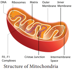
Mitochondria contain 73% of proteins, 25-30% of lipids, 5-7% of RNA, DNA (in traces) and enzymes (about 60 types). Mitochondria are called Power house of a cell, as they produce energy rich ATP.

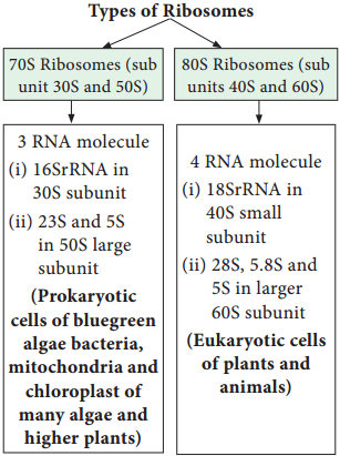
All the enzymes of Kreb’s cycle are found in the matrix except succinate dehydrogenase. Mitochondria consist of circular DNA and 70S ribosome. They multiply by fission and replicates by strand displacement model.

Because of the presence of DNAs it is semiautonomous organelle. Unique characteristic of mitochondria is that they are inherited from female parent only. Mitochondrial DNA comparisons are used to trace human origins. It is also used to track and date recent evolutionary time because it mutates 5 to 10 time faster than DNA in the nucleus.

Plastids

The term plastid is derived from the Greek word Platikas (formed/moulded) and used by A.F.U. Schimper in 1885. He classified plastids into following types according to their structure, pigments and function. Plastids multiply by fission.
Cell Organelles img 13

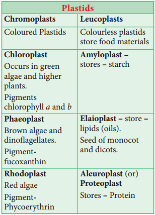
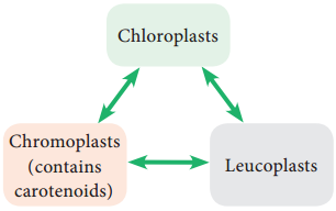
According to Schimper, different kind of plastids can transform into one another.

Chloroplast

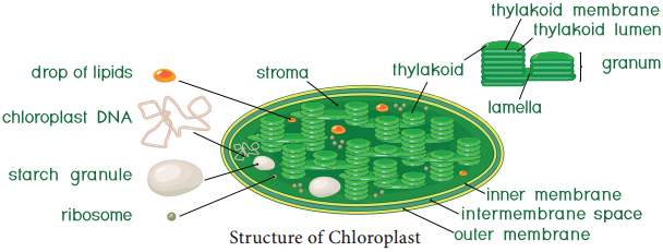
Chloroplasts are vital organelle found in green plants. Chloroplast has a double membrane the outer membrane and the inner membrane separated by a space called periplastidial space. The space enclosed by the inner membrane of chloroplast is filled with gelatinous matrix, lipo-proteinaceous fluid called stroma. Inside the stroma there are flat interconnected sacs called thylakoid. The membrane of thylakoid enclose a space called thylakoid lumen.
Cell Organelles img 5

Grana (singular: Granum) are formed when many of these thylakoids are stacked together like pile of coins. Light is absorbed and converted into chemical energy in the granum, which is used in stroma to prepare carbohydrates. Thylakoid contain chlorophyll pigments. The chloroplast contains osmophilic granules, 70s ribosomes, DNA (circular and non histone) and RNA.

These chloroplast genome encodes approximately 30 proteins involved in photosynthesis including the components of photosystem I & II, cytochrome bf complex and ATP synthase. One of the subunits of RuBisco is encoded by chloroplast DNA.

It is the major protein component of chloroplast stroma, single most abundant protein on earth. The thylakoid contain small, rounded photosynthetic units called quantosomes. Chloroplast is a semi-autonomous organelle and divides by fission (Figure 6.16).
Cell Organelles img 6

Functions:

  • Photosynthesis
  • Light reactions takes place in granum
  • Dark reactions take place in stroma
  • Chloroplast is involved in photorespiration.

Ribosome

Ribosomes were first observed by George Palade (1953) as dense particles or granules in the electron microscope. Electron microscopic observation reveals that ribosomes are composed of two rounded sub units, united together to form a complete unit.

Mg2+ is required for structural cohesion of ribosomes. Biogenesis of ribosome is a de nova formation, auto replication and nucleolar origin. Each ribosome is made up of one small and one large sub-unit Ribosomes are the sites of protein synthesis in the cell. Ribosome is not a membrane bound organelle (Figure 6.17).
Cell Organelles img 7

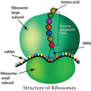
Ribosome Consists of RNA and Protein:

RNA 60% and protein 40%. During protein synthesis, many ribosomes are attached to the single mRNA and is called polysomes or polyribosomes. The function of polysomes is the formation of several copies of a particular polypeptide during protein synthesis. They are free in non-protein synthesising cells. In protein synthesising cells they are linked together with the help of Mg2+ ions.
Cell Organelles img 8

Lysosomes (Suicidal Bags of Cell)

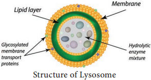
Lysosomes were discovered by Christian de Duve (1953), these are known as suicidal bags. They are spherical bodies enclosed by a single unit membrane. They are found in eukaryotic cell. Lysosomes are small vacuoles formed when small pieces of golgi body are pinched off from its tubules.

They contain a variety of hydrolytic enzymes, that can digest material within the cell. The membrane around lysosome prevent these enzymes from digesting the cell itself (Figure 6.18).
Cell Organelles img 9

Functions:

Intracellular Digestion:
They digest carbohydrates, proteins and lipids present in cytoplasm.

Autophagy:
During adverse condition they digest their own cell organelles like mitochondria and endoplasmic reticulum.

Autolysis:
Lysosome causes self destruction of cell.

Ageing:
Lysosomes have autolytic enzymes that disrupts intracellular molecules.

Phagocytosis:
Large cells or contents are engulfed and digested by macrophages, thus forming a phagosome in cytoplasm. These phagosome fuse with lysosome for further digestion.
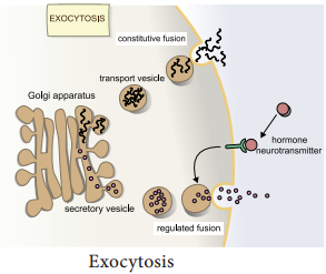
Exocytosis:
Lysosomes release their enzymes outside the cell to digest other cells (Figure 6.19).
Cell Organelles img 10

Microbodies

Eukaryotic cells contain many enzyme bearing membrane enclosed vesicles called microbodies. They are single unit membrane bound cell organelles. Example: Peroxisomes and glyoxysomes.
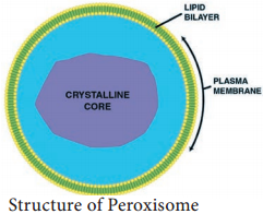
Peroxisomes

Peroxisomes were identified as organelles by Christian de Duve (1967). Peroxisomes are small spherical bodies and single membrane bound organelle. It takes part in photorespiration and associated with glycolate metabolism. In plants, leaf cells have many peroxisomes. It is also commonly found in liver and kidney of mammals. These are also found in cells of protozoa and yeast (Figure 6.20).
Cell Organelles img 11

Glyoxysomes

Glyoxysome was discovered by Harry Beevers (1961). It is a single membrane bound organelle. It is a sub cellular organelle and contains enzymes of glyoxylate pathway. β-oxidation of fatty acid occurs in glyoxysomes of germinating seeds Example: Castor seeds.

Sphaerosomes

It is spherical in shape and enclosed by single unit membrane. Example: Storage of fat in the endosperm cells of oil seeds.
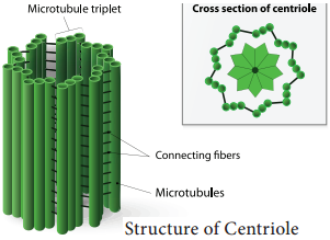
Centrioles

Centrioles consists of nine triplet peripheral fibrils made up of tubulin. The central part of the centriole is called hub, is connected to the tubules of the peripheral triplets by radial spokes (9+0 pattern). The centriole form the basal body of cilia or flagella and spindle fibers which forms the spindle apparatus in animal cells. The membrane is absent in centriole (non-membranous organelle) (Figure 6.21).
Cell Organelles img 12

Vacuoles

In plant cells vacuoles are large, bounded by a single unit membrane called Tonoplast. The Vacuoles contain cell sap, which is a solution of sugars, amino acids, mineral salts, waste chemical and anthocyanin pigments. Beetroot cells contain anthocyanin pigments in their vacuoles.

Vacuoles accumulate products like tannins. The osmotic expansion of a cell kept in water is chiefly regulated by vacuole and the water enters the vacuole by osmosis. The major function of plant vacuole is to maintain water pressure known as turgor pressure, which maintains the plant structure. Vacuoles organises itself into a storage/sequestration compartment. Example: Vacuoles store, most of the sucrose of the cell.

  • Sugar in Sugar beet and Sugar cane.
  • Malic acid in Apple.
  • Acids in Citrus fruits.
  • Flavonoid pigment cyanidin 3 rutinoside in the petals of Antirrhinum.