Plant Water Relations and its Different Issues

Learninsta presents the core concepts of Biology with high-quality research papers and topical review articles.

Plant Water Relations and its Different Issues

Water plays an essential role in the life of the plant. The availability of water influences the external and internal structures of plants as protoplasm is made of 60-80% water. Water is a universal solvent since most of the substances get dissolved in it and the high tensile strength of water molecule is helpful in the ascent of sap. Water maintains the internal temperature of the plant as well as the turgidity of the cell.

Imbibition

Colloidal systems such as gum, starch, proteins, cellulose, agar, gelatin when placed in water, will absorb a large volume of water and swell up. These substances are called imbibants and the phenomenon is imbibition.

Examples:

  1. The swelling of dry seeds
  2. The swelling of wooden windows, tables, doors due to high humidity during the rainy season.

Significance of Imbibition

  1. During germination of seeds, imbibition increases the volume of seed enormously and leads to bursting of the seed coat.
  2. It helps in the absorption of water by roots at the initial level.

Water Potential (Ψ)

The concept of water potential was introduced in 1960 by Slatyer and Taylor. Water potential is potential energy of water in a system compared to pure water when both temperature and pressure are kept the same. It is also a measure of how freely water molecules can move in a particular environment or system. Water potential is denoted by the Greek symbol Ψ (psi) and measured in Pascal (Pa).

At standard temperature, the water potential of pure water is zero. Addition of solute to pure water decreases the kinetic energy thereby decreasing the water potential. Comparatively a solution always has low water potential than pure water. In a group of cells with different water potential, a water potential gradient is generated. Water will move from higher water potential to lower water potential.

Water Potential (Ψ) Can be Determined by,

  1. Solute concentration or Solute potential (ΨS)
  2. Pressure potential (ΨP)

By correlating two factors, water potential is written as,
ΨW = ΨS + ΨP

1. Solute Potential (ΨS)

Solute potential, otherwise known as osmotic potential denotes the effect of dissolved solute on water potential. In pure water, the addition of solute reduces its free energy and lowers the water potential value from zero to negative. Thus the value of solute potential is always negative. In a solution at standard atmospheric pressure, water potential is always equal to solute potential (ΨW = ΨS).

2. Pressure Potential (ΨP)

Pressure potential is a mechanical force working against the effect of solute potential. Increased pressure potential will increase water potential and water enters cell and cells become turgid. This positive hydrostatic pressure within the cell is called Turgor pressure. Likewise, withdrawal of water from the cell decreases the water potential and the cell becomes flaccid.

3. Matric Potential (ΨM)

Matric potential represents the attraction between water and the hydrating colloid or gel-like organic molecules in the cell wall which is collectively termed as matric potential. Matric potential is also known as imbibition pressure. The matric potential is maximum (most negative value) in a dry material. Example: The swelling of soaked seeds in water.

Osmotic Pressure and Osmotic Potential

When a solution and its solvent (pure water) are separated by a semipermeable membrane, a pressure is developed in the solution, due to the presence of dissolved solutes. This is called osmotic pressure (OP). Osmotic pressure is increased with the increase of dissolved solutes in the solution.

More concentrated solution (low Ψ or Hypertonic) has high osmotic pressure. Similarly, less concentrated solution (high Ψ or Hypotonic) has low osmotic pressure. The osmotic pressure of pure water is always zero and it increases with the increase of solute concentration. Thus osmotic pressure always has a positive value and it is represented as π.

Osmotic potential is defined as the ratio between the number of solute particles and the number of solvent particles in a solution. Osmotic potential and osmotic pressure are numerically equal. Osmotic potential has a negative value whereas on the other hand osmotic pressure has a positive value.

Turgor Pressure and Wall Pressure

When a plant cell is placed in pure water (hypotonic solution) the diffusion of water into the cell takes place by endosmosis. It creates a positive hydrostatic pressure on the rigid cell wall by the cell membrane. Henceforth the pressure exerted by the cell membrane towards the cell wall is Turgor Pressure (TP).

The cell wall reacts to this turgor pressure with equal and opposite force, and the counter-pressure exerted by the cell wall towards cell membrane is wall pressure (WP). Turgor pressure and wall pressure make the cell fully turgid. TP + WP = Turgid.

Diffusion Pressure Deficit (DPD) or Suction Pressure (SP)

Pure solvent (hypotonic) has higher diffusion pressure. Addition of solute in pure solvent lowers its diffusion pressure. The difference between the diffusion pressure of the solution and its solvent at a particular temperature and atmospheric pressure is called as Diffusion Pressure Deficit (DPD) termed by Meyer (1938). DPD is increased by the addition of solute into a solvent system.

Increased DPD favours endosmosis or it sucks the water from hypotonic solution; hence Renner (1935) called it as Suction pressure. It is equal to the difference of osmotic pressure and turgor pressure of a cell. The following three situations are seen in plants:

  • DPD in normal cell: DPD = OP – TP.
  • DPD in fully turgid cell: Osmotic pressure is always equal to turgor pressure in a fully turgid cell.
  • OP = TP or OP-TP =0. Hence DPD of fully turgid cell is zero.
  • DPD in flaccid cell: If the cell is in flaccid condition there is no turgor pressure or TP = 0. Hence DPD = OP.

Osmosis

Osmosis (Latin: Osmos-impulse, urge) is a special type of diffusion. It represents the movement of water or solvent molecules through a selectively permeable membrane from the place of its higher concentration (high water potential) to the place of its lower concentration (low water potential).

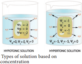
Types of Solutions Based on Concentration

(i) Hypertonic (Hyper = High; tonic = solute):

This is a strong solution (low solvent/ high solute/ low Ψ) which attracts solvent from other solutions.

(ii) Hypotonic (Hypo = low; tonic = solute):

This is a weak solution (high solvent/ low or zero solute/ high Ψ) and it diffuses water out to other solutions (Figure 11.7).

(iii) Isotonic (Iso = identical; tonic = soute):

It refers to two solutions having same concentration. In this condition the net movement of water molecule will be zero. The term hyper, hypo and isotonic are relative terms which can be used only in comparison with another solution.
Plant Water Relations img 1

1. Types of Osmosis

Based on the direction of movement of water or solvent in an osmotic system, two types of osmosis can occur, they are Endosmosis and Exosmosis.

(i) Endosmosis:

Endosmosis is defined as the osmotic entry of solvent into a cell or a system when it is placed in a pure water or hypotonic solution. For example, dry raisins (high solute and low solvent) placed in the water, it swells up due to turgidity.

(ii) Exosmosis:

Exosmosis is defined as the osmotic withdrawal of water from a cell or system when it is placed in a hypertonic solution. Exosmosis in a plant cell leads to plasmolysis.

2. Plasmolysis (Plasma = cytoplasm; lysis = breakdown)

When a plant cell is kept in a hypertonic solution, water leaves the cell due to exosmosis. As a result of water loss, protoplasm shrinks and the cell membrane is pulled away from the cell wall and finally, the cell becomes flaccid.

This process is named as plasmolysis. Wilting of plants noticed under the condition of water scarcity is an indication of plasmolysis. Three types of plasmolysis occur in plants:

  • Incipient Plasmolysis
  • Evident Plasmolysis and
  • Final Plasmolysis.

Differences among them are given in table 11.2.
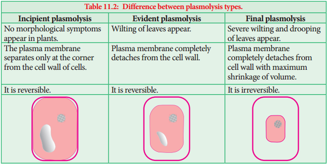
Plant Water Relations img 2

Significance

Plasmolysis is exhibited only by living cells and so it is used to test whether the cell is living or dead.

3. Deplasmolysis

The effect of plasmolysis can be reversed, by transferring them back into water or hypotonic solution. Due to endosmosis, the cell becomes turgid again. It regains its original shape and size. This phenomenon of the revival of the plasmolysed cell is called deplasmolysis. Example: Immersion of dry raisin in water.

4. Reverse Osmosis

Reverse Osmosis follows the same principles of osmosis, but in the reverse direction. In this process movement of water is reversed by applying pressure to force the water against a concentration gradient of the solution. In regular osmosis, the water molecules move from the higher concentration (pure water = hypotonic) to lower concentration (salt water = hypertonic).

But in reverse osmosis, the water molecules move from the lower concentration (salt water = hypertonic) to higher concentration (pure water = hypotonic) through a selectively permeable membrane (Figure 11.9).
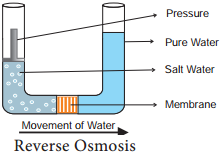
Plant Water Relations img 3

Uses:
Reverse osmosis is used for purification of drinking water and desalination of sea water.

Cell to Cell Transport Significance and its Types

Learninsta presents the core concepts of Biology with high-quality research papers and topical review articles.

Cell to Cell Transport Significance and its Types

Cell to cell or short distance transport covers the limited area and consists of few cells. They are the facilitators or tributaries to the longdistance transport. The driving force for the cell to cell transport can be passive or active (Figure 11.1). The following chart illustrate the various types of cell to cell transport:
Cell to Cell Transport img 1

Passive Transport

1. Diffusion

When we expose a lightened incense stick or mosquito coil or open a perfume bottle in a closed room, we can smell the odour everywhere in the room. This is due to the even distribution of perfume molecules throughout the room. This process is called diffusion. In diffusion, the movement of molecules is continuous and random in order in all directions (Figure 11.2).

Characteristics of Diffusion

  1. It is a passive process, hence no energy expenditure involved.
  2. It is independent of the living system.
  3. Diffusion is obvious in gases and liquids.
  4. Diffusion is rapid over a shorter distance but extremely slow over a longer distance.
  5. The rate of diffusion is determined by temperature, concentration gradient and relative density.

Significance of Diffusion in Plants

  1. Gaseous exchange of O2 and CO2 between the atmosphere and stomata of leaves takes place by the process of diffusion. O2 is absorbed during respiration and CO2 is absorbed during photosynthesis.
  2. In transpiration, water vapour from intercellular spaces diffuses into atmosphere through stomata by the process of diffusion.
  3. The transport of ions in mineral salts during passive absorption also takes place by this process.

2. Facilitated Diffsion

Cell membranes allow water and nonpolar molecules to permeate by simple diffusion. For transporting polar molecules such as ions, sugars, amino acids, nucleotides and many cell metabolites is not merely based on concentration gradient. It depends on,

(i) Size of Molecule:
Smaller molecules diffuse faster.

(ii) Solubility of the Molecule:
Lipid soluble substances easily and rapidly pass through the membrane. But water soluble substances are difficult to pass through the membrane. They must be facilitated to pass the membrane.

In facilitated diffusion, molecules cross the cell membrane with the help of special membrane proteins called transport proteins, without the expenditure of ATP. There are two types of transport proteins present in the cell membrane. They are channel protein and a carrier protein.

I. Channel Protein

Channel protein forms a channel or tunnel in the cell membrane for the easy passage of molecules to enter the cell. The channels are either open or remain closed. They may open up for specific molecules. Some channel proteins create larger pores in the outer membrane. Examples: Porin and Aquaporin.

(i) Porin

Porin is a large transporter protein found in the outer membrane of plastids, mitochondria and bacteria which facilitates smaller molecules to pass through the membrane.

(ii) Aquaporin

Aquaporin is a water channel protein embedded in the plasma membrane. It regulates the massive amount of water transport across the membrane (Figure 11.3). Plants contain a variety of aquaporins. Over 30 types of aquaporins are known from maize. Currently, they are also recognized to transport substrates like glycerol, urea, CO2, NH3, metalloids, and Reactive Oxygen Species (ROS) in addition to water. They increase the permeability of the membrane to water. They confer drought and salt stress tolerance.
Cell to Cell Transport img 3

II. Carrier Protein

Carrier protein acts as a vehicle to carry molecules from outside of the membrane to inside the cell and vice versa (Figure 11.4). Due to association with molecules to be transported, the structure of carrier protein gets modified until the dissociation of the molecules.
Cell to Cell Transport img 4

There are 3 types of carrier proteins classified on the basis of handling of molecules and direction of transport (Figure 11.5). They are:-

  1. Uniport
  2. Symport
  3. Antiport

1. Uniport:
In this molecule of a single type move across a membrane independent of other molecules in one direction.

2. Symport or Co-Transport:
The term symport is used to denote an integral membrane protein that simultaneously transports two types of molecules across the membrane in the same direction.

3. Antiport or Counter Transport:
An antiport is an integral membrane transport protein that simultaneously transports two different molecules, in opposite directions, across the membrane.
Cell to Cell Transport img 5

Active Transport

The main disadvantage of passive transport processes like diffusion is the lack of control over the transport of selective molecules. There is a possibility of harmful substances entering the cell by a concentration gradient in the diffusion process. But selective permeability of cell membrane has a great control over entry and exit of molecules.

Active transport is the entry of molecules against a concentration gradient and an uphill process and it needs energy which comes from ATP. Passive transport uses kinetic energy of molecules moving down a gradient whereas, active transport uses cellular energy to move them against a gradient.

The transport proteins discussed in facilitated diffusion can also transport ions or molecules against a concentration gradient with the expenditure of cellular energy as an active process. Pumps use a source of free energy such as ATP or light to drive the thermodynamically uphill transport of ions or molecules. The pump action is an example of active transport. Example: Na+-K+-ATPase pump (Table 11.1).
Cell to Cell Transport img 6

Types of Transport

Learninsta presents the core concepts of Biology with high-quality research papers and topical review articles.

Types of Transport

Transport is the process of moving water, minerals and food to all parts of the plant body. Conducting tissues such as xylem and phloem play an important role in this. What is the need for transport? Water absorbed from roots must travel up to leaves by xylem for food preparation by photosynthesis. Likewise, food prepared from leaves has to travel to all parts of the plant including roots. Both the processes are interconnected and depend on each other.

Based on the distance travelled by water (sap) or food (solute) they are classified as:-

  1. Short Distance (Cell to cell transport) and
  2. Long Distance Transport.

1. Short-distance (Cell to Cell Transport):

Involvement of few cells, mostly in the lateral direction. They are the connecting link to xylem and phloem from root hairs or leaf tissues respectively. Examples: Diffusion, Imbibition, and Osmosis.

2. Long-Distance Transport:

Transport within the network of xylem or phloem is an example for long-distance transport. Examples: Ascent of Sap and Translocation of Solutes.

Based on energy expenditure during transport, they are classified as:-

  1. Passive Transport and
  2. Active Transport

1. Passive Transport:

It is a downhill process which utilizes physical forces like gravity and concentration. No energy expenditure is required. It includes diffusion, facilitated diffusion, imbibition, and osmosis.

2. Active Transport:

It is a biological process and it runs based on the energy obtained from respiration. It is an uphill process. The different modes of transport are air, water, and land transport, which includes Rails or railways, road and off-road transport. Other modes also exist, including pipelines, cable transport, and space transport.

Transport modes are the means of supporting the mobility of passengers and freight. They are mobile transport assets and fall into three basic types; land (road, rail, pipelines), water (shipping), and air.

Water transport is the slowest means of transport and, therefore, important for transporting the bulky raw materials which does not care of the speed of movement of commodities.

Among different modes of transport, Railways are the cheapest. Trains cover the distance in less time and comparatively, the fare is also less to other modes of transportation. Therefore, Railways is the cheapest mode of transportation.

There are two major types of cell transport: passive transport and active transport. Passive transport requires no energy. It occurs when substances move from areas of higher to lower concentration. Types of passive transport include simple diffusion, osmosis, and facilitated diffusion.

The bicycle is a tremendously efficient means of transportation. In fact cycling is more efficient than any other method of travel-including walking! The one billion bicycles in the world are a testament to its effectiveness. The engine for this efficient mode of transport is the human body.

The air travel, today, is the fastest, most comfortable and prestigious mode of transport. It has reduced distances by minimising the travel time. It is very essential for a vast country like India, where distances are large and the terrain and climatic conditions are diverse.

The cheapest means of transport for a long distance is Waterways. The amount for loading and unloading goods is much cheaper if it has to travel a long distance. If one has to travel physically to a short distance then it is advisable to take a train. Railways are both cheaper and comfortable to travel.

Modes of transport include air, land (rail and road), water, cable, pipeline and space. The field can be divided into infrastructure, vehicles and operations. Transport is important because it enables trade between people, which is essential for the development of civilizations.

The four elements of transport are:

  1. The Way
  2. The Unit of Carriage
  3. The Motive Power unit, and the Terminal

Natural ways are cheap and free, and have no maintenance costs unless we try to improve them artificially. The sea, the air, the rivers, and footpaths are all natural ways.

Transportation is the movement of goods and logistics is the management of the inward and outward transportation of goods from the manufacturer to the end user. Logistics and transportation deals with getting products and services from one location to another.

There are two major types of cell transport: passive transport and active transport. Passive transport requires no energy. It occurs when substances move from areas of higher to lower concentration. Types of passive transport include simple diffusion, osmosis, and facilitated diffusion.

The different modes of transport are air, water, and land transport, which includes Rails or railways, road and off-road transport. Other modes also exist, including pipelines, cable transport, and space transport.
Types of Transport img 1

Secondary Growth in Dicot Root

Learninsta presents the core concepts of Biology with high-quality research papers and topical review articles.

Secondary Growth in Dicot Root

Secondary growth in dicot roots is essential to provide strength to the growing aerial parts of the plants. It is similar to that of the secondary growth in dicot stem. However, there is marked diffrence in the manner of the formation of vascular cambium.

The vascular cambium is completely secondary in origin. It originates from a combination of conjunctive tissue located just below the phloem bundles, and as a portion of pericycle tissue present above the protoxylem to form a complete and continuous wavy ring. This wavy ring later becomes circular and produces secondary xylem and secondary phloem similar to the secondary growth in stems.
Secondary Growth in Dicot Root img 1

Differences Between Secondary Growth in Dicot Stem and Root

Secondary Growth in Dicot Stem

Secondary Growth in Dicot Root

1. The cambial ring formed is circular in cross section from the beginning1. The cambial ring formed is wavy in the beginning and later becomes circular
2. The cambial ring is partially primary (fasicular cambium) and partially secondary (Interfasicular cambium) in origin2. The cambial ring is completely secondary in origin
3. Generally, periderm originates from the cortical cells (extrastelar in origin)3. Generally, periderm originates from the pericycle.(intrastealar in origin)
4. More amount of cork is produced as stem is aboveground4. Generally, less amount of cork is produced as root is underground
5. Lenticels of periderm are prominent5. Lenticels of periderm are not very prominent

Most of the dicotyledonous roots show secondary growth in thickness, similar to that of dicotyledonous stems. Certain dicotyledonous roots do not show secondary growth. The secondary vascular tissues originate as a result of the cambial activity. The phellogen gives rise to the periderm.

Secondary growth takes place in all dicotyledonous woody plants. The root increases in girth by the activity of stelar and extrastelar cambium.

This is followed by periclinal division of the cells of pericycle present against protoxylem to form multiple layers of cells, which are joined by cambial cells derived from conjunctive tissues and together they make a complete cambium ring. Thus, the correct answer is option D.

In a dicot stem, secondary growth occurs both in the stele and cortex. The process occurs simultaneously but is caused by separate strips of secondary meristem. In the stele, secondary growth is initiated by vascular cambium, while in the cortex, it is initiated by cork cambium.

In botany, secondary growth is the growth that results from cell division in the cambia or lateral meristems and that causes the stems and roots to thicken, while primary growth is growth that occurs as a result of cell division at the tips of stems and roots, causing them to elongate, and gives rise to primary tissue. There are two types of lateral tissues involved in secondary growth, namely, vascular cambium and cork cambium.

In general, monocots do not undergo secondary growth. If they do increase in girth (like palm trees and yucca plants), it does not result in the development of a secondary xylem and phloem, since monocots don’t have vascular cambium.

Difference Between the Secondary Growth in Dicot Stem and Dicot Root. The growth in thickness by the activity of secondary tissues is called secondary thickening. It involves stelar growth by the activity of vascular cambial ring and extra stelar growth by the activity of cork cambium.

Initiation of secondary growth takes place in the zone of maturation soon after the cells stop elongating there. The vascular cambium differentiates between the primary xylem and phloem in this zone and pericycle cells divide simultaneously with the procambium initials.

A process of formation of secondary tissues due to activity of vascular cambium and cork cambium for increasing thickness or girth or diameter of plant is termed as secondary growth.

Bougainvillea is a member of the Nyctaginaceae and is an example of a dicotyledonous stem which displays anomalous secondary growth. In this TS, near the centre of the stem, you will see some primary vascular bundles embedded in lignified pith parenchyma.

Secondary growth is characterized by an increase in thickness or girth of the plant, and is caused by cell division in the lateral meristem. Secondary vascular tissue is added as the plant grows, as well as a cork layer. The bark of a tree extends from the vascular cambium to the epidermis.

The increase in length of the shoot and the root is referred to as primary growth. It is the result of cell division in the shoot apical meristem. Secondary growth is characterized by an increase in thickness or girth of the plant.

The primary root grows vertically downwards into the soil. Smaller lateral roots known as the secondary roots are produced on the primary root. The secondary roots in turn produce tertiary roots. These roots grow in various directions and help in fixing the plant firmly into the soil.

Secondary growth is the growth in thickness due to the formation of secondary tissues by lateral meristems. Secondary growth does not occur in monocots because monocots do not possess vascular cambium in between the vascular bundles.

Secondary growth is the outward growth of the plant, making it thicker and wider. Secondary growth is important to woody plants because they grow much taller than other plants and need more support in their stems and roots. Lateral meristems are the dividing cells in secondary growth, and produce secondary tissues.

The process of secondary growth is controlled by the lateral meristems, and is similar in both stems and roots. Lateral meristems include the vascular cambium and, in woody plants, the cork cambium (cambium is another term for meristem).

Secondary xylem is a complex tissue that consists not only of non-living supporting and conducting cells but also of important living components (rays and axial wood parenchyma) which, with those in the secondary phloem, comprise a three-dimensional symplastic pathway through which photosynthate and other essential.

Lateral meristems are known as secondary meristems because they are responsible for secondary growth, or increase in stem girth and thickness. Meristems form anew from other cells in injured tissues and are responsible for wound healing.

Plant growth from lateral meristems such as the vascular cambium and cork cambium. This growth thickens plants and creates wood and bark (only in woody plants). Allows for taller, stronger plants, more branching and reproduction, and more conduction of fluids.

An example of a secondary meristem is the lateral meristem (e.g. cork cambium and accessory cambia). Being meristematic, the secondary meristem is comprised of undifferentiated (or partially differentiated), actively dividing cells.

The vascular cambium is responsible for increasing the diameter of stems and roots and for forming woody tissue. The cork cambium produces some of the bark. Cell division by the cambium produces cells that become secondary xylem and phloem.

In botany, secondary growth is the growth that results from cell division in the cambia or lateral meristems and that causes the stems and roots to thicken, while primary growth is growth that occurs as a result of cell division at the tips of stems and roots, causing them to elongate, and gives rise to primary tissue.

Secondary growth is the growth in thickness due to the formation of secondary tissues by lateral meristems. Secondary growth does not occur in monocots because monocots do not possess vascular cambium in between the vascular bundles.

In particular, secondary growth is substantial for constant plant growth and the remodeling of body structures.

As an important meristem involved, the vascular cambium forms a cylindrical domain below the organ surface producing tissues for long-distance transport and mechanical support: wood (xylem) and bast (phloem). Pericycle forms the boundary of stele and encloses vascular bundles and pith. It is a primary structure and is not formed as a result of secondary growth.

Secondary Growth in Dicot Stem and its Overview

Learninsta presents the core concepts of Biology with high-quality research papers and topical review articles.

Secondary Growth in Dicot Stem and its Overview

Vascular Cambium

The vascular cambium is the lateral meristem that produces the secondary vascular tissues. i.e., secondary xylem and secondary phloem.

Origin and Formation of Vascular Cambium

A strip of vascular cambium that is believed to originate from the procambium is present between xylem and phloem of the vascular bundle. This cambial strip is known as intrafascicular or fascicular cambium. In between the vascular bundles, a few parenchymatous cells of the medullary rays that are in line with the fascicular cambium become meristematic and form strips of vascular cambium. It is calledinterfascicular
cambium.

This interfascicular cambium joins with the intrafascicular cambium on both sides to form a continuous ring. It is called a vascular cambial ring. The differences between interfascicular and intrafascicular cambia are summarised below:

Intrafascicular Cambium

Interfascicular Cambium

1. Present inside the vascular bundles1. Present in between the vascular bundles
2. Originates from the procambium2. Originates from the medullary rays
3. Initially it forms a part of the primary
meristem.
3. From the beginning it forms a part of the secondary meristem

Organization of Vascular Cambium

The cells of vascular cambium do not fit into the usual description of meristems which have isodiametric cells, with a dense cytoplasm and large nuclei. While the active vascular cambium possesses cells with large central vacuole (or vacuoles) surrounded by a thin, layers of dense cytoplasm.

Further, the most important character of the vascular cambium is the presence of two kinds of initials, namely, fusiform initials and ray initials.

Fusiform Initials

These are vertically elongated cells. They give rise to the longitudinal or axial system of the secondary xylem (treachery elements, fires, and axial parenchyma) and phloem (sieve elements, fiers, and axial parenchyma). Based on the arrangement of the fusiform initials, two types of vascular cambium are recognized.

Storied (Stratifid cambium) and Non-Storied (Non-stratified cambium)

If the fusiform initials are arranged in horizontal tiers, with the end of the cells of one tier appearing at approximately the same level, as seen in tangential longitudinal section (TLS), it is called storied (stratified) cambium. It is the characteristic of the plants with short fusiform initials. Whereas in plants with long  fusiform initials, they strongly overlap at the ends, and this type of cambium is called non-storied (nonstartified) cambium.
Secondary Growth in Dicot Stem and its Overview img 1

Ray Initials

These are horizontally elongated cells. They give rise to the ray cells and form the elements of the radial system of secondary xylem and phloem.

Activity of Vascular Cambium

The vascular cambial ring, when active, cuts of new cells both towards the inner and outer side. The cells which are produced outward form secondary phloem and inward secondary xylem.
Secondary Growth in Dicot Stem and its Overview img 2

At places, cambium forms some narrow horizontal bands of parenchyma which passes through secondary phloem and xylem. These are the rays. Due to the continued formation of secondary xylem and phloem through vascular cambial activity, both the primary xylem and phloem get gradually crushed.

Secondary Xylem

The secondary xylem, also called wood, is formed by a relatively complex meristem, the vascular cambium, consisting of vertically (axial) elongated fusiform initials and horizontally (radially) elongated ray initials.

The axial system consists of vertical files of treachery elements, fiers, and wood parenchyma. Whereas the radial system consists of rows of parenchymatous cells oriented at right angles to the longitudinal axis of xylem elements.

The secondary xylem varies very greatly from species to species with reference to relative distribution of the different cell types, density and other properties. It is of two types.

Porous Wood or Hard Wood

Generally, the dicotyledonous wood, which has vessels is called porous wood or hard wood. Example: Morus rubra.

Non – Porous Wood or Sof Wood

Generally, the gymnosperm wood, which lacks vessels is known as non – porous wood or sof wood. Example: Pinus.

Differences between Porous Wood and Non-porous Wood

Porous wood or Hard wood, Example: Morus  

Non porous wood or soft wood, Example: Pinus

1. Common in angiosperms1. Common in gymnosperms
2. Porous because it contains vessels2. Non-porous because it does not contain vessels

Secondary Growth in Dicot Stem and its Overview img 3
Secondary Growth in Dicot Stem and its Overview img 4

Annual Rings

The activity of vascular cambium is under the control of many physiological and environmental factors. In temperate regions, the climatic conditions are not uniform throughout the year. In the spring season, cambium is very active and produces a large number of xylary elements having vessels/tracheids with wide lumen.

The wood formed during this season is called spring wood or early wood. The tracheary elements are fairly thin walled. In winter, the cambium is less active and forms fewer xylary elements that have narrow vessels/tracheids and this wood is called autumn wood or late wood. The treachery elements are with narrow lumen, very thick walled.

The spring wood is lighter in colour and has a lower density whereas the autumn wood is darker and has a higher density. The annual ring denotes the combination of early wood and late wood and the ring becomes evident to our eye due to the high density of late wood. Sometimes annual rings are called growth rings but it should be remembered all the growth rings are not annual. In some trees more than one growth ring is formed with in a year due to climatic changes.

Additional growth rings are developed within a year due to adverse natural calamities like drought, frost, defoliation, flood, mechanical injury and biotic factors during the middle of a growing season, which results in the formation of more than one annual ring.

Such rings are called pseudoor false – annual rings. Each annual ring corresponds to one year’s growth and on the basis of these rings, the age of a particular plant can easily be calculated. The determination of the age of a tree by counting the annual rings is called dendrochronology.

Dendroclimatology

It is a branch of dendrochronology concerned with constructing records of past climates and climatic events by analysis of tree growth characteristics, especially growth rings.
Secondary Growth in Dicot Stem and its Overview img 5

Differences Between Spring Wood and Autumn Wood

Spring wood or Early wood

Autumn wood or Late wood

1. The activity of cambium is faster1. Activity of cambium is slower
2. Produces large number of xylem elements2. Produces a fewer xylem elements
3. Xylem vessels/trachieds have wider lumen3. Xylem vessels/trachieds have narrow lumen
4. Wood is lighter in colour and has lower density4. Wood is darker in colour and has a higher density

Another feature of wood related to seasonal changes is the diffuse porous and ring porous condition. On the basis of diameter of xylem vessels, two main types of angiosperm woods are recognized.

Diffuse Porous Woods

Diffuse porous woods are woods in which the vessels or pores are rather uniform in size and distribution throughout an annual ring. Example: Acer

Ring Porous Woods

The pores of the early wood are distinctly larger than those of the late wood. This rings of wide and narrow vessels occur. Example: Quercus
Secondary Growth in Dicot Stem and its Overview img 6

Differences Between Diffuse Porous Wood and Ring Porous Wood

Diffuse porous wood

Ring porous wood

1. This type of wood is formed where the
climatic conditions are uniform
1. This type of wood is formed where the
climatic conditions are not uniform
2. The vessels are more or less equal
in diameter in any annual ring
2. The vessels are wide and narrow within
any annual ring
3. The vessels are uniformly distributed
throughout the wood
3. The vessels are not uniformly distributed throughout the wood

Tyloses

In many dicot plants, the lumen of the xylem vessels is blocked by many balloonlike ingrowths from the neighbouring parenchymatous cells. These balloon-like structures are called tyloses.
Secondary Growth in Dicot Stem and its Overview img 7

Usually, these structures are formed in secondary xylem vessels that have last their function i.e., in heart wood. In fully developed tyloses, starchy crystals, resins, gums, oils, tannins or coloured substances are found. Wood is also classifid into sap wood and heart wood.

Sap Wood and Heart Wood

Sap wood and heart wood can be distinguished in the secondary xylem. In any tree the outer part of the wood, which is paler in colour, is called sap wood or alburnum. The centre part of the wood, which is darker in colour is called heart wood or duramen.

The sap wood conducts water while the heart wood stops conducting water. As vessels of the heart wood are blocked by tyloses, water is not conducted through them. Due to the presence of tyloses and their contents the heartwood becomes coloured, dead and the hardest part of the wood.

From the economic point of view, generally the heartwood is more useful than the sapwood. The timber from the heartwood is more durable and more resistant to the attack of microorganisms and insects than the timber from sapwood.
Secondary Growth in Dicot Stem and its Overview img 8

Differences Between Sap Wood (alburnum) and Heart Wood (duramen)

Sap Wood (Alburnum)

Heart Wood (Duramen)

1. Living part of the wood1. Dead part of the wood
2. It is situated on the outer side of wood2. It is situated in the centre part of wood
3. It is pale coloured3. It is dark coloured
4. Very soft in nature4. Hard in nature
5. Tyloses are absent5. Tyloses are present
6. It is not durable and not resistant to
microorganisms
6. It is more durable and resists
microorganisms

Secondary Phloem

The vascular cambial ring produces secondary phloem or bast on the outer side of the vascular bundle. Just as the secondary xylem, the secondary phloem also has two tissue systems – the axial (vertical) and the radial (horizontal) systems derived respectively from the vertically elongated fusiform initials and horizontally elongated ray initials of vascular cambium.

While sieve elements, phloem fire, and phloem parenchyma represent the axial system, phloem rays represent the radial system. Life span of secondary phloem is less compared to secondary xylem. Secondary phloem is a living tissue that transports soluble organic compounds made during photosynthesis to various parts of plant. Some commercially important phloem or bast fires are obtained from the following plants.

  1. Flax-Linum usitatissimum
  2. Hemp-Cannabis sativa
  3. Sun hemp-Crotalaria juncea
  4. Jute-Corchorus capsularis

Periderm

Whenever stems and roots increase in thickness by secondary growth, the periderm, a protective tissue of secondary origin replaces the epidermis and often primary cortex. The periderm consists of phellem, phellogen, and phelloderm.

Phellem (Cork)
Secondary Growth in Dicot Stem and its Overview img 9

It is the protective tissue composed of nonliving cells with suberized walls and formed centrifugally (outward) by the phellogen (cork cambium) as part of the periderm. It replaces the epidermis in older stems and roots of many seed plants. It is characterized by regularly arranged tiers and rows of cells. It is broken here and there by the presence of lenticels.

Phellogen (Cork Cambium)

It is a secondary lateral meristem. It comprises homogenous meristematic cells unlike vascular cambium. It arises from epidermis, cortex, phloem or pericycle (extrastelar in origin). Its cells divide periclinally and produce radially arranged fies of cells. The cells towards the outer side diffrentiate into phellem (cork) and those towards the inside as phelloderm (secondary cortex).

Phelloderm (Secondary Cortex)

It is a tissue resembling cortical living parenchyma produced centripetally (inward) from the phellogen as a part of the periderm of stems and roots in seed plants.

Differences Between Phellem and Phelloderm

Phellem (Cork)

Phelloderm (Secondary cortex)

1. It is formed on the outer side of
phellogen
1. It is formed on the inner side of
phellogen
2. Cells are compactly arranged in regular
tires and rows without intercellular
spaces
2. Cells are loosely arranged with
intercellular spaces
3. Protective in function3. As it contains chloroplast, it
synthesises and stores food
4. Consists of nonliving cells with
suberized walls.
4. Consists of living cells, parenchymatous
in nature and does not have suberin
5. Lenticels are present5. Lenticels are absent

Bark

The term ‘bark’ is commonly applied to all the tissues outside the vascular cambium of stem (i.e., periderm, cortex, primary phloem and secondary phloem). Bark protects the plant from parasitic fungi and insects, prevents water loss by evaporation and guards against variations of external temperature. It is an insect repellent, decay proof, fieproof and is used in obtaining drugs or spices.

The phloem cells of the bark are involved in conduction of food while secondary cortical cells involved in storage. If the phellogen forms a complete cylinder around the stem, it gives rise to ring barks. Example: Quercus. When the bark is formed in overlapping scale like layers, it is known as scale bark. Example: Guava. While ring barks normally do not peeled off scale barks peeled off, scale barks peeled off.
Secondary Growth in Dicot Stem and its Overview img 10

Lenticel

Lenticel is raised opening or pore on the epidermis or bark of stems and roots. It is formed during secondary growth in stems. When phellogen is more active in the region of lenticels, a mass of loosely arranged thin-walled parenchyma cells are formed. It is called complementary tissue or filing tissue. Lenticel is helpful in exchange of gases and transpiration called lenticular transpiration.
Secondary Growth in Dicot Stem and its Overview img 11