Heredity And Variation

Learninsta presents the core concepts of Biology with high-quality research papers and topical review articles.

Heredity And Variation

Genetics is oftn described as a science which deals with heredity and variation.

Heredity:
Heredity is the transmission of characters from parents to off springs.

Variation:
The organisms belonging to the same natural population or species that shows a diffrence in the characteristics is called variation. Variation is of two types

  1. Discontinuous variation and
  2. Continuous variation

1. Discontinuous Variation:

Within a population there are some characteristics which show a limited form of variation. Example: Style length in Primula, plant height of garden pea. In discontinuous variation, the characteristics are controlled by one or two major genes which may have two or more allelic forms. These variations are genetically determined by inheritance factors.

Individuals produced by this variation show diffrences without any intermediate form between them and there is no overlapping between the two phenotypes. The phenotypic expression is unaffcted by environmental conditions. This is also called as qualitative inheritance.

2. Continuous Variation:

This variation may be due to the combining effects of environmental and genetic factors. In a population most of the characteristics exhibit a complete gradation, from one extreme to the other without any break. Inheritance of phenotype is determined by the combined effects of many genes, (polygenes) and environmental factors. This is also known as quantitative inheritance. Example: Human height and skin color.

Importance of variations

  • Variations make some individuals better fited in the struggle for existence.
  • They help the individuals to adapt themselves to the changing environment.
  • It provides the genetic material for natural selection.
  • Variations allow breeders to improve better yield, quicker growth, increased resistance and lesser input.
  • They constitute the raw materials for evolution.

Asexual and Sexual Reproduction of Parthenocarpy

Learninsta presents the core concepts of Biology with high-quality research papers and topical review articles.

Asexual and Sexual Reproduction of Parthenocarpy

As mentioned earlier, the ovary becomes the fruit and the ovule becomes the seed after fertilization. However in a number of cases, fruit like structures may develop from the ovary without the act of fertilization. Such fruits are called parthenocarpic fruits. Invariably they will not have true seeds. Many commercial fruits are made seedless. Examples: Banana, Grapes and Papaya.

Signifiance

  • The seedless fruits have great signifiance in horticulture.
  • The seedless fruits have great commercial importance.
  • Seedless fruits are useful for the preparation of jams, jellies, sauces, fruit drinks etc.
  • High proportion of edible part is available in parthenocarpic fruits due to the absence of seeds.

Polyembryony Types and its Occurence

Learninsta presents the core concepts of Biology with high-quality research papers and topical review articles.

Polyembryony Types and its Occurence

Occurrence of more than one embryo in a seed is called polyembryony (Figure 1.24). The first case of polyembryony was reported in certain oranges by Anton von Leeuwenhoek in the year 1719. Polyembryony is divided into four categories based on its origin.
Polyembryony Types and its Occurence img 1

1. Cleavage polyembryony (Example: Orchids)

2. Formation of embryo by cells of the Embryo sac other than egg (Synergids – Aristolochia; antipodals – Ulmus and endosperm – Balanophora)

3. Development of more than one Embryo sac within the same ovule.
(Derivatives of same MMC, derivatives of two or more MMC – Casuarina)

4. Activation of some sporophytic cells of the ovule (Nucellus / integuments-Citrus and Syzygium).

Practical applications

The seedlings formed from the nucellar tissue in Citrus are found better clones for Orchards. Embryos derived through polyembryony are found virus free.

Apomixis Definition and its Types

Learninsta presents the core concepts of Biology with high-quality research papers and topical review articles.

Apomixis Definition and its Types

Reproduction involving fertilization in flowering plants is called amphimixis and wherever reproduction does not involve union of male and female gametes is called apomixis.

The term Apomixis was introduced by Winkler in the year 1908. It is defied as the substitution of the usual sexual system (Amphimixis) by a form of reproduction which does not involve meiosis and syngamy. Maheswari (1950) classifid Apomixis into two types – Recurrent and Non recurrent

Recurrent apomixis:
It includes vegetative reproduction and agamospermy.

Non recurrent apomixis:
Haploid embryo sac developed aftr meiosis, develops into a embryo without fertilization. The outline classifiation of Recurrent apomixis is given below.
Apomixis Definition and its Types img 1

Vegetative reproduction:
Plants propagate by any part other than seeds

Bulbils – Fritillaria imperialis; Bulbs – Allium; Runner – Mentha arvensis; Sucker Chrysanthemum

Agamospermy:
It refers to processes by which Embryos are formed by eliminating meiosis and syngamy.

Adventive embryony:
An Embryo arises directly from the diploid sporophytic cells either from nucellus or integument. It is also called sporophytic budding because gametophytic phase is completely absent. Adventive embryos are found in Citrus and Mangifera.

Diplospory (Generative apospory):
A diploid embryo sac is formed from megaspore mother cell without a regular meiotic division Examples. Eupatorium and Aerva.

Apospory:
Megaspore mother cell (MMC) undergoes the normal meiosis and four megaspores formed gradually disappear. A nucellar cell becomes activated and develops into a diploid embryo sac. This type of apospory is also called somatic apospory. Examples Hieracium and Parthenium.

Post Fertilization Structure and Events

Learninsta presents the core concepts of Biology with high-quality research papers and topical review articles.

Post Fertilization Structure and Events

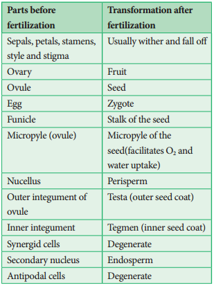
After fertilization, several changes take place in the floral parts up to the formation of the seed (Figure 1.20).
Post Fertilization Structure and Events img 1

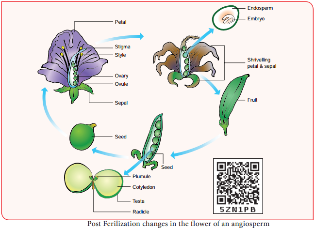
The events after fertilization (endosperm, embryo development, formation of seed, fruits) are called post fertilization changes.
Post Fertilization Structure and Events img 2

Endosperm

The primary endosperm nucleus (PEN) divides immediately after fertilization but before the zygote starts to divide, to form the endosperm. The primary endosperm nucleus is the result of triple fusion (two polar nuclei and one sperm nucleus) and thus has 3n number of chromosomes. It is a nutritive tissue and regulatory structure that nourishes the developing embryo.

Depending upon the mode of development three types of endosperm are recognized in angiosperms. They are nuclear endosperm, cellular endosperm and helobial endosperm (Figure 1.21).
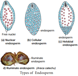
Post Fertilization Structure and Events img 3

Nuclear endosperm:
Primary Endosperm Nucleus undergoes several mitotic divisions without cell wall formation thus a free nuclear condition exists in the endosperm. Examples: Coccinia, Capsella and Arachis

Cellular endosperm:
Primary endosperm nucleus divides into 2 nuclei and it is immediately followed by wall formation. Subsequent divisions also follow cell wall formation. Examples: Adoxa, Helianthus and Scoparia

Helobial endosperm:
Primary Endosperm Nucleus moves towards base of embryo sac and divides into two nuclei. Cell wall formation takes place leading to the formation of a large micropylar and small chalazal chamber. The nucleus of the micropylar chamber undergoes several free nuclear division whereas that of chalazal chamber may or may not divide. Examples: Hydrilla and Vallisneria.

The endosperms may either be completely consumed by the developing embryo or it may persist in the mature seeds. These seeds without endosperms are called non-endospermous or ex-albuminous seeds. Examples: Pea, Groundnut and Beans. These seeds with endosperms are called endospermous or albuminous seeds. The endosperms in these seeds supply nutrition to the embryo during seed germination.
Examples: Paddy, Coconut and Castor.

Ruminate endosperm:
The endosperm with irregularity and unevenness in its surface forms ruminate endosperm. Examples: Areca catechu, Passiflra and Myristica

Functions of endosperm:

  • It is the nutritive tissue for the developing embryo.
  • In majority of angiosperms, the zygote divides only after the development of endosperm.
  • Endosperm regulates the precise mode of embryo development.

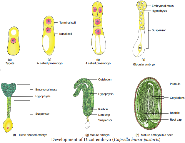
Development of Dicot embryo

The Stages involved in the development of Dicot embryo (Capsella bursa-pastoris – Onagrad or crucifer type) is given in Figure 1.22. The embryo develops at micropylar end of embryo sac. The zygote undergoes transverse division to form upper or terminal cell and lower or basal cell.

Further divisions in the zygote during the development lead to the formation of embryo. Embryo undergoes globular, heart shaped stages before reaching a mature stage. Th mature embryo has a radicle, two cotyledons and a plumule.
Post Fertilization Structure and Events img 4

Seed

The fertilized ovule is called seed and possesses an embryo, endosperm and a protective coat. Seeds may be endospermous (wheat, maize, barley and sunflower) or non endospermous. (Bean, Mango, Orchids and cucurbits).

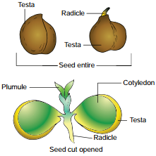
Cicer seed (example for Dicot seed)

The mature seeds are attached to the fruit wall by a stalk called funiculus. The funiculus disappears leaving a scar called hilum. Below the hilum a small pore called micropyle is present. It facilitates entry of oxygen and water into the seeds during germination.

Each seed has a thick outer covering called seed coat. The seed coat is developed from integuments of the ovule. The outer coat is called testa and is hard whereas the inner coat is thin, membranous and is called tegmen.

In Pea plant the tegmen and testa are fused. Two cotyledons laterally attached to the embryonic axis and store the food materials in pea whereas in other seeds like castor the endosperm contains reserve food and the Cotyledons are thin. The portion of embryonal axis projecting beyond the cotyledons is called radicle or embryonic root.

The other end of the axis called embryonic shoot is the plumule. Embryonal axis above the level of cotyledon is called epicotyl whereas the cylindical region between the level of cotyledon is called hypocotyl (Figure 1.23 a).
Post Fertilization Structure and Events img 5

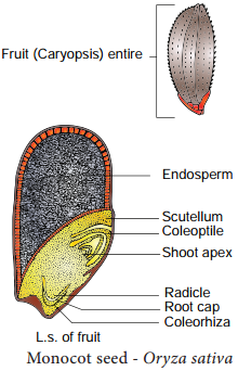
Oryza seed (example for Monocot seed)

The seed of paddy is one seeded and is called Caryopsis. Each seed remains enclosed by a brownish husk which consists of glumes arranged in two rows. The seed coat is a brownish, membranous layer closely adhered to the grain.

Endosperm forms the bulk of the grain and is the storage tissue. It is separated from embryo by a defiite layer called epithelium. The embryo is small and consists of one shieldshaped cotyledon known as scutellum present towards lateral side of embryonal axis.

A short axis with plumule and radicle protected by the root cap is present. The plumule is surrounded by a protective sheath called coleoptile. The radicle including root cap is also covered by a protective sheath called coleorhiza. The scutellum supplies the growing embryo with food material absorbed from the endosperm with the help of the epithelium (Figure 1.23 b).
Post Fertilization Structure and Events img 6