Earthworm – Lampito Mauritii

Learninsta presents the core concepts of Biology with high-quality research papers and topical review articles.

Earthworm – Lampito Mauritii

Earthworm is a terrestrial invertebrate that inhabits the upper layers of the moist soil, rich in decaying organic matter. It is nocturnal and during the day it lives in burrows made by burrowing and swallowing the soil. In gardens, they can be traced by their faecal deposits known as worm castings on the soil surface.
Earthworm - Lampito mauritii img 1

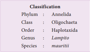
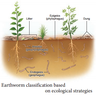
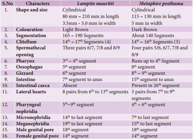
Earthworms are considered as “Friends of Farmers”. The common Indian earthworms are Lampito mauritii (Syn. Megascolex mauritii), Perioynx excavatus and Metaphire posthuma (Syn. Pheretima posthuma). Earthworms are also conveniently classified based on their ecological strategies as epigeics, anecics and endogeics (Figure 4.1).

Epigeics (Greek for “up on the earth”) are surface dwellers, eg. Perionyx excavatus and Eudrilus eugeniae. Anecics (Greek for “outer layer of the earth”) are found in upper layers of the soil, eg. Lampito mauritii, Lumbricus terrestris. Endogeics (Greek for “within the earth”) are found in deeper layers of the soil eg. Octochaetona thurstoni.
Earthworm - Lampito mauritii img 2

Morphology

Lampito mauritii is commonly found in Tamil Nadu. It has a long and cylindrical narrow body which is bilaterally symmetrical. L. mauritii is 80 to 210 mm in length with a diameter of 3.5 – 5 mm, and is light brown in colour, with purplish tinge at the anterior end. This colour of the earthworm is mainly due to the presence of porphyrin pigment.

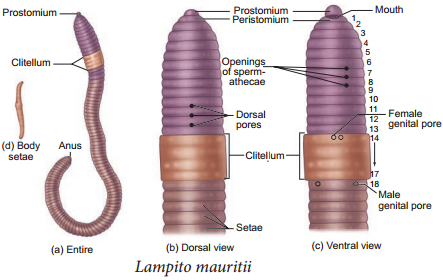
The body of the earthworm is encircled by a large number of grooves which divides it into a number of compartments called segments or metameres (Figure 4.2). L. mauritii consists of about 165 – 190 segments. The dorsal surface of the body is marked by a dark mid dorsal line (dorsal blood vessel) along the longitudinal axis of the body.

The ventral surface is distinguished by the presence of genital openings. The mouth is found in the centre of the first segment of the body, called the peristomium. Overhanging the mouth is a small flap called the upper lip or prostomium. The last segment has the anus called the pygidium. In mature worms, segments 14 to 17 may be found swollen with a glandular thickening of the skin called the clitellum. This helps in the formation of the cocoon.

Due to the presence of clitellum, the body of an earthworm is divided into pre clitellar region (1st – 13th segments), clitellar region (14th – 17th segments) and the post – clitellar region (after the 17th segment). In all the segments of the body except the first, last and clitellum, there is a ring of chitinous body setae. This body setae arises from a setigerous sac of the skin and it is curved as S – shaped. Setae can be protruded or retracted and their principal role is in locomotion.

The external apertures are the mouth, anus, dorsal pores, spermathecal openings, genital openings and nephridiopores. The dorsal pores are present from the 10th segment onwards. The coelomic fluid communicates to the exterior through these pores and keeps the body surface moist and free from harmful microorganisms.

Spermathecal openings are three pairs of small ventrolateral apertures lying intersegmentally between the grooves of the segments 6/7, 7/8 and 8/9.  A pair of female genital apertures lie on the ventral side in the 14th segment and a pair of male genital apertures are situated latero-ventrally in the 18th segment. Nephridiopores are numerous and found throughout the body of the earthworm except a few anterior segments, through which the metabolic wastes are eliminated.
Earthworm - Lampito mauritii img 3
Earthworm - Lampito mauritii img 4

Anatomy

The body wall of the earthworm is very moist, thin, soft, skinny, elastic and consists of the cuticle, epidermis, muscles and coelomic epithelium. The epidermis consists of supporting cells, gland cells, basal cells and sensory cells.

A spacious body cavity called the coelom is seen between the alimentary canal and the body wall. The coelom contains the coelomic fluid and serves as a hydrostatic skeleton, in which the coelomocytes are known to play a major role in regeneration, immunity and wound healing. The coelomic fluid of the earthworm is milky and alkaline, which consists of granulocytes or eleocytes, amoebocytes, mucocytes and leucocytes.

Digestive System

The digestive system of the earthworm consists of the alimentary canal and the digestive glands. The alimentary canal runs as a straight tube throughout the length of the body from the mouth to anus (Figure 4.3).
Earthworm - Lampito mauritii img 5

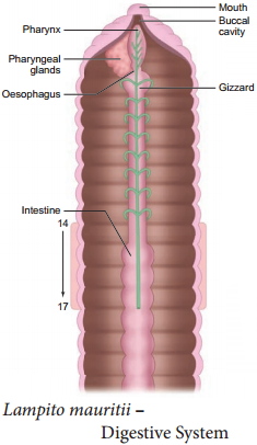
The mouth opens into the buccal cavity which occupies the 1st and 22nd segments. The buccal cavity leads
into a thick muscular pharynx, which occupies the 3rd and 4th segments and is surrounded by the pharyngeal glands. A small narrow tube, oesophagus lies in the 5th segment and continues into a muscular gizzard in the 6th segment.

The gizzard helps in the grinding of soil particles and decaying leaves. Intestine starts from the 7th segment and continues till the last segment. The dorsal wall of the intestine is folded into the cavity as the typhlosole. This fold contains blood vessels and increases the absorptive area of the intestine. The inner epithelium consists of columnar cells and glandular cells. The alimentary canal opens to the exterior through the anus.

The ingested organic rich soil passes through the digestive tract where digestive enzymes breakdown complex food into smaller absorbable units. The simpler molecules are absorbed through the intestinal membrane and are utilized.

The undigested particles along with earth are passed out through the anus, as worm castings or vermicasts. The pharyngeal or salivary gland cells and the glandular cells of the intestine are supposed to be the digestive glands which secrete digestive enzymes for digestion of food.

Respiratory System

The earthworm has no special respiratory organs like lungs or gills. Respiration takes place through the body wall. The outer surface of the skin is richly supplied with blood capillaries which aid in the diffusion of gases. Oxygen diffuses through the skin into the blood while carbon dioxide from the blood diffuses out. The skin is kept moist by mucous and coelomic fluid and facilitates exchange of gases.

Circulatory System

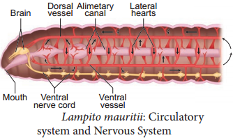
Lampito mauritii exhibits a closed type of blood vascular system consisting of blood vessels, capillaries and lateral hearts (Figure 4.4). Two median longitudinal vessels run above and below the alimentary canal as dorsal and ventral vessels of the earthworm. There are paired valves in the dorsal vessels which prevent the backward flow of the blood.

The ventral vessel has no valves and is non contractile, allowing the backward flow of blood. In the anterior part of the body the dorsal vessel is connected with the ventral vessel by eight pairs of commissural vessels or the lateral hearts lying in the 6th to 13th segments. These vessels run on either side of the alimentary canal and pump blood from the dorsal vessel to the ventral vessel.

The dorsal vessel receives blood from various organs in the body. The ventral vessel supplies blood to the various organs. Blood glands are present in the anterior segments of the earthworm. They produce blood cells and haemoglobin which is dissolved in the plasma and gives red colour to the blood.
Earthworm - Lampito mauritii img 6

Nervous System

The bilobed mass of nervous tissue called supra – pharyngeal ganglia, lies on the dorsal wall of the pharynx in the 3rd segment, is referred as the “brain”. The ganglion found below the pharynx in the 4th segment is called the sub-pharyngeal ganglion (Figure. 4.4). The brain and the sub – pharyngeal ganglia are connected by a pair of circum-pharyngeal connectives.
Earthworm - Lampito mauritii img 7

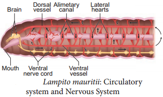
They run one on each side of the pharynx. Thus a nerve ring is formed around the anterior region of the alimentary canal. The double ventral nerve cord runs backward from the sub – pharyngeal ganglion. The brain along with other nerves in the ring integrates sensory inputs and command muscular responses of the body.

The earthworm’s receptors are stimulated by a group of slender columnar cells connected with nerves. The Photoreceptors (sense of light) are found on the dorsal surface of the body. Gustatory (sense of taste) and oldfactory receptors (sense of smell) are found in the buccal cavity. Tactile receptors (sense of touch), chemoreceptors (detect chemical changes) and thermoreceptors (changes in temperature) are present in
the prostomium and the body wall.

Excretory System

Excretion is the process of elimination of metabolic waste products from the body. In earthworm, excretion is effected by segmentally arranged, minute coiled, paired tubules called nephridia. There are three types of nephridia; (i) pharyngeal or tufted nephridia – present as paired tufts in the 5th – 9th segments (ii) Micronephridia or Integumentary nephridia – attached to the lining of the body wall from the 14th segment to the last which open on the body surface (iii) Meganephridia or septal nephridia – present as pair on both sides of intersegmental septa of the 19th segment to the last and open into intestine (Figure 4.5).
Earthworm - Lampito mauritii img 8

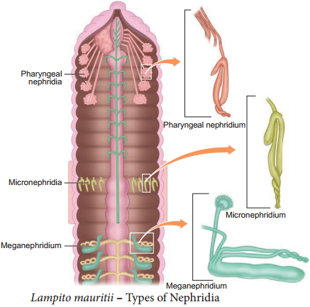
The meganephridium has an internal funnel like opening called the nephrostome, which is fully ciliated. The nephrostome is in the preceding segment and the rest of the tube is in the succeeding segment. This tube consists of three distinct divisions, the ciliated, the glandular and the muscular region.

The waste material collected through the ciliated funnel is pushed into the muscular part of nephridium by the ciliated region. The glandular part extracts the waste from the blood and finally the wastes exit out through the nephridiopore.

Besides nephridia, special cells on the coelomic wall of the intestine, called chloragogen cells are present. They extract the nitrogenous waste from the blood of the intestinal wall, into the body cavity to be sent out through the nephridia.

Reproductive System

Earthworms are hermaphrodites or monoecious i.e. male and female reproductive organs are found in the same individual (Figure 4.6). Self fertilization is avoided because two sex organs mature at different times, which means the sperm develops earlier than the production of ova (Protandrous). Thus cross fertilization takes place.
Earthworm - Lampito mauritii img 9

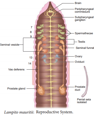
In the male reproductive system, two pairs of testes are present in the 10th and 11th segments. The testes
give rise to the germ cells or spermatogonia, which develops into spermatozoa in the two pairs of seminal vesicles. Two pairs of seminal funnels called ciliary rosettes are situated in the same segments as the testes.

The ciliated funnels of the same side are connected to a long tube called vas deferens. The vasa deferentia run upto the 18th segment where they open to the exterior through the male genital aperture. The male genital aperture contains two pairs of penial setae for copulation. A pair of prostate glands lies in the 18th – 19th segments. The secretion of the prostate gland serves to cement the spermatozoa into bundles known as spermatophores.

The female reproductive system consists of a pair of ovaries lying in the 13th segment. Each ovary has finger like projections which contain ova in linear series. Ovarian funnels are present beneath the ovaries which continue into the oviducts.

They open as a pair of genital apertures on the ventral side of the 14th segment. Spermathecae or seminal receptacles are three pairs lying in segments 7th, 8th and 9th, opening to the exterior on the ventral side between 6th & 7th, 7th & 8th and 8th & 9th segments. They receive spermatozoa from the partner and store during copulation.

A mutual exchange of sperms occurs between two worms during mating. One worm has to find another worm and they mate juxtaposing opposite gonadal openings, exchanging the sperms. Mature egg cells in the nutritive fluid are deposited in the cocoons produced by the gland cells of the clitellum which also collects the partner’s sperms from the spermthecae. Fertilization and development occurs within the cocoons, which are deposited in the soil. After about 2 – 3 weeks, each cocoon produces baby earthworms. Development is direct and no larva is formed during development.

Life Cycle

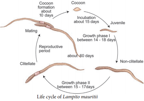
Lampito mauritii begins its life cycle, from the fertilized eggs. The eggs are held in a protective cocoon. These cocoons have an incubation period of about 14 – 18 days after which they hatch to release juveniles (Figure 4.7).

The juveniles undergo changes into non-clitellate forms in phase – I after about 15 days, which then develops a clitellum, called the clitellate at the end of the growth phase – II taking 15 – 17 days to complete. During the reproductive stage, earthworms copulate, and later shed their cocoons in the soil after about 10 days. The life cycle of Lampito mauritii takes about 60 days to complete.
Earthworm - Lampito mauritii img 10

Neural Tissue Definition and its Uses

Learninsta presents the core concepts of Biology with high-quality research papers and topical review articles.

Neural Tissue Definition and its Uses

Nervous tissue exerts the greatest control over the body’s responsiveness to changing conditions. Neurons, the unit of neural system are excitable cells (Figure 3.8).
Neural Tissue img 1

The neuroglial cells which constitute the rest of the neural system protect and support the neurons. Neuroglia makes up more than one-half of the volume of neural tissue in our body. When a neuron is suitably stimulated, an electrical disturbance is generated which swifty travels along its plasma membrane. Arrival of the disturbance at the neuron’s endings, or output zone, triggers events that may cause stimulation or inhibition of adjacent neurons and other cells (You will study in detail in Chapter 10)

Neurons, or nerves, transmit electrical impulses, while neuroglia do not; neuroglia have many other functions including supporting and protecting neurons.

Integration and communication are the two major functions of nervous tissue. Nervous tissue contains two categories of cells – neurons and neuroglia. Neurons are highly specialized nerve cells that generate and conduct nerve impulses.

Nervous tissue contains two major cell types, neurons and glial cells. Neurons are the cells responsible for communication through electrical signals.

Nervous tissue is made up of different types of neurons, all of which have an axon. Bundles of axons make up the nerves in the PNS and tracts in the CNS. Functions of the nervous system are sensory input, integration, control of muscles and glands, homeostasis, and mental activity.

The function of muscle tissue (smooth, skeletal, and cardiac) is to contract, while nervous tissue is responsible for communication.

Brain, spinal cord and nerves constitute nervous tissue. Tendon is a fibrous connective tissue connecting bones to muscles. Nervous tissue is absent in tendon. These are made up of collagen.

Neurons, also known as nerve cells, send and receive signals from your brain. While neurons have a lot in common with other types of cells, they’re structurally and functionally unique. Specialized projections called axons allow neurons to transmit electrical and chemical signals to other cells.

Although the nervous system is very complex, there are only two main types of cells in nerve tissue. The actual nerve cell is the neuron. It is the “conducting” cell that transmits impulses and the structural unit of the nervous system. The other type of cell is neuroglia, or glial, cell.

Muscle Tissue Function and Its Types

Learninsta presents the core concepts of Biology with high-quality research papers and topical review articles.

Muscle Tissue Function and Its Types

Each muscle is made of many long, cylindrical fires arranged in parallel arrays. These fibres are composed of numerous fine firils, called myofirils. Muscle fires contract (shorten) in response to stimulation, then relax (lengthen) and return to their uncontracted state in a coordinated fashion. In general muscles play an active role in all the movements of the body.

Muscles are of three types, skeletal, smooth and cardiac. Skeletal muscle tissue is closely attached to skeletal bones. In a typical muscle such as the biceps, the striated (striped) skeletal muscle fires are bundled together in a parallel fashion. A sheath of tough connective tissue encloses several bundles of muscle fires (You will learn more about this in Chapter 9).

The smooth muscle fires taper at both ends (fusiform) and do not show striations (Figure 3.7). Cell junctions hold them together and they are bundled together in a connective tissue sheath. The walls of internal organs such as the blood vessels, stomach and intestine contain this type of muscle tissue. Smooth muscles are ‘involuntary’ as their functions cannot be directly controlled. Unlike the smooth muscles, skeletal muscles can be controlled by merely thinking.

Cardiac muscle tissue is a contractile tissue present only in the heart. Cell junctions fuse the plasma membranes of cardiac muscle cells and make them stick together. Communication junctions (intercalated discs) at some fusion points allow the cells to contract as a unit, i.e., when one cell receives a signal to contract, its neighbours are also stimulated to contract.
Muscle Tissue img 1

An Overview Of Connective Tissue

Learninsta presents the core concepts of Biology with high-quality research papers and topical review articles.

An Overview Of Connective Tissue

Connective tissue develops from the mesoderm and is widely distributed in the body. There are three main classes namely Loose connective tissue, Dense connective tissue and Specialized connective tissue. Major functions of connective tissues are binding, support, protection, insulation and transportation.
An Overview Of Connective Tissue img 1

Components of Connective Tissue

All connective tissues consist of three main components namely fires, ground substance and cells. The ‘Fibres’ of connective tissue provide support. Three types of fires are found in the connective tissue matrix. They are collagen, elastic and reticular fires.

Connective tissues are of three types namely, Loose connective tissues (Areolar, Adipose and Reticular) and Dense connective tissues (dense regular, dense irregular and elastic) and Specialized connective tissues (cartilage, bone and blood).

Loose Connective Tissues

In this tissue the cells and fires are loosely arranged in a semi fluid ground substances. For example the Areolar connective tissue beneath the skin acts as a support framework for epithelium and acts as a reservoir of water and salts for the surrounding body tissues, hence apply called tissue fluid. It contains firoblasts, macrophages, and mast cells (Figure 3.5).
An Overview Of Connective Tissue img 2

Adipose tissue is similar to areolar tissue in structure and function and located beneath the skin. Adipocytes commonly called adipose or fat cells predominate and account for 90% of this tissue mass. The cells of this tissue store fats and the excess nutrients which are not utilised immediately are converted to fats and are stored in tissues. Adipose tissue is richly vascularised indicating its high metabolic activity. While fasting, these cells maintain life by producing and supplying energy as fuel.

Adipose tissues are also found in subcutaneous tissue, surrounding the kidneys, eyeball, heart, etc. Adipose tissue is called ‘white fat’ or white adipose tissue. The adipose tissue which contains abundant mitochondria is called ‘Brown fat’ or Brown adipose tissue. White fat stores nutrients whereas brown fat is used to heat the blood stream to warm the body. Brown fat produces heat by nonshivering thermogenesis in neonates.

Reticular connective tissue resembles areolar connective tissue, but, the matrix is filled with firoblasts called reticular cells. It forms an internal framework (stroma) that supports the blood cells (largely lymphocytes) in the lymph nodes, spleen and bone marrow. Dense connective tissues (connective tissue proper) Fibres and firoblasts are compactly packed in the dense connective tissues. Orientation of fires show a regular or irregular pattern and is called dense regular and dense irregular tissues.

Dense regular connective tissues primarily contain collagen fires in rows between many parallel bundles of tissues and a few elastic fires. The major cell type is firoblast. It attaches muscles and bones and withstands great tensile stress when pulling force is applied in one direction. This connective tissue is present in tendons, that attach skeletal muscles to bones and ligaments attach one bone to another. Dense irregular connective tissues have bundles of thick collagen fires and firoblasts which are arranged irregularly.

The major cell type is the firoblast. It is able to withstand tension exerted in many directions and provides structural strength. Some elastic fires are also present. It is found in the skin as the leathery dermis and forms firous capsules of organs such as kidneys, bones, cartilages, muscles, nerves and joints.

Elastic connective tissue contains high proportion of elastic fires. It allows recoil of tissues following stretching. It maintains the pulsatile flow of blood through the arteries and the passive recoil of lungs following inspiration. It is found in the walls of large arteries; ligaments associated with vertebral column and within the walls of the bronchial tubes.

Specialised connective tissues are classified as cartilage, bones and blood. The intercellular material of cartilage is solid and pliable and resists compression. Cells of this tissue (chondrocytes) are enclosed in small cavities within the matrix secreted by them (Figure 3.6). Most of the cartilages in vertebrate embryos are replaced by bones in adults. Cartilage is present in the tip of nose, outer ear joints, ear pinna, between adjacent bones of the vertebral column, limbs and hands in adults.
An Overview Of Connective Tissue img 3

Bones have a hard and non-pliable ground substance rich in calcium salts and collagen fires which gives strength to the bones. It is the main tissue that provides structural frame to the body. Bones support and protect softer tissues and organs.

The bone cells (osteocytes) are present in the spaces called lacunae. Limb bones, such as the long bones of the legs, serve weightbearing functions. They also interact with skeletal muscles attached to them to bring about movements.

The bone marrow in some bones is the site of production of blood cells. Blood is the fluid connective tissue containing plasma, red blood cells (RBC), white blood cells (WBC) and platelets. It functions as the transport medium for the cardiovascular system, carrying nutrients, wastes, respiratory gases throughout the body. You will learn more about blood in Chapter 7.

Organisation of Epithelial Tissue

Learninsta presents the core concepts of Biology with high-quality research papers and topical review articles.

Organisation of Epithelial Tissue

Epithelial tissue is a sheet of cells that covers the body surface or lines the body cavity. It occurs in the body as a covering, as a lining epithelium and as glandular, epithelium. The functions of epithelium includes protection, absorption, filtration, excretion, secretion and sensory reception.

Based on the structural modification of the cells, the epithelial tissues are classified into simple epithelium and compound epithelium or stratified epithelium. Simple epithelium is composed of a single layer of cells. They are found in the organs of absorption, secretion and filtration. Simple epithelial tissue is further classified into squamous epithelium, cuboidal epithelium, columnar epithelium, ciliated epithelium and pseudostratifid epithelium (Figure 3.2).

The squamous epithelium is made of a single thin layer of flattened cells with irregular boundaries. They are found in the kidney glomeruli, air sacs of lungs, lining of heart, blood vessels and lymphatic vessels and are involved in functions like forming a diffusion boundary and filtration in sites where protection is not important.

The cuboidal epithelium is made of a single layer of cube like cells. This tissue is commonly found in the kidney tubules, ducts and secretory portions of small glands and surface of the ovary. Its main functions are secretion and absorption.

The columnar epithelium is composed of single layer of tall cells with round to oval nuclei at the base. It lines the digestive tract from the stomach to the rectum. The two modifications of this lining are the presence of microvilli on the apical surface of the absorptive cells and Goblet cell which secretes the protective lubricating mucus. The functions of this epithelium include absorption, secretion of mucus, enzymes and other substances.

If the columnar cells bear cilia on their free surfaces they are called ciliated epithelium. This ciliated type propels mucus by ciliary actions and it lines the small bronchioles, fallopian tubes and uterus. Nonciliated type lines most of the digestive tract, gall bladder and secretory ducts of glands.

Pseudo-stratified epithelial cells are columnar, but unequal in size. Although the epithelium is single layered yet it appears to be multi-layered because the nuclei lie at different levels in different cells. Hence, it is also called pseudostratified epithelium and its functions are protection, secretion and absorption. Ciliated forms line the trachea and the upper respiratory tract. The non ciliated forms, line the epididymis, large ducts of a glands and tracts of male urethra.
Epithelial Tissue img 1

Glandular Epithelium

Some of the cuboidal or columnar cells get specialized for secretion and are called glandular epithelium (Figure 3.3). They are mainly of two types: unicellular, consisting of isolated glandular cells (goblet cells of the alimentary canal), and multicellular, consisting of cluster of cells (salivary gland). On the basis of the mode of pouring of their secretions, glands are divided into two categories namely exocrine and endocrine
glands. Exocrine glands secrete mucus, saliva, earwax, oil, milk, digestive enzymes and other cell products.

These products are released through ducts or tubes. In contrast endocrine glands do not have ducts. Their secretions called hormones are secreted directly into the fluid bathing the gland. The exocrine glands are classified as unicellular and multicelluar glands.

The multicelluar glands are further classified based on the structure as simple and compound glands, based on their secretory units as tubular, alveolar (Acinus) and tubulo alveolar. Based on the mode of secretion exocrine glands are classified as merocrine, holocrine and apocrine.
Epithelial Tissue img 2

Compound epithelium is made of more than one layer (multi-layered) of cells and thus has a limited role in secretion and absorption(Figure 3.4). The compound epithelia may be stratified and transitional. Their main function is to provide protection against chemical and mechanical stresses. They cover the dry surface of the skin, the moist surface of buccal cavity, pharynx, inner lining of ducts of salivary glands and of pancreatic ducts.

There are four types of compound epithelium namely, stratified squamous epithelium, cuboidal epithelium, columnar epithelium and transitional epithelium. Stratified squamous epithelium is of two types called keratinized type which forms the dry epidermis of the skin and the non keratinized type forms the moist lining of the oesophagus, mouth, conjunctiva of the eyes and vagina.

Stratified cuboidal epithelium mostly found in the ducts of sweat glands and mammary glands. Stratified columnar epithelium has limited distribution in the body, found around the lumen of the pharynx, male urethra and lining of some glandular ducts.

Transitional Epithelium is found lining the ureters, urinary bladder and part of the urethra. This epithelium allows stretching and is protective in function. All cells of the epithelium are held together with little intercellular material. In most of the animal tissues, specialized junctions provide both structural and functional links between its individual cells.

Three types of cell junctions are found in the epithelium and other tissues. These are called as tight, adhering and gap junctions. Tight junctions help to stop substances from leaking across a tissue. Adhering junctions perform cementing to keep neighbouring cells together. Gap junctions facilitate the cells to communicate with each other by connecting the cytoplasm of adjoining cells, for rapid transfer of ions, small molecules and sometimes big molecules.